I. INTRODUCTION
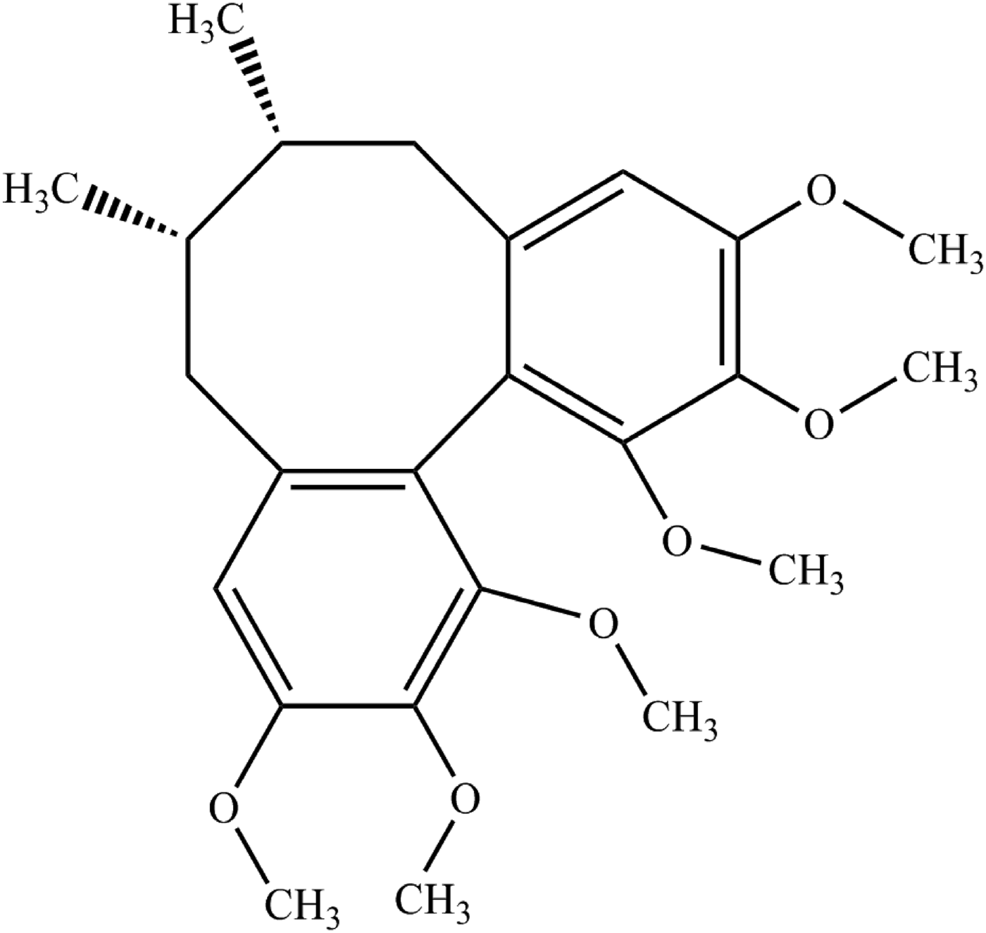
Deoxyschisandrin, systematic name (6R, 7S, 12R)-5,6,7,8-tetrahydro-1,2,3,10,11,12-hexamethoxy-6,7-dimethyl-dibenzo[a, c]cyclooctene, is an important lignan for anti-hepatotoxicity of Schisandra chinensis (Turcz.) Baill, which was described by Ikeya et al. (Reference Ikeya, Taguchi, Sasaki, Nakajima and Yosioka1980). Deoxyschisandrin (Figure 1) has strong biological activities and pharmacological properties, such as elevating the content of hepatic mitochondrial glutathione and protecting liver against CCl4 damage (Huang et al., Reference Huang, Shen and Shen2005; Chien et al., Reference Chien, Wu and Tsai2011), enhancing renal mitochondrial antioxidant status, promoting the generation of liver glycogen evidently.
Structural formula of deoxyschisandrin.

At present, the crystal structure of deoxyschisandrin has not been reported.
II. EXPERIMENTAL
A. Sample preparation
The title compound was extracted, isolated, purified from S. chinensis, and the natural sample was characterized by high-performance liquid chromatography (HPLC), as well as by UV, IR, and MS (positive electrospray, [M﹢H]+ = 403.2121 Da). Then, pure deoxyschisandrin was re-crystallized in petroleum ether–ethyl acetate (8:1, v/v).
B. Diffraction data collection and reduction
The diffraction pattern for the title compound was collected at room temperature using an X'Pert PRO diffractometer (PANalytical) with an X'celerator detector and CuKα 1 radiation (λ = 1.540 56 Å, generator setting: 40 kV, 30 mA). The diffraction data were collected over the angular range from 5 to 50° 2θ with a step size of 0.013 13° 2θ and a counting time of 10.16 ms/step. Data evaluation was performed using the software package Material Studio 4.2 (Accelrys Co., Ltd., USA).
Indexing was carried out using peak positions obtained from the powder diffraction profiles by the X-Cell method and then the indexing result was refined with the type of Pawley (Pan et al., Reference Pan, Guo, Duan, Cheng and Li2012). MC/SA search algorithm in Powder Solve package (Engel et al., Reference Engel, Wilke, König, Harris and Leusen1999) was used to constantly adjust the conformation, position, and orientation of the trial model in the unit cell of deoxyschisandrin, and then the results of Powder Solve were refined by Rietveld refinement techniques based on the experimental X-ray powder diffraction pattern. In the Rietveld refinement (Young, Reference Young1993), variables defining the structural model and the powder diffraction profile were adjusted by least-squares methods in order to obtain an optimal fit between the experimental and the calculated patterns. After Rietveld refinement, the final R wp of the structure was converged at 5.07%.
III. RESULTS
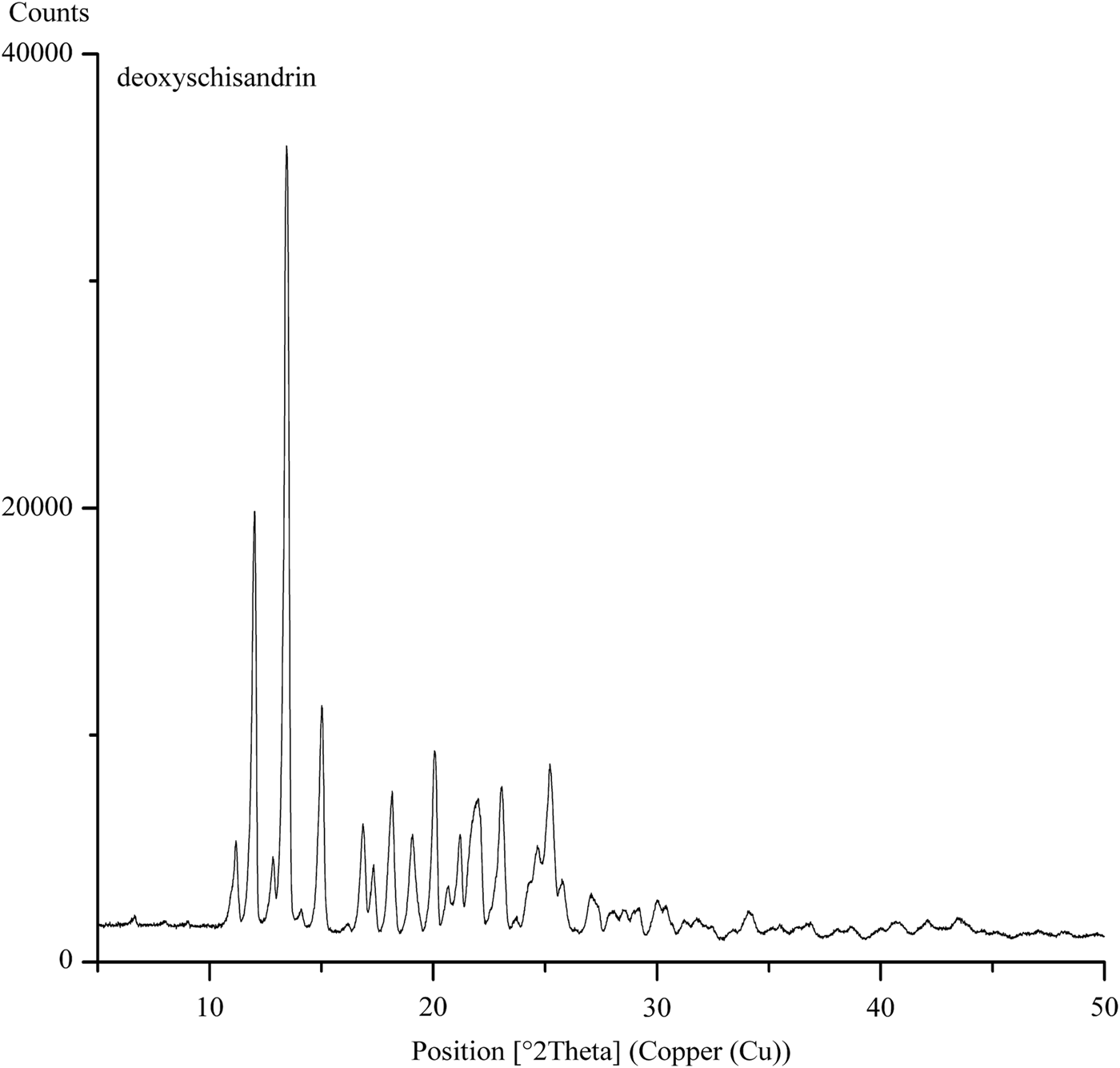
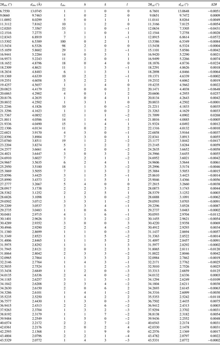
The experimental powder diffraction pattern is depicted in Figure 2. Indexing results show that deoxyschisandrin is monoclinical with space group P21 and unit-cell parameters: a = 13.083(3) Å, b = 19.563(9) Å, c = 8.805(6) Å, β = 90.472(0)°, unit-cell volume V = 2253.82 Å3, Z = 4, and space group P21 (Table I). After Rietveld refinement, the structure of deoxyschisandrin was solved. The final results were compared with single-crystal data which were collected on an Oxford Diffraction Xcalibur Nova system with MoKα radiation (λ = 0.710 73 Å) at room temperature and the θ from 3.05 to 28.77°, the deviations of the two methods were between 0.543 and 0.808%. All lines of powder and single-crystal data were indexed and are consistent with the P21 space group.
X-ray powder diffraction pattern of deoxyschisandrin, using CuKα 1 radiation (λ = 1.540 56 Å).
Indexed X-ray powder diffraction data of deoxyschisandrin, C24H32O6. Only the peaks with I rel of 1 or greater are presented [a = 13.083(3) Å, b = 19.563(9) Å, c = 8.805(6) Å, β = 90.472(0)°, unit-cell volume V = 2253.82 Å3, Z = 4, and space group P21]. All measured lines were indexed and are consistent with the P21 space group. The d-values were calculated using CuK α 1 radiation (λ = 1.540 56 Å).