The term myofibrillar myopathies (MFMs) encompasses a group of clinically heterogeneous but morphologically homogeneous muscle disorders defined by the presence of foci of myofibril dissolution, accumulation of the products of myofibrillar degradation and ectopic expression of multiple proteins (Refs Reference Nakano1, Reference De Bleecker, Engel and Ertl2, Reference Nakano3). These diseases were previously known as desmin-related myopathy or desmin-storage myopathy, because they all show desmin accumulation in muscle fibres (Refs Reference Goebel4, Reference Goebel and Fardeau5, Reference Goebel, Goldfarb and Karpati6, Reference Goldfarb7, Reference Olivé8, Reference Olivé9, Reference Ferrer10, Reference Selcen, Ohno and Engel11, Reference Goldfarb12). However, in addition to desmin, a wide variety of proteins have been found in excess or to be aberrantly expressed in muscle cells in MFMs (Refs Reference De Bleecker, Engel and Ertl2, Reference Nakano3, Reference Olivé9, Reference Ferrer10, Reference Selcen, Ohno and Engel11); furthermore, desmin mutations are the cause of a limited number of cases (Refs Reference Goldfarb12, Reference Muñoz-Mármol13, Reference Dalakas14), whereas mutations in several other genes have been identified in a number of patients (Refs Reference Vicart15, Reference Selcen and Engel16, Reference Selcen and Engel17, Reference Vorgerd18).
MFMs belong to a group of muscle conditions known as protein-surplus myopathies. The presence of protein aggregates in muscle fibres distinguishes MFMs from muscular dystrophies and other muscle disorders in which mutations in a particular gene result in an absence or reduction of the respective protein (Refs Reference Goebel and Fardeau5, Reference Goebel and Warlo19, Reference Goebel and Warlo20, Reference Goebel and Muller21). MFMs are considered the paradigm of conformational protein disorders affecting skeletal and cardiac muscles. Conformational protein disorders affecting the nervous system include Alzheimer disease, tauopathies, amyloidopathies, Parkinson disease and related synucleinopathies, Huntington disease, several autosomic dominant spinocerebellar atrophies due to expanded CAG triplets, and prion diseases (Refs Reference Carrell and Lomas22, Reference Sherman and Goldberg23, Reference Ellis and Pinheiro24, Reference Crowther25).
Genetics
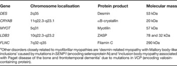
Although MFMs share common morphological features under optical and electron microscopy, the wide spectrum of clinical manifestations associated with the disease, and different patterns of inheritance observed in familial cases, predicted genetic heterogeneity (Ref. Reference Nakano1). This has been confirmed in recent years by the identification of causative mutations in several genes encoding proteins that are components of the Z-disc or that play major roles in the maintenance of sarcomere assembly. These include desmin (DES) (Refs Reference Goldfarb7, Reference Goldfarb12, Reference Muñoz-Mármol13, Reference Dalakas14, Reference Dagvadorj26, Reference Olivé27, Reference Goldfarb and Laing28), αB-crystallin (CRYAB) (Refs Reference Vicart15, Reference Selcen and Engel29), myotilin (MYOT, TTID) (Refs Reference Selcen and Engel16, Reference Olivé30), ZASP (LDB3) (Ref. Reference Selcen and Engel17) and filamin C (FLNC) (Ref. Reference Vorgerd18) (Table 1). In addition, mutations in selenoprotein N (SEPN1), an enzyme involved in cell defence against oxidative stress, are the cause of desmin-related myopathy with Mallory body-like inclusions (Ref. Reference Ferreiro31). The relative frequency of mutations in the causative genes is currently unknown. In a series of patients with MFMs studied in the Mayo clinics, 6% showed mutations in DES, 3% had a mutation in CRYAB, 10% in MYOT, 15% in LDB3, and 3% in FLNC (see myofibrillar myopathy entry at http://www.genereviews.org/). Further genetic heterogeneity is suspected since the causative gene remains to be discovered in the majority of patients suffering from MFM (Ref. Reference Dalakas14).
Molecular genetics of myofibrillar myopathies: causative genesa

a Other disorders closely related to myofibrillar myopathies are ‘desmin-related myopathy with Mallory body-like inclusions’ caused by mutations in SENP1 (encoding selenoprotein N) and ‘inclusion body myopathy associated with Paget disease of the bone and frontotemporal dementia’ due to mutations in VCP (encoding valosin-containing protein).
An additional disorder – IBMPFD (‘inclusion body myopathy associated with Paget disease of the bone and frontotemporal dementia’) – which is caused by mutations in the valosin-containing protein (VCP) gene (Ref. Reference Watts32) has several links with MFM since protein aggregates containing VCP, ubiquitin, desmin and αB-crystallin have been identified in affected muscles in IBMPFD. Furthermore, granulofilamentous material, which represents the ultrastructural hallmark of desminopathies, has been found in skeletal muscle in IBMPFD (Ref. Reference Hübbers33). VCP has been implicated in multiple essential cell pathways, including the cell cycle, membrane fusion, and ubiquitin-dependent protein degradation.
This review focuses on MFMs caused by mutations in desmin, αB-crystallin, myotilin, ZASP and filamin C genes. A shematic representation of the main Z-disc proteins, including those mutated in different subgroups of MFMs, and their relations with other components of the sarcomere and sarcolemma is shown in Figure 1.
Schematic drawing of components of the Z-disc. The Z-disc forms the borders of individual sarcomeres (see boxed diagram). In addition to the well-known thin filaments (F-actin) and thick filaments (myosin) of the sarcomere, two other filament complexes are present at the Z-disc, made up of the giant proteins titin and nebulin. The Z-discs largely consist of α-actinin homodimers organised in an antiparallel fashion and providing a backbone for the insertions of actin filaments, as well as titin and nebulin. Proteins whose genes are mutated in myofibrillar myopathies are shown in red. Desmin filaments surround the Z-disc; they interconect the filaments to each other, and link the myofibrils to the sarcolemma and nuclear membrane. αB-crystallin binds to desmin. Myotilin interacts with several Z-disc proteins, including α-actinin, filamin C, F-actin, and FATZ. Filamin C binds to actin filaments, and interacts with several proteins, including γ and δ sarcoglycans (not shown), myotilin, FATZ and β1 integrin. ZASP binds directly to α-actinin. For further details, see Ref. Reference Frank145. Abbreviations: FATZ, ‘filamin-, actinin-, and telethonin-binding protein of the Z-disc’; MLP, muscle LIM protein; ZASP, ‘Z-band alternatively spliced PDZ-motif-containing protein’.

Z-disc proteins mutated in MFMs
Desmin
Desmin is the muscle-specific member of the intermediate filament family of cytoskeletal proteins. In mature striated muscle, desmin filaments surround the Z-discs, interconnect them to each other, and link the entire contractile apparatus to the sarcolemma, cytoplasmic organelles and the nuclear membrane, probably through plectin (Refs Reference Lazarides34, Reference Price35, Reference Hermannn and Aebi36). In cardiac muscle, desmin is increased at intercalated discs and is the major component of Purkinje fibres. Desmin is encoded by a single-copy gene (DES) that encompasses nine exons and codes for 476 amino acids (Ref. Reference Li37). Desmin comprises three domains: the N- and C-terminal (head and tail) structures and the central α-helical core. The central core is composed in turn of four consecutive helical segments: 1A, 1B, 2A and 2B (Refs Reference Weber and Geisler38, Reference Brown, Cohen and Parry39, Reference Herrmann40, Reference Strelkov41). Mice lacking desmin have severe cardiovascular lesions and skeletal myopathy (Refs Reference Li42, Reference Milner43, Reference Tornell44).
αB-crystallin
αB-crystallin is a chaperone that binds to unfolded proteins under stress conditions to prevent aberrant secondary structures and aggregation. In skeletal muscle, αB-crystallin binds to desmin and actin. αB-crystallin is encoded by the gene CRYAB; it encompasses three exons and codes for 175 amino acids (Refs Reference Brakenhoff45, Reference Golenhofen46, Reference Horwitz47).
Myotilin
Myotilin is expressed exclusively in skeletal and cardiac muscle and is a component of sarcomeric Z-disc (Ref. Reference Salmikangas48); it interacts with several other Z-disc proteins including α-actinin, filamin C, F-actin and FATZ (filamin-, α-actinin-, and telethonin-binding protein of the Z-disc) (Refs Reference Salmikangas48, Reference van der Ven49, Reference Salmikangas50, Reference von Nandelstadh51, Reference Gontier52). The myotilin gene (MYOT) encompasses ten exons and codes for 498 amino acids (Ref. Reference Salmikangas48). Mice lacking myotilin have no apparent morphological skeletal or cardiac muscle disorders, and they develop normally. These observations suggest either that myotilin does not participate in muscle development and basal function maintenance or that other proteins serve as structural and functional compensatory molecules when myotilin is absent (Ref. Reference Moza53).
ZASP
ZASP (Z-band alternatively spliced PDZ-motif-containing protein) is a sarcomeric protein that plays a major role in maintaining Z-disc stability in striated and cardiac muscle. ZASP directly binds to the C-terminal region of α-actinin. The ZASP gene (LDB3) encompasses 18 exons, which are differentially spliced to form several isoforms: exon 4 is expressed without exon 6 in three cardiac isoforms, and exon 6 is expressed without exon 4 in skeletal muscle isoforms (Refs Reference Faulkner54, Reference Au55, Reference Huang56). Zebrafish lacking cypher/ZASP show severe developmental defects, including thinning of the cardiac ventricular wall (Ref. Reference Van der Meer57). Drosophila larvae lacking ZASP do not form Z-discs and do not recruit α-actinin to the Z-disc (Ref. Reference Jani and Schöck58). Targeted deletion of ZASP in mouse causes skeletal and cardiac myopathy and premature death from failure of multiple striated muscles (Ref. Reference Zhou59). The yeast two-hybrid assay and pull-down assay demonstrated that the Asp626Asn mutation of Cypher/ZASP, which causes dilated cardiomyopathy in Japanese patients, increased the affinity of the LIM domain for protein kinase C, thus suggesting a novel mechanism for impaired cardiac muscle function (Ref. Reference Arimura60).
Filamin C
Filamin C is a muscle-specific member of the filamin family of proteins (Refs Reference Himmel61, Reference van der Flier and Sonnenberg62). Filamins have an actin-binding domain at their N-terminus, and a C-terminal Ig-like domain that enables the protein to dimerise. In addition to its role in organising actin filaments, filamin C interacts with several other proteins, including γ and δ sarcoglycans, myotilin, calsarcin and β1 integrin. These interactions suggest that filamin C plays an important role during myofibrillogenesis (Refs Reference Thompson63, Reference van der Ven64, Reference Takada65, Reference Stossel66). The filamin C gene (FLNC) encompasses 46 exons and codes for 2705 amino acids (Refs Reference Xie67, Reference van der Ven68). Filamin-C-deficient mice have defects in primary myogenesis resulting in reduced myotubes and reduced muscle fibres; as a result, animals die shortly after birth (Ref. Reference Dalkilic69).
Clinical aspects
Clinical phenotypes associated with MFMs are widely heterogeneous. The disease may present in infancy or late in adulthood. Distal weakness is seen at the disease onset in most of the patients, but some present with proximal muscle weakness. Cardiopathy, peripheral neuropathy, and respiratory insufficiency may be present or absent. A summary of MFMs is presented in Table 2.
Summary of myofibrillar myopathies, with causative genes and main phenotypes

aNumber of identified mutations.
Abbreviations: AD, autosomal dominant; AR, autosomal recessive; CRYAB, αB-crystallin; DES, desmin; FLNC, filamin C; LDB3, LIM-domain-binding 3 [gene encoding ZASP; LIM is named from the first three proteins identified with this domain (LIN-11, Is11 AND MEC-3)]; LGMD1A, limb-girdle muscular dystrophy type 1A; MYOT, myotilin.
Desminopathies
MFMs caused by desmin mutations are disabling conditions that most often present with distal muscle weakness, often associated with a cardiopathy, in young adults (Refs Reference Goldfarb7, Reference Goldfarb12, Reference Muñoz-Mármol13, Reference Dalakas14, Reference Dagvadorj26, Reference Olivé27, Reference Goldfarb and Laing28, Reference Sjoberg70, Reference Li71, Reference Sjoberg72, Reference Park73, Reference Dagvadorj74, Reference Fidzianska75, Reference Vrabie76, Reference Bar77, Reference Dalakas78). Age at onset usually varies between 15 and 40 years. Autosomal dominant inheritance occurs in the majority of cases, but de novo mutations are also frequently found. By contrast, autosomal recessive mutations have been reported in only a few cases (Refs Reference Goldfarb12, Reference Muñoz-Mármol13).
Muscular involvement may be manifested as a distal myopathy with later involvement of the proximal muscles, or as limb-girdle myopathy with distal progression. In some families the disease presents as a scapuloperoneal syndrome (Ref. Reference Walter79). Respiratory impairment is frequently observed during the course of the illness. Involvement of bulbar and facial muscles is a late complication in some patients, or may even be present at the disease onset (Refs Reference Olivé27, Reference Vrabie76). Cardiomyopathy may be manifested as hypertrophic, dilated or restrictive. In addition, atrioventricular conduction abnormalities with recurrent episodes of syncopes and arrhythmias are frequently seen in desminopathies.
The variable clinical spectrum of desminopathies depends on several factors, including the position of the mutation in the desmin gene. Patients with mutations in the 2B segment of DES tend to show primarily skeletal muscle pathology, whereas patients with mutations in the 1B segment and tail region show predominant cardiopathy (Ref. Reference Goldfarb and Laing28). The prognosis is usually poor; pacemaker requirement and/or cardiac transplantation are indicated in cases with severe cardiopathy.
αB-crystallinopathies
The first reported family with crystallinopathy presented with proximal and distal myopathy, involvement of the velopharyngeal muscles, respiratory failure, cardiopathy and cataracts, with age of onset in the 30s. The disease was transmitted with an autosomal dominant inheritance (Refs Reference Vicart15, Reference Fardeau80, Reference Fardeau81). Two further cases were later identified: one presented with cervical and limb-girdle weakness and respiratory failure; the other had proximal and distal limb weakness. Both patients had peripheral neuropathy; cardiac involvement and lens opacities were absent (Ref. Reference Selcen and Engel29). Another patient, bearing a different mutation in CRYAB, suffered from dilated cardiomyopathy without apparent skeletal muscle involvement (Ref. Reference Inagaki, Hayashi and Arimura82).
Myotilinopathies
The first MYOT-associated disease mutations were identified in two families suffering from limb-girdle muscular dystrophy type 1A (LGMD1A) (Refs Reference Hauser83, Reference Hauser84). Affected individuals presented between the ages of 18 and 58 years with proximal weakness in the lower extremities, later spreading to involve distal muscles. In some cases, nasal speech was a prominent symptom. The pattern of inheritance was autosomal dominant (Refs Reference Hauser83, Reference Hauser84).
More recently, MYOT-associated disease mutations were identified in a subset of patients with familial or sporadic MFM (Refs Reference Selcen and Engel16, Reference Olivé30). The pattern of inheritance was autosomal dominant in some cases but other patients did not have a family history of disease. The age of disease onset varied from 42 to 77 years (Ref. Reference Olivé30). Distal muscle weakness of the lower limbs was the commonest presentation but other cases presented with proximal weakness. In the majority of patients, foot drop due to anterior tibialis weakness was the first symptom of the disease. However, results of muscle computed tomography studies indicate that the disease actually started in the posterior leg muscles (Ref. Reference Olivé30). In the upper extremities, wrist and finger extensors and deltoids were the most affected muscles. Peripheral neuropathy and cardiomyopathy was frequently observed in some cases (Ref. Reference Selcen and Engel16), but not in others (Ref. Reference Olivé30). A MYOT mutation has been shown to be the cause of spheroid body myopathy in a large family presenting at a mean age of 40 with proximal muscle weakness (Ref. Reference Foroud85). A few other patients with myotilin mutations have been identified (Refs Reference Fischer86, Reference Berciano87).
Considering all these data, late-onset distal or mixed distal and proximal myopathy is the most frequent clinical phenotype associated with myotilin mutations, whereas peripheral neuropathy, cardiomyopathy and respiratory insufficiency are observed only occasionally.
ZASPopathies
Mutations in LDB3 were identified after screening a series of 54 cases with MFM (Ref. Reference Selcen and Engel17). Three heterozygous mutations were detected in 11 cases; two were found in exon 6, whereas another was found in exon 9. Dominant inheritance occurred in seven patients. The age at onset was between 44 and 73 years. Furthermore, the Ala165Val ZASP mutation has been recently identified as the cause of autosomal dominant distal myopathy described by Markesbery et al. Haplotype studies in this family and in five other unrelated families of European ancestry carrying the identical mutation revealed common markers at the locus, suggesting the existence of a founder mutation (Ref. Reference Griggs88). In addition, mutations in exon 4 and exon 6 of the ZASP gene have been identified in patients suffering from dilated cardiomyopathy (Ref. Reference Arimura60).
Patients with zaspopathy present after the age of 40 with distal leg weakness, with the disorder slowly progressing to involve intrinsic hand muscles and mild proximal weakness. As for myotilinopathies, muscle imaging studies indicate early involvement of the posterior calf muscles. Cardiopathy and peripheral neuropathy are additional features in some patients.
Filaminopathy
Members of a large German pedigree suffering from limb-girdle myopathy were shown to carry a heterozygous nonsense mutation in the filamin C gene (Ref. Reference Vorgerd18). Four additional families carrying the same mutation were later identified. Haplotype analyses strongly indicate a founder mutation. The mean age at onset of clinical symptoms was 44 years (with a range of 24 to 57 years). Muscular involvement was slowly progressive, following a pattern of LGMD. Patients frequently developed respiratory muscle weakness, and about a third suffered from cardiac abnormalities comprising conduction blocks, tachycardia, diastolic dysfunction and left ventricular hypertrophy (Ref. Reference Kley89).
Pathology: optic and electron microscopy
Myofibrillar inclusions containing protein aggregates are the morphological hallmark of MFMs. These are observed on routine histochemical studies as abnormal fibre regions containing amorphous, granular or hyaline inclusions best visualised on trichrome-stained sections as dark-blue or purple inclusions. Additional pathological features are the presence of rimmed and unrimmed vacuoles filled with membranous material, and areas devoid of oxidative enzyme activity.
Myotilinopathy
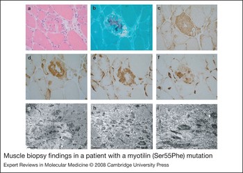
Muscle biopsies in myotilinopathies show variable myopathic changes including variation in fibre size, fibre splitting, and internal nuclei, as well as variable degrees of fatty and fibrous tissue replacement. Typically, muscle fibres contain nonhyaline and dense hyaline deposits of various shape and size. These structures are best visualised with modified trichrome stain as dark blue or red-blue inclusions and are eosinophilic on haematoxylin and eosin staining. Small cytoplasmic bodies and nemaline-like bodies often clustered in groups are frequently observed. The hyaline inclusions show strong congophilia (Refs Reference Selcen and Engel16, Reference Olivé30). Spheroid bodies may be present. Rimmed and nonrimmed vacuoles are found in the majority of cases. Oxidative enzyme and ATPase activities are absent in the fibre areas containing the inclusions (Refs Reference Selcen and Engel16, Reference Olivé30). Immunohistochemical studies reveal accumulation of myotilin, desmin, dystrophin and many other proteins in muscle fibres containing cytoplasmic inclusions (Refs Reference Selcen and Engel16, Reference Olivé30, Reference Fischer86). Under electron microscopy, streaming of the Z-line is a prominent feature of early lesions in myotilinopathy. More-damaged fibres show major myofibrillar abnormalities with disruption of the sarcomeres, which are replaced by remnants of myofibrils, and electron-dense material. Mitochondrial abnormalities are usually observed at the periphery of disrupted sarcomeres. Nuclear and sarcoplasmic filamentous structures, such as those described in inclusion body myositis, can be found in myotilinopathies (Ref. Reference Fischer86). Autophagic vacuoles filled with polymorphous and dense osmiophilic material, myelin-like inclusions and membranous structures are common in myotilinopathy (Refs Reference Selcen and Engel16, Reference Olivé30). Characteristic myopathology in myotilinopathies is shown in Figure 2.
Muscle biopsy findings in a patient with a myotilin (Ser55Phe) mutation. Serial cryostat consecutive sections stained with haematoxylin and eosin (a) and modified trichrome stain (b) showing polymorphous hyaline inclusions and nonrimmed vacuoles. The abnormal fibres display strong desmin (c), αB-crystallin (d), myotilin (e) and ubiquitin immunoreactivity (f). Under electron microscopy, areas of sarcomeric disorganisation are replaced by remnants of electron-dense granular and filamentous material (g–i). These findings have been previously reported (see Ref. 30). Magnifications: a–h, ×400. Bars: g, 2 µm; h, 0.5 µm; i, 0.2 µm.

Desminopathy
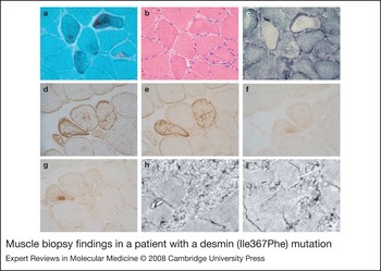
Muscle biopsies of affected muscles in desminopathy show nonspecific variation in the fibre size, small atrophic fibres, increased numbers of internal nuclei, and variable numbers of rimmed vacuoles. The most characteristic feature is the presence of amorphous eosinophilic regions that stain dark green or bluish with modified trichrome and show strong immunoreactivity for desmin, αB-crystallin and many other proteins. These areas are typically devoid of oxidative enzymes and ATPase activities. Necrosis and inflammatory infiltrates are rare. Electron microscopy reveals accumulation of granulofilamentous material under the sarcolemma or among the myofibrils, and small aggregates of electron-dense material between the myofibrils. Areas in the vicinity of these aggregates show variable streaming of the Z-line. In addition, disorganisation of the myofibrillar network is evident in some cases. As in myotilinopathies, autophagic vacuoles with polymorphous and myelin-like debris, and accumulation of mitochondria, may be present (Refs Reference Goebel4, Reference Olivé8, Reference Dalakas14, Reference Park73, Reference Fidzianska75, Reference Vrabie76, Reference Bar77, Reference Schröder90). Characteristic myopathology in desminopathies is shown in Figure 3.
Muscle biopsy findings in a patient with a desmin (Ile367Phe) mutation. Cryostat consecutive sections stained with modified trichrome stain (a), haematoxylin and eosin (b) and NADH (c), showing several fibres containing bluish, amorphous cytoplasmic inclusions devoid of oxidative enzyme activity. Abnormal fibre regions display strong desmin (d), αB-crystallin (e), and faint myotilin (f) and ubiquitin (g) immunoreactivity. Under electron microscopy, a matrix of granulofilamentous material is observed among the myofibrils (h and i). These findings have been previously reported (see Ref. 27). Magnifications: a–g, ×400. Bars h and I, 1 µm.

αB-crystallinopathy
Optical and electron microscopy findings in muscle biopsies in αB-crystallinopathy were indistinguishable from desminopathy; for example, desmin and αB-crystallin-immunoreactive inclusions and granulofilamentous material are present in both disorders (Refs Reference Vicart15, Reference Selcen and Engel29, Reference Fardeau81). However, immunohistochemical studies in two cases have shown an apparent lack of ubiquitin, gelsolin and α1-antichymotrypsin in αB-crystallinopathy (Ref. Reference Selcen and Engel29).
ZASPopathies
Muscle biopsies of affected muscles in cases associated with mutations in the ZASP gene show similar abnormalities to those seen in myotilinopathies (Ref. Reference Selcen and Engel17). Interestingly, only mild ZASP accumulation is observed (Ref. Reference Griggs88).
Filaminopathy
The histological findings in patients with MFM-associated mutation in the filamin C gene studied at different stages of the disease vary from mild myopathic changes to distinct muscular-dystrophy-like features. Rimmed and nonrimmed vacuoles are observed in a few cases. Some fibres contain pleomorphic hyaline, granular and amorphous deposits, ranging from red-blue to dark blue in trichrome stain. Most of them display congophilia and show strong filamin C, desmin, myotilin, αB-crystallin, dystrophin, Xin and sarcoglycan immunoreactivity. Core-like features are additional frequent findings (Refs Reference Vorgerd18, Reference Kley89).
At the ultrastructural level, major myofibrillar abnormalities including abnormal myofibrillar alignment, rod-body formation, and accumulation of granulofilamentous material similar to that seen in desminopathies are observed. In addition, sarcoplasmic paired helical filaments have been found (Refs Reference Vorgerd18, Reference Kley89).
Molecular pathogenesis
Protein aggregates
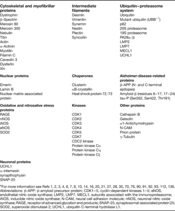
A major morphological hallmark of MFMs is the presence of cytoplasmic accumulation of proteins forming insoluble aggregates in muscle cells. In addition to disease-specific mutant proteins (i.e. desmin in desminopathies, αB-crystallin in αB-crystallinopathies, myotilin in myotilinopathies), immunohistochemical studies have shown that protein aggregates in MFMs contain a wide variety of proteins (Box 1) (Refs Reference Nakano1, Reference De Bleecker, Engel and Ertl2, Reference Nakano3, Reference Goebel4, Reference Goebel, Goldfarb and Karpati6, Reference Goldfarb7, Reference Olivé8, Reference Ferrer10, Reference Dalakas14, Reference Selcen and Engel16, Reference Goebel and Warlo20, Reference Goebel and Muller21, Reference Olivé27, Reference Goldfarb and Laing28, Reference Olivé30, Reference Sjoberg70, Reference Vrabie76, Reference Schröder90, Reference Bar91, Reference Howman92, Reference Olivé93; see also Refs Reference Ferrer112, Reference Janué, Olivé and Ferrer136). The mechanisms leading to protein aggregation are not fully understood, but most mutant proteins are prone to aggregation, as revealed by studies in transgenic mice and in transfected cells (Ref. Reference Sherman and Goldberg23).
Proteins that accumulate in myofibrillar myopathiesa

aFor more information see Refs Reference Nakano1, Reference De Bleecker, Engel and Ertl2, Reference Nakano3, Reference Goebel4, Reference Goebel, Goldfarb and Karpati6, Reference Goldfarb7, Reference Olivé8, Reference Ferrer10, Reference Dalakas14, Reference Selcen and Engel16, Reference Goebel and Warlo20, Reference Goebel and Muller21, Reference Olivé27, Reference Goldfarb and Laing28, Reference Olivé30, Reference Sjoberg70, Reference Vrabie76, Reference Schröder90, Reference Bar91, Reference Howman92, Reference Olivé93, Reference Ferrer112, Reference Janué, Olivé and Ferrer136.
Abbreviations: β-APP: β-amyloid precursor protein; CDK1–5, cyclin-dependent kinases 1–5; eNOS, endothelial nitric oxide synthase; LMP2, LMP7, MECL1, subunits associated with the immunoproteasome; iNOS, inducible nitric oxide synthase; N-CAM, neural cell adhesion molecule; nNOS, neuronal nitric oxide synthase; RAGE, receptor of advanced glycation end products; SNAP-25, synaptosomal-associated protein 25; SOD2, superoxide dismutase 2; UCHL1, ubiquitin C-terminal hydrolase L1.
Desmin transgenic mice expressing a deletion of seven amino acids (Arg173 to Glu179) show disruption of the cytoskeleton and accumulation of desmin and other proteins in the myocardium (Ref. Reference Wang94). Truncated desmin induces desmin–vimentin–cytokeratin coprecipitation, involution of intermediate filament networks, and nuclear fragmentation (Ref. Reference Yu95), and aberrant desmin attracts other proteins and produces high molecular weight insoluble aggregates (Ref. Reference Yu95). Cells transfected with mutant desmin may show variable disorganisation of filament assembly and abnormal protein deposits (Refs Reference Park73, Reference Schröder90, Reference Bar96, Reference Kaminska97, Reference Bar98).
Transgenic mice expressing mutant myotilin (Thr57Ile) show myofibrillar aggregation, streaming of the Z-line, and vacuolisation. Mutant myotilin protein localises in the Z-disc and in the aggregates, together with other proteins associated with the Z-disc (Ref. Reference Garvey99). Overexpression of wild-type myotilin in LGMD1A mutant transgenic mice results in more-severe muscle degeneration, enhanced myofibrillar aggregation, and earlier onset of aggregation when compared with single-transgenic mutant mice (Ref. Reference Garvey100). However, expression of myotilin missense mutations Ser55Phe, Ser60Phe and Ser60Cys in transfected cells does not lead to gross changes in the total amount of myotilin or to aberrant post-translational modifications. These forms of mutated myotilin do not disrupt the endogenous desmin cytoskeleton or lead to pathological protein aggregation in transfected cells (Ref. Reference Fischer86).
Transgenic mice expressing mutant αB-crystallin show abnormal αB-crystallin and desmin aggregates in cardiomyocytes (Ref. Reference Wang101). Muscle cells transfected with mutant αB-crystallin also exhibit abnormal aggregates containing both proteins (Ref. Reference Vicart15).
The MFM-associated mutation Trp2710X in the filamin C gene produces a mutated filamin C polypeptide in which the mutant dimerisation domain is less stable and more susceptible to proteolysis. As a result, it does not dimerise properly and forms aggregates in vitro (Ref. Reference Löwe102).
Neural proteins in aggregates; role of neuron-restrictive silencer factor
Neural proteins are not present in significant amounts in normal muscles but they are components of protein aggregates in MFMs, particularly in myotilinopathies. mRNA and protein levels of certain neural proteins such as synaptophysin, SNAP25 (‘synaptosome-associated protein of 25 kDa’), α-internexin and UCHL1 (‘ubiquitin C-terminal hydrolase L1’) are increased in myotilinopathies (Ref. Reference Barrachina103). The reasons for this phenomenon are not clear but recent studies have shown that abnormal expression of certain transcription factors may be the cause.
NRSF/REST is a neuronal gene repressor in non-neuronal cells (Ref. Reference Schoenherr and Anderson104). Several studies have shown that synaptophysin and SNAP25 are NRSF/REST target genes (Ref. Reference Bruce105). UCHL1 and α-internexin are also regulated by REST (Ref. Reference Barrachina103). Interestingly, mRNA and protein NRSF/REST levels are reduced in myotilinopathy muscles when compared with control muscles, and this is accompanied by increased aberrant expression levels of several neuronal proteins such as UCHL1, SNAP25, synaptophysin and α-internexin (Ref. Reference Barrachina103).
The proteasome and immunoproteasome in MFMs
The ubiquitin–proteasomal pathway plays a crucial role in nonlysosomal protein degradation (Ref. Reference Glickman and Ciechanover106). Misfolded proteins or unassembled subunits of larger protein complexes and retrotranslocated proteins from the endoplasmic reticulum are subjected to proteasomal degradation (Ref. Reference Kostova and Wolf107). The ubiquitin–proteasome pathway is initiated by activation of ubiquitin by ubiquitin-activation enzyme E1; then active ubiquitin is transferred to a substrate-dependent ubiquitin-conjugating enzyme, E2. Finally, ubiquitin is transferred to a member of the ubiquitin-ligase family, E3. This is followed by polyubiquitination of the substrate, produced via a series of isopeptide linkages between a lysine residue of the attached ubiquitin and the C-terminal glycine of the next ubiquitin molecule (Ref. Reference Glickman and Ciechanover106).
The 20S proteasome is organised into a structure resembling a hollow cylinder composed of four stacked rings, each composed of seven subunits. In the presence of ATP, the 20S proteasome can associate with two caps or 19S complexes, thus forming the 26S proteasome complex. The 20S proteasome has three main peptidase activities: chymotrypsin-like, trypsin-like, and peptidylglutamyl-peptide-hydrolysing activities (Ref. Reference Glickman and Ciechanover106). The 20S proteasome can also bind to the PA28α/β activator (11S regulator) to form the PA28–proteasome complex, which appears to increase antigen processing and presentation. In the presence of certain stimuli, the β subunits of the 20S proteasome are replaced by homologous proteins called LMP2, LMP7 and MECL1, thus forming the immunoproteasome. Hybrid proteasomes formed by 19S, 20S and PA28 are involved in the immune response (Refs Reference Glickman and Ciechanover106, Reference Sijts, Zaiss and Kloetzel108).
Several proteasome subunits are abnormally expressed in MFMs, as revealed by immunohistochemistry and western blotting. Subunits of 20S, 19S and PA28 colocalise with ubiquitin and other proteins, including desmin, αB-crystallin, gelsolin and phosphorylated tau in abnormal protein aggregates in myotilinopathies (Ref. Reference Ferrer10). Moreover, fibres with protein aggregates express LMP2, LMP7 and MECL1 (Ref. Reference Ferrer10). However, this is accompanied by preserved or even increased proteolytic activity in vitro in myotilinopathies (Ref. Reference Ferrer10). Studies in heart muscle from a transgenic mouse model of desminopathy have also demonstrated overexpression of the 20S subunit and downregulation of the 19S subunit of the proteasome together with increased peptidase activity (Ref. Reference Liu109). Together, these observations strongly suggest that abnormalities of the ubiquitin–proteasome system in MFMs are not the result of defective peptidase activity per se, but rather arise from a failure in the entry of ubiquitinated proteins into the 20S proteasome.
Clusterin in MFMs
Impaired protein proteasomal degradation probably plays a role in protein accumulation in MFMs (Ref. Reference Ferrer10). However, abnormal proteasomal expression and activity do not completely explain why several proteins cluster and aggregate into abnormal protein deposits in MFMs. As shown in transgenic mice and transfected cell lines, mutation of target proteins facilitates protein aggregation (Refs Reference Vicart15, Reference Sjoberg70, Reference Bar91, Reference Wang94, Reference Bar96, Reference Bar98, Reference Garvey99, Reference Wang101, Reference Löwe102). Yet additional complementary proteins may be involved in the process.
One important complementary protein is clusterin (apolipoprotein J, SP40), a sialoglycoprotein with a nearly ubiquitous tissue distribution. Although its functions are not well known, clusterin has been considered as a type of molecular chaperone. Clusterin is assembled as a heterodimeric molecule formed by the antiparallel alignment of α- and β-chains, in which the cysteine-rich centres, linked by five disulphide bridges, are flanked by two predicted coiled α-helices and three predicted amphipathic β-helices (Ref. Reference Jones and Jomary110). Clusterin is associated with abnormal protein accumulation in conformational protein disorders affecting the central nervous system including Alzheimer disease, Parkinson disease and prion diseases (Ref. Reference Freixes111).
Clusterin immunoreactivity is present in association with myotilin, αB-crystallin and ubiquitin in abnormal protein deposits in MFMs (Ref. Reference Ferrer112). Furthermore, western blots have shown several clusterin-immunoreactive bands probably corresponding to heterodimers and aggregates with other proteins in control and MFM cases (Ref. Reference Ferrer112).
The aggresome in MFMs
Aggresomes are microtubule-based inclusion bodies composed of aggregates of misfolded proteins near the centrosome. They are formed in response to cellular overload of misfolded proteins after the ubiquitin–proteasome system and the unfolded-protein response become overwhelmed. Aggregated proteins translocate to the aggresome by active transport (Ref. Reference Kopito113). γ-Tubulin is thought to mediate the link between microtubules and the centrosome and functions as a regulator of the microtubule-organising centre. Interestingly, the expression of γ-tubulin – considered as a marker for aggresomes – is increased and colocalises with aberrant protein accumulations in several conformational diseases characterised by the production of specific inclusions. Therefore, aggresomes have been implicated in the biogenesis of several intracellular inclusions composed of abnormal protein aggregates, including Mallory bodies, expanded polyglutamine inclusions and Lewy bodies in Parkinson disease (Refs Reference Waelter114, Reference Riley115, Reference Olanow116).
Protein inclusions in MFM share many features with aggresomes as indicated by double-labelling immunofluorescence and confocal microscopy; γ-tubulin colocalises with clusterin and with αB-crystallin in individual muscle fibres in MFMs. Moreover, γ-tubulin protein expression levels are increased in MFMs (Ref. Reference Ferrer112). Furthermore, the Arg120Gly mutant of αB-crystallin, which causes cardiomyopathy, forms aggresomes in vitro (Ref. Reference Chávez-Zobel117), and is accompanied by aggresome-related dense bodies containing desmin and αB-crystallin in transgenic mice (Ref. Reference Sanbe118).
Mutant ubiquitin in protein aggregates
A frequent event in some but not all degenerative diseases with abnormal protein aggregates is ‘molecular misreading’, where acquired, non-DNA-encoded dinucleotide deletions occur within mRNA, resulting in the production of potentially toxic mutant proteins. Molecular misreading of the ubiquitin-B (UBB) gene results in a dinucleotide deletion in UBB mRNA in the vicinity of GAGAG sequence motifs. Downstream from the deletion, the mutant RNAs are translated in the +1 reading frame, giving rise to a protein with an aberrant C-terminus known as UBB+1 (Ref. Reference van Leeuwen119). UBB+1 accumulates in neurons with neurofibrillary tangles in Alzheimer disease and related tauopathies, as well as in intranuclear inclusions in Huntington disease, but not in Lewy bodies in α-synucleinopathies (Ref. Reference de Pril, Fischer and van Leeuwen120). UBB+1 has also been found in non-neuronal cells including hepatocytes with Mallory bodies (Ref. Reference French121), as well as in affected muscles in inclusion body myositis (Ref. Reference Fratta122). It is important to point out that at certain expression levels UBB+1 is unable to ubiquitinate proteins since UBB+1 itself might be a substrate for ubiquitination. Moreover, ubiquitinated UBB+1 inhibits the proteasome (Ref. Reference de Pril, Fischer and van Leeuwen120).
Expression of mutant ubiquitin UBB+1 has been observed in myotilinopathies and, although in lower amounts, also in desminopathies (Ref. Reference Olivé123). Complete or partial colocalisation of UBB+1 and myotilin is found in selected fibres in myotilinopathies, as revealed in double-labelled sections stained for UBB+1 and myotilin (Ref. Reference Olivé123).
p62 in MFMs
Sequestosome 1, also known as p62, is a 62 kDa protein encoded by an immediate-early response gene activated by a variety of extracellular signals (Ref. Reference Geetha and Wooten124). p62 possesses a ubiquitin-associated (UBA) domain at its C-terminus that binds polyubiquitin chains, and that may regulate ubiquitin-mediated processes (Ref. Reference Seibenhener, Geetha and Wooten125). Moreover, p62 participates actively in the formation of protein aggregates by recruiting polyubiquitinated proteins through its UBA domain (Ref. Reference Seibenhener126).
p62 binds specifically to protein aggregates or to misfolded and ubiquitinated proteins in several human diseases, including neuronal and glial ubiquitinated inclusions in Alzheimer disease, Pick disease, Parkinson disease, dementia with Lewy bodies, and multiple system atrophy. Furthermore, p62 has been shown to be a major component of Mallory bodies (Refs Reference Zatloukal127, Reference Kuusisto, Parkkinen and Alafuzoff128), and forms cytoplasmic aggregates in affected muscles in hereditary myopathy with early respiratory failure (HMERF) arising from titin mutations (Ref. Reference Lange129).
Strong p62 C-terminal and moderate p62 N-terminal immunoreactivities decorating myofibrillar inclusions are found in myotilinopathies; p62 colocalises with myotilin. By contrast, p62 immunoreactivity occurs in less that 1% of fibres in a given section in desminopathies (Ref. Reference Olivé123). The different amount of p62 in myotilinopathies versus desminopathies may be related to the different degree of compaction of myofibrillary inclusions in these disorders, as well as with the less common formation of inclusion bodies in desminopathies.
Oxidative and nitrosative stress in MFMs
Free radical production is a widespread phenomenon occurring under physiological aerobic metabolism in eukaryotic cells. Skeletal muscles are particularly prone to accumulating oxidative damage to DNA, lipids and proteins over time (Refs Reference Stagsted, Bendixen and Andersen130, Reference Yan131). Increased protein carbonyl levels in muscles and increased protein oxidative damage have been described in various muscle diseases including Duchenne and other muscular dystrophies (Ref. Reference Rando132), mitochondrial myopathies (Ref. Reference Esposito133), and inflammatory myopathies (Ref. Reference Haslbeck134). Free radical damage to DNA has also been observed in Duchenne and Becker muscular dystrophies (Ref. Reference Rodriguez and Tarnapolsky135).
Increased levels of the advanced glycation end products (AGEs) N-carboxymethyl-lysine and N-carboxyethyl-lysine (glycoxidation markers), and increased levels of malondialdehyde-lysine and 4-hydroxynonenal adducts (lipoxidation markers), occur in muscle samples in myotilinopathies and desminopathies, as revealed by western blotting, immunohistochemistry, and double-labelling immunofluorescence and confocal microscopy (Ref. Reference Janué, Olivé and Ferrer136). In addition, RAGE (AGE receptor) immunoreactivity is found in the sarcolemma, as well as within the cytoplasm of abnormal muscle fibres in myotilinopathies and desminopathies. Superoxide dismutase (SOD2) expression is also activated in both groups of MFMs (Ref. Reference Janué, Olivé and Ferrer136).
Redistribution of nitric oxide synthases (NOS) has been demonstrated in dystrophinopathies, and it is likely related to the production of nitric oxide (NO) as a mediator to reduce cellular susceptibility to metabolic stress (Ref. Reference Buchwalow137). Recent studies point to the synergistic interaction between mechanical and oxidative/nitrosative stresses of sarcolemmal damage in dystrophinopathies (Ref. Reference Dudley138). Western blots of nitrotyrosine (N-Tyr) have shown increased nitration in myotilinopathies and desminopathies when compared with controls (Ref. Reference Janué, Olivé and Ferrer136). In addition, strong endothelial NOS (eNOS) and neuronal NOS (nNOS) immunoreactivities are observed at the sarcolemma as well as within the cytoplasm of muscle fibres containing protein deposits in myotilinopathies and desminopathies (Ref. Reference Janué, Olivé and Ferrer136).
A link between oxidative damage and abnormal protein aggregation?
Recent studies have identified markers of oxidative stress colocalising with myotilin and with desmin in damaged muscle fibres in myotilinopathies and desminopathies, respectively. Interestingly, oxidative adducts are present not only in muscle fibres containing myotilin aggregates in myotilinopathies but also in fibre areas devoid of myotilin deposits (Ref. Reference Janué, Olivé and Ferrer136). By contrast, sections double-immunostained for RAGE and myotilin reveal almost complete colocalisation of RAGE with myotilin in affected fibres in myotilinopathies and desminopathies. Double-labelling of eNOS and myotilin also reveals complete colocalisation in protein aggregates in MFMs (Ref. Reference Janué, Olivé and Ferrer136).
Oxidised proteins are removed by the ubiquitin–proteasome system (Refs Reference Poppek and Grune139, Reference Divald and Powell140). However, excessive oxidative stress may render the proteolytic capacity of this system insufficient, and thereby facilitate the accumulation of abnormal proteins through covalent crosslinking reactions and increased surface hydrophobicity (Refs Reference Davies141, Reference Grune142). Triple-labelling for AGE, ubiquitin and p62 and confocal microscopy has revealed colocalisation in several but not all aggregates in MFMs, thus suggesting a link between protein oxidation and protein aggregation in myotilinopathies and desminopathies (Ref. Reference Janué, Olivé and Ferrer136).
Desmin is a main target of oxidation and nitration in myotilinopathies and desminopathies
Detection of oxidised and nitrated proteins in MFMs has been performed with bidimensional gel electrophoresis, in-gel digestion and mass spectrometry. Several spots were identified in blots processed for AGEs and for N-Tyr in control and diseased cases. Oxidised and nitrated spots of about 50 kDa were identified as desmin or mutant desmin in desminopathies. Similar analysis in myotilinopathies has identified oxidised and nitrated spots as desmin (Ref. Reference Janué143). In addition to desmin, pyruvate kinase muscle splice form M1 was oxidised in myotilinopathies. Damage to these proteins was variable from one case to another, with no particular association with gender, course or disease phenotype (Ref. Reference Janué143).
The causes of oxidative stress leading to oxidative damage are not fully known in MFMs, but several concomitant factors may contribute to the progression of oxidative and nitrosative injuries in MFMs. Abnormalities in mitochondria are common in MFMs. This is morphologically manifested as increased numbers of mitochondria in the vicinity of abnormal protein aggregates and isolated changes in individual mitochondria morphology. Mitochondrial dysfunctions have also been reported in MFMs, involving low respiratory chain complex I activity (Ref. Reference Reimann144). Moreover, redox proteomic studies have shown that, in addition to desmin, the oxidation of mitochondrial pyruvate kinase is a putative component of impaired mitochondrial function, at least in some cases of myotilinopathy (Ref. Reference Reimann144).
Concluding comments
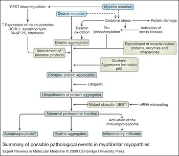
MFMs are the paradigm of conformational protein diseases of the skeletal (and cardiac) muscles. Recent studies have provided new insights into the genetic and molecular basis of the still-emerging group of MFMs and have permitted a better delineation of phenotypes resulting from mutations in DES, CRYAB, MYOT, LDB3 and FLNC genes. Protein aggregation is now recognised as an important pathogenic factor in MFMs. A summary of the proposed pathogenic events in MFMs is illustrated in Figure 4.
Summary of possible pathological events in myofibrillar myopathies. Since mutant proteins are prone to aggregate, mutation is a principal factor in protein aggregation. However, protein aggregates contain not only mutated proteins but also a range of other protein components, including cytoskeletal and myofibrillar proteins, intermediate filaments, nuclear proteins, chaperones, Alzheimer-disease-related proteins, kinases, neuronal proteins, stress-associated proteins, and proteins related to proteasome and immunoproteasome function. The causes of protein accumulation are poorly understood but ectopic expression of neuronal-related proteins as UCHL1, synaptophysin, SNAP-25 and α-internexin appears to be the result of decreased expression of NRSF/REST, a neuronal repressor factor in non-neuronal cells. Proteins involved in protein aggregation and ubiquitination are also expressed in aggresomes. Importantly, mutant ubiquitin UBB+1 accumulates in protein aggregates: UBB+1 is unable to ubiquitinate proteins although UBB+1 is a target of ubiquitination and ubiquitinated UBB+1 inhibits the proteasome. Abnormal post-translational protein modifications also occur in myofibrillar myopathies, particularly as a result of oxidation and nitration. Increased lipoxidative, glycoxidative and nitrosative damage occurs within and around aberrant protein aggregates. A main target of oxidation and nitration in myotilinopathies and desminopathies is desmin. Other oxidised proteins are components of the respiratory chain and energy metabolism. Mutant and recruited proteins cannot be eliminated by the proteasome and accumulate in the cytoplasm. Organelles and debris are further incorporated in autophagosomes and lysosomes, eventually forming membrane-bound inclusions and rimmed vacuoles. Abbreviations: NRSF/REST, neuron restrictive silencing factor; SNAP-25, synaptosome-associated protein of 25 kDa; UBB+1, ubiquitin translated in the +1 reading frame, giving rise to a protein with an aberrant C-terminus; UCHL1; ubiquitin C-terminal hydrolase L1.

Despite this progress, many aspects of MFMs require further study. The genetic background of many MFM patients is still unresolved, and it is likely that new causative genes will be identified in the future. Epigenetic factors (regulating transcription and translation) involved in muscle vulnerability in the different types of MFMs also require investigation. Furthermore, the roles of microRNA and transcription factors in regulation of the ectopically expressed proteins need to be investigated. Other key research areas in understanding pathogenic events in MFMs are the causes of oxidative stress (e.g. mitochondrial function) and post-translational modifications of the accumulating proteins. Looking ahead to therapeutic implications, identification and use of factors that would permit normal proteasome and autophagosome functions are an interesting possibility. However, major research is necessary to develop specific therapies. While no effective treatment is currently available for patients with MFM, life-threatening cardiac and pulmonary complications are important points that should be addressed.
Acknowledgements and funding
The authors’ work is funded by grants from the Spanish Ministry of Health, Instituto de Salud Carlos III PI05/1213 and PI05/1570. We thank T. Yohannan for editorial assistance. We are grateful for the comments and suggestions of the peer reviewers.