Vasodilator stress perfusion cardiovascular magnetic resonance has been shown to be a useful non-invasive tool in the assessment of myocardial ischaemia with high specificity and sensitivity.Reference Crean and Merchant 1 – Reference Sharples, Hughes and Crean 5 Although there are current recommendations that recognise the value of stress perfusion cardiovascular magnetic resonance in the adult congenital heart disease population,Reference Kilner, Geva, Kaemmerer, Trindade, Schwitter and Webb 6 there is limited experience with its use in this context, with only small cohorts reported in paediatric populationsReference Buechel, Balmer, Bauersfeld, Kellenberger and Schwitter 7 – Reference Manso, Castellote, Dos and Casaldáliga 9 and no published data in a pure adult congenital heart disease population. Vasodilator stress perfusion cardiovascular magnetic resonance is a comprehensive technique allowing for assessment of ventricular size and function, myocardial perfusion, and scar characterisation. We follow a large adult congenital heart disease population of over 8000 active patients and perform ∼900 cardiovascular magnetic resonance studies for congenital indications per year. The purpose of our study was to review our 5-year experience with the use of a stress perfusion cardiovascular magnetic resonance protocol in a referral population from our large adult congenital heart disease centre.
Materials and methods
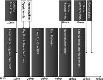
We undertook a retrospective review of our institutional database for all patients referred by the adult congenital heart disease clinic undergoing stress perfusion cardiovascular magnetic resonance from 2007 to 2012. All patients were instructed to abstain from caffeine and caffeine-containing substances for 24 hours before the scan. Stress cardiovascular magnetic resonance protocol overview is shown in Figure 1. Slow infusion of 0.56 mg/kg of vasodilator agent was performed over 4 minutes. Dipyridamole was diluted to at least 1:1 ratio with 5% dextrose solution, hand injected via an antecubital vein, and flushed with 20 ml of 0.9% saline solution to reduce the risk of phlebitis. During and following the slow 4-minute injection, symptoms and haemodynamic parameters were recorded to confirm appropriate symptomatic and physiological response – heart rate increase of 10–15 beats/minute; blood pressure decrease of 10–15 mmHg. The vectorcardiogram was monitored throughout for signs of heart block. The technologist waited 3 minutes following completion of dipyridamole injection before initiating the stress perfusion sequence to allow adequate physiological effect of the vasodilator. Following completion of the stress perfusion sequence, dipyridamole effects were reversed with 1 mg/kg intravenous aminophylline.
Adult congenital heart disease stress perfusion CMR imaging protocol. The basic stress CMR protocol can be completed in an hour. Optional sequences can be added in between segments of the basic protocol as shown in the example above. Coronary magnetic resonance angiography is usually performed after late gadolinium enhancement imaging, when all the gadolinium has been delivered, to maximise T1 signal from coronary arteries. CMR = cardiovascular magnetic resonance.

Scans were performed on a 1.5 T (MAGNETOM Avanto; Siemens Healthcare, Erlangen, Germany) or on a 3 T (MAGNETOM Verio; Siemens Healthcare) equipped with 32-element cardiac array coils. For planning, end-systolic four-, three-, and two-chamber long-axis cine slices were reviewed and three cardiac short-axis slices, 8–10 mm thick, were then evenly spaced throughout the left ventricle – avoiding the left ventricular outflow tract on the three-chamber slice – to obtain perfusion data from basal, mid, and apical myocardial segments. A inversion-recovery, gradient-recalled echo perfusion sequence was used, with an individual pre-pulse per slice – typical parameters: time to echo 1.11 ms, temporal resolution 150 ms, inversion recovery pre-pulse 110 ms, twofold acceleration, slice thickness 8 mm, acquired in-plane resolution 1.7 × 1.7 mm, all three slices acquired every heart beat for 60 seconds.
Patients received 0.2 mmol/kg gadobutrol (Gadovist; Bayer Healthcare, Berlin, Germany) given in divided boluses. Patients received 0.05 mmol/kg gadolinium into an antecubital vein at 5 ml/second for stress and again for rest followed in each case by a 30-ml saline flush delivered by power injector (MEDRAD, Pennsylvania, United States of America). A final 0.1 mmol/kg of gadolinium – ‘top-up’ – was injected after completion of the rest perfusion sequence. Late gadolinium enhancement imaging was performed with either segmented gradient recalled echo or single-shot steady-state free precession inversion-recovery sequences 8–10 minutes after intravenous administration of the ‘top-up’ dose.
Whole-heart navigated magnetic resonance angiography was performed for assessment of coronary artery origins, course, and abnormalities where appropriate – typical isotropic spatial resolution of 1.5 mm. Contiguous slices from the atrioventricular groove to the apex were acquired for left ventricular function using cine steady-state free precession images in the short-axis plane – typical parameters: thickness/inter-slice gap 6mm/2 mm; in-plane resolution 1.5 × 1.5 mm; temporal resolution 35–50 ms.
Perfusion abnormalities were assessed qualitatively by visual comparison of the stress and rest perfusion and the late gadolinium enhancement images. A significant stress-induced perfusion defect was defined as present if gadolinium wash-in into myocardium was delayed, with a corresponding reduction in the myocardial signal relative to adjacent normal myocardium in a typical distribution, either subendocardial or transmural in at least two contiguous segments and associated with persistence for five or more heartbeats.
The results of all additional studies performed for evaluation of myocardial ischaemia were recorded, including invasive coronary angiography; coronary computed tomographic angiography; single-photon emission computed tomography; dobutamine stress echocardiography; treadmill stress echocardiogram; and treadmill exercise test. Invasive coronary angiography, coronary computed tomographic angiography, or surgical findings, where available, were considered the reference standard for analysis of sensitivity and specificity of cardiovascular magnetic resonance myocardial perfusion imaging for the detection of myocardial ischaemia.
Institutional Research and Ethics Board approval was obtained for this retrospective study and the requirement for individual patient consent was waived.
Results
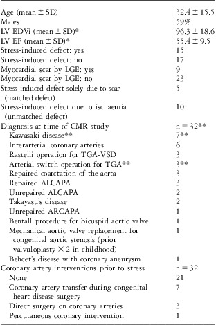
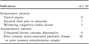
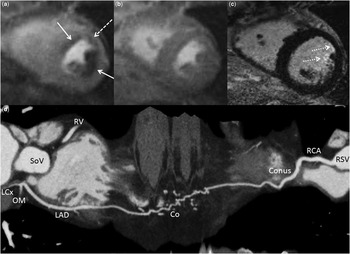
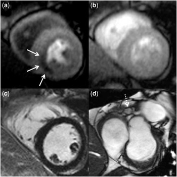
In all, 32 adults (mean age 30.7 ± 14.4 years, 63% males) underwent 34 stress perfusion cardiovascular magnetic resonance studies over a 5-year period. A total of 21 studies were performed at 1.5 T and 13 studies were performed at 3 T. All 34 studies were performed with vasodilator stress agents – 31 dipyridamole and 3 adenosine. There were two patients who underwent a repeat stress perfusion cardiovascular magnetic resonance study – one to confirm a positive stress after an interval of some months and one to confirm resolution of ischaemia following surgery. Population characteristics and scan indication are given in Tables 1 and 2, respectively. The majority of patients (84%) had other investigations in addition to cardiovascular magnetic resonance: single-photon emission computed tomography 56%; coronary computed tomography 47%; invasive coronary angiography 44%; treadmill exercise test 41%; treadmill stress echocardiogram 22%; and dobutamine stress echocardiogram 6%. Of the 34 scans, 11 – 10 patients, 1 repeat scan – were positive, demonstrating significant stress-induced perfusion defects (Figs 2 and 3, Supplementary Video S1).
Population characteristics.

ALCAPA = Anomalous left coronary artery from pulmonary artery; ARCAPA = Anomalous right coronary artery from pulmonary artery; CMR = cardiovascular magnetic resonance; LV EDVi = left ventricular end-diastolic volume indexed to body surface area; LV EF = left ventricular ejection fraction; LGE = late gadolinium enhancement; SD = standard deviation; TGA = Transposition of the great arteries; VSD = ventricular septal defect.
*Volumetric data based on 31 of 32 patients – one patient could not have formal quantification due to irregular heart rate.
**One patient with a history of arterial switch operation for TGA, subsequently suffered Kawasaki disease.
Indication for stress cardiovascular magnetic resonance.

Proximal occlusion of ALCAPA repair with subsequent collateralisation – dipyridamole stress perfusion cardiovascular magnetic resonance. Images from dipyridamole stress perfusion cardiovascular magnetic resonance of a 23-year-old man who had previously had repair of ALCAPA. Stress perfusion sequences ( a ) demonstrate reversible subendocardial perfusion defects in the LAD and circumflex territories (solid arrows) not seen on the rest perfusion sequence ( b ) and significantly greater in volume than small subendocardial anterior wall and anterolateral papillary muscle infarcts (dotted arrows) on late gadolinium enhancement sequences ( c ). There is also mild peri-infarct ischaemia (dashed arrow) in the anterior wall ( a ). These stress perfusion defects were demonstrated to be secondary to restricted flow to the left anterior descending coronary artery and circumflex territories via a complex network of narrow calibre collaterals from the right coronary artery. Curved multiplanar reconstruction images from gated contrast-enhanced coronary computed tomography ( d ) demonstrate absence of the left main coronary artery due to remote occlusion following ALCAPA repair. A complex network of narrow calibre collaterals connect a dilated conus branch of the right coronary artery to the distal LAD supplying the LAD territory and circumflex territories retrogradely. ALCAPA = anomalous left coronary from pulmonary artery; Co = collaterals; Conus = conus branch; LAD = left anterior descending coronary artery; LCx = circumflex coronary artery; OM = large calibre obtuse marginal; RCA = right coronary artery; RSV = right sinus of Valsalva; RV = right ventricle; SoV = sinus of Valsalva.

RCA territory stress perfusion defect in a patient with Kawasaki disease and proximal RCA aneurysm. Images from dipyridamole stress perfusion cardiovascular magnetic resonance of a 30-year-old male with Kawasaki disease and known aneurysm of the proximal RCA demonstrating a reversible RCA territory perfusion defect (white solid arrow) on stress perfusion sequences ( a ) with no defect on rest perfusion sequences ( b ) or evidence of infarct on late gadolinium sequences ( c ). The proximal right coronary artery aneurysm (dotted arrow) is visualised on cine steady-state free precession imaging ( d ). RCA = right coronary artery.

Vasodilator stress cardiovascular magnetic resonance
In all, 10 patients (31%) had stress-induced perfusion defects (Figs 2 and 3, Supplementary Video S1). Cardiovascular magnetic resonance perfusion defects were proved to be due to a significant lesion on catheter angiography or coronary computed tomography in nine cases. Out of the 10 patients with positive studies, three have undergone successful surgical correction of their coronary artery abnormality; three have been recommended for surgery/intervention; two have confirmed disease, but are awaiting final treatment decision; one has declined further investigation because of cessation of symptoms; and one has undergone optimisation of medical management.
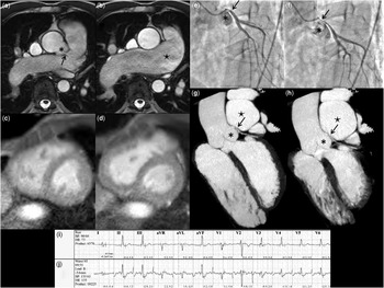
Out of the 23 negative stress perfusion studies, the findings were either concordant with single-photon emission computed tomography or confirmed by catheter angiography and/or coronary computed tomography in 15. In two of the negative stress cardiovascular magnetic resonance cases, catheter angiography demonstrated significant lesions that required surgery. In both of these, there was dynamic extrinsic compression of the left main coronary artery rather than intrinsic luminal disease (Fig 4). In the remaining six patients with negative stress cardiovascular magnetic resonance studies, the clinicians were sufficiently confident in the negative test result to reassure the patient and continue conservative management/follow-up. All 32 patients are under continuing medical follow-up without any major adverse cardiac events reported to date (median follow-up 258.5 days, interquartile range 140.5–583.0 days).
Coronary compression in repaired TOF-APV and free pulmonary regurgitation and extrinsic compression of left main coronary artery by aneurysmal main pulmonary artery – false negative stress perfusion CMR. Stress CMR, invasive coronary angiograms, coronary CT, and exercise tolerance test in a 56-year-old male with repaired TOF-APV and free pulmonary regurgitation who presented with angina on exertion. Axial cine steady-state free precession sequences from a single axial level demonstrate the origin of the left main coronary artery (black arrow) in diastole ( a ) and its displacement by a dilated main pulmonary artery (black star) in systole ( b ). The main pulmonary artery (black star) dilated significantly from 47 mm in diastole to 57 mm in systole. Normal stress perfusion CMR study with no perfusion defect at stress ( c ) or rest ( d ). Invasive coronary angiography demonstrating a normal calibre proximal left main coronary artery (black arrow) in diastole ( e ) and subsequent compression by the dilated main pulmonary artery during systole ( f ). Maximum intensity projection images from gated CT angiogram acquired throughout the cardiac cycle in an oblique plane in diastole ( g ) and systole ( h ) demonstrating the significant systolic dilation of the main pulmonary artery (black star) causing compression of the proximal left main coronary artery (black arrow) against the left sinus of Valsalva (asterisk). Exercise tolerance test with sample electrocardiograms at rest ( i ), and at peak exercise ( j ) in the period of worst ST depression during which the patient experienced typical angina. CMR = cardiovascular magnetic resonance; CT = computed tomography; TOF-APV = tetralogy of Fallot with absent pulmonary valve.

Diagnostic performance of cardiovascular magnetic resonance including vasodilator stress
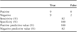
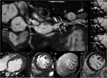
In the 19 patients for whom a reference standard was available – surgical findings, catheter angiography, or coronary computed tomography – the sensitivity and specificity of stress perfusion cardiovascular magnetic resonance were 82% and 100%, respectively, and the positive and negative predictive values were 100% and 82%, respectively (Table 3). Out of the 14 patients in whom whole-heart navigator steady-state free precession acquisition was performed, abnormal coronary findings were identified in 11 cases [interarterial course × 3; coronary artery aneurysms × 2 (Fig 5); anomalous left coronary artery from pulmonary artery × 1; significant left main coronary artery stenosis × 1; reimplanted but occluded coronary artery with collaterals × 1; subtotal occlusion of both coronary arteries × 1; single coronary artery system × 1; stable pericardial hood augmentation of coronary artery × 1]. There were two studies that were non-diagnostic.
Diagnostic performance of vasodilator stress CMR.

CMR = cardiovascular magnetic resonance.
The diagnostic performance of stress CMR based on 20 scans; each with an individual reference standard – 19 patients. One patient had a pre-operative stress CMR with intra-operative findings as the reference standard and a post-operative stress CMR with a coronary computed tomography as the post-operative reference standard.
Kawasaki aneurysms with mural thrombus and transmural infarction due to distal embolisation of thrombus. Curved multiplanar reconstruction ( a ) and axial ( e ) images from gated navigator steady-state free precession whole-heart coronary magnetic resonance angiogram in a 20-year-old male with Kawasaki disease-related fusiform aneurysms in the proximal and mid-circumflex coronary artery (asterisk). The axial, curved multiplanar reconstruction images and short-axis sections through the vessel ( b – d ) reveal mural thrombus (white solid arrow) and eccentric wall calcification (black dashed arrow) in the mid-circumflex artery aneurysm on this contrast-enhanced study. Cardiac short-axis cine steady-state free precession ( f ) and late gadolinium enhancement images ( h ) demonstrate thinning and transmural circumflex territory infarction (black arrows) likely because of distal embolisation of thrombus from the aneurysm. The stress perfusion cardiovascular magnetic resonance image demonstrates a matched perfusion defect ( g ) due to the infarction. Note that the aneurysmal circumflex coronary artery (asterisk) is almost as large as the descending thoracic aorta (star). LA = left atrium; LAD = left anterior descending coronary artery; LM = left main coronary artery; LV = left ventricle; RA = right atrium; RCA = right coronary artery; SoV = sinuses of Valsalva.

Discussion
Our study contributes data demonstrating the feasibility of stress perfusion cardiovascular magnetic resonance in the care of adult congenital heart disease patients, an area with a sparse existing literature. We have demonstrated an excellent positive predictive value for coronary lesions causing significant ischaemia. The sensitivity of the technique appears similar to many other forms of non-invasive ischaemia testing although a little lower than published cardiovascular magnetic resonance perfusion data from a recent meta-analysis.Reference Hamon, Fau, Née, Ehtisham, Morello and Hamon 10 , Reference Salerno and Beller 11 However, it should be noted that published data were derived entirely from an adult coronary heart disease population in which intrinsic atherosclerosis was the cause of the flow-limitation – a mechanism that was rarely the cause of ischaemia in our cohort. In our series, two of these false negatives were in patients with classic angina histories with positive exercise testing and surgically confirmed evidence of extrinsic left main stem compression. It is possible that this compression may have been dynamically worsened by exercise but remained relatively unaffected by vasodilator stress. Another possibility is false negative stress due to caffeine intake. Despite suggestions that a small amount of caffeine intake 1 hour before a stress test need not necessitate cancellation of a scan,Reference Kovacs, Pivonka, Khosla and Khosla 12 it has also been acknowledged that caffeine content in coffee is highly variableReference Gilbert, Marshman, Schwieder and Berg 13 and patients may not always be willing to disclose its consumption when screened at the time of the scan.
Cardiovascular magnetic resonance stress perfusion studies can be reliably undertaken in under an hour. However, additional sequences including magnetic resonance angiography of the aorta,Reference Prince 14 phase contrast analysis – shunt fraction calculation,Reference Brenner, Caputo and Mostbeck 15 valvular analysis,Reference Firmin, Nayler, Klipstein, Underwood, Rees and Longmore 16 and coronary flow analysisReference Edelman, Manning, Gervino and Li 17 – or gated navigator whole-heart coronary magnetic resonance angiographyReference Weber, Martin and Higgins 18 – for assessment of coronary aneurysms, reimplanted coronary arteries, coronary fistulas, and anomalous coronary arteries – can be added to the scan as clinically indicated. In addition, volumetric measurement routinely performed as part of the complete stress perfusion cardiovascular magnetic resonance protocol is the reference standard in ventricular functional analysis.Reference Niwa, Uchishiba and Aotsuka 19 , Reference Semelka, Tomei and Wagner 20 This is especially true in adult congenital heart disease, in which the anatomy can be atypical and accurate assessment of ventricular volumes and function is crucial.Reference Crean, Maredia and Ballard 21
Congenital heart patients may have lesions that place them at risk for myocardial ischaemia. These include anomalous coronary arteries and late presentations of unrepaired anomalous connection of a coronary artery to the pulmonary artery. Many routine congenital surgical operations such as the arterial switch, Ross and Bentall procedures involve coronary transfer and/or reimplantation with the potential for coronary ostial distortion and subsequent ischaemia. In addition, paediatric pathologies such as Kawasaki and Takayasu's disease are not uncommonly looked after by congenital heart physicians and both may have long-term significant coronary sequelae.
Historically, myocardial perfusion single-photon emission computed tomography has been the mainstay of non-invasive investigation, often used to triage those in need of further investigation by invasive coronary angiography. This is not ideal as single-photon emission computed tomography involves exposure to a non-trivial dose of ionising radiation, and the cancer induction risk is higher in younger patientsReference Korenman 22 , Reference Tokunaga, Norman and Asano 23 Owing to the fact that the adult congenital heart disease population is one subject to repeated examinations with a lifetime of follow-up, minimising radiation is desirable.Reference Gerber, Carr and Arai 24 In addition, there are strong signals in the data from patients with ischaemic heart disease to suggest a significantly lower diagnostic accuracy for single-photon emission computed tomography compared with stress cardiovascular magnetic resonance.Reference Greenwood, Maredia and Younger 2 , Reference Schwitter, Wacker and Wilke 25
Limitations
The current study is limited by its retrospective nature and the relatively small number of patients. In addition, the case mix was heterogeneous and included both repaired and unrepaired pathologies. However, this reflects the “real-world” patient population presenting to a large but typical adult congenital heart disease service. A minor limitation is that studies were performed at two different field strengths. Although there is emerging data to support the superiority of perfusion imaging performed at 3 T, the majority of perfusion studies worldwide continue to be performed at 1.5 T given the greater flexibility of this platform for general cardiac imaging. Finally, a reference standard of coronary assessment was not available in all patients. However, in keeping with the prognostic literature on stress cardiovascular magnetic resonance in the adult world,Reference Jahnke, Nagel and Gebker 26 we have also seen a very low event rate (zero) in those in our cohort with a negative stress result but no confirmatory coronary imaging.
In summary, we have demonstrated stress perfusion cardiovascular magnetic resonance to be a safe and useful tool in the clinical assessment of adult patients with coronary artery abnormalities both congenital and those acquired in childhood. Cardiovascular magnetic resonance is the natural choice in the assessment and follow-up of adult congenital heart disease and it is likely that stress perfusion cardiovascular magnetic resonance for adult congenital heart disease patients – with symptoms or for surveillance in patients with previous coronary surgery – will gain increasing acceptance. Additional sequences, particularly navigator steady-state free precession whole-heart coronary magnetic resonance angiography, which enables visualisation of the coronary arteries, further enhance the versatility of a cardiovascular magnetic resonance-based approach. A positive stress perfusion cardiovascular magnetic resonance study had a high positive predictive value for coronary ischaemia in our cohort. Our experience suggests that in situations where extrinsic coronary compression is suspected, a negative vasodilator cardiovascular magnetic resonance stress should be interpreted with caution. However, there was no instance in which a coronary abnormality identified at cardiovascular magnetic resonance was subsequently disproven. The comprehensive stress perfusion cardiovascular magnetic resonance examination appears to have a valuable role in the investigation of an evolving subset of adult congenital heart disease patients without exposure to ionising radiation.
Acknowledgements
The authors express thanks to cardiovascular magnetic resonance and cardiovascular computed tomography technologists of Toronto General Hospital past and present whose expertise and dedication to excellent image quality made this research possible.
Supplementary materials
To view supplementary material for this article, please visit http://dx.doi.org/10.1017/S104795111300111X