Atrioventricular septal defects with a common atrioventricular junction, or atrioventricular canal malformations, are a group of anomalies that, owing to the deficiencies of atrioventricular septation, also show abnormalities of the atrioventricular valves.Reference Cetta, Minichi, Edwards, Dearani and Puga 1 The spectrum of lesions thus produced has provoked significant controversy in terms of optimal classification, and still leave problems in description.Reference Valdes-Cruz and Cayre 2 Patients falling within the spectrum typically have the common junction shared between the ventricles, although the junction itself can be guarded by a valve with a common orifice, or with separate orifices for the two ventricles.Reference Aiello, Afiune, Menahem and Anderson 3 The common junction is the consequence of the lack of contiguity between the atrial and ventricular septal structures, with these features also underscoring that lack of atrioventricular septation.Reference Anderson, Ho, Macartney and Becker 4 Occasionally, the common atrioventricular junction may be exclusively connected to one of the ventricles. When the junction is exclusively connected to the left ventricle, the right atrium can be blind-ending, as seen typically in tricuspid atresia, but with the obligatory right-to-left shunt occurring across an ostium primum type.Reference Aiello, Afiune, Menahem and Anderson 3 We describe here the case of an infant with similar haemodynamics, but in our case due to the presence of an imperforate right component of the common atrioventricular valve.
Case report
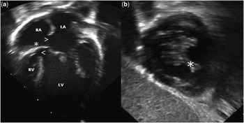
A male infant, aged 7 months, was diagnosed prenatally as having tricuspid atresia with normally related great arteries, a small muscular ventricular septal defect, and a hypoplastic right ventricle. The infant was born at 39 6/7 weeks via Caesarian section, owing to reduced foetal heart rate. Apgar scores were 8 at 1 minute and 9 at 5 minutes. Transcutaneous pulse oxymetry showed oxygen saturations of 70–80% in room air. On examination, the patient was not in acute distress, and showed no dysmorphic features. Cardiac examination revealed normal and regular heart sounds, with a systolic murmur graded at 2 from 6 audible over the left sternal border. The electrocardiogram showed left axis deviation (QRS axis of −9°), with left ventricular hypertrophy and right atrial enlargement. The initial chest X-ray showed a heart of normal size, with mildly decreased pulmonary vasculature. The postnatal echocardiogram revealed an unusual intracardiac anatomy that had not previously been appreciated in the prenatal studies. What had initially been interpreted as tricuspid atresia due to the absence of the right atrioventricular connection was now seen to be produced by an imperforate right-sided component of a common atrioventricular valve (Fig 1a). The imperforate valve interposed between the cavity of the right atrium and the inlet component of the hypoplastic right ventricle.
The four-chamber view ( a ) showed an imperforate right-sided component of the common atrioventricular valve (*) interposing between the floor of the RA and the hypoplastic RV inlet. The RA communicates with the LA through a large ostium primum defect. A ventricular septal defect, inlet type is also seen (arrows). ( b ) There is fusion of the quadrifolate right sided component of the common atrioventricular valve (*). LA = left atrium; LV = left ventricle; RA = right atrium; RV = right ventricular.

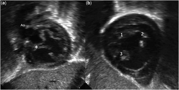
An ostium primum defect provided the communication with the left atrium, which in turn connected to the left ventricle through a trifoliate left atrioventricular valve (Fig 2b). The aorta arose from the dominant left ventricle, being displaced anteriorly and superiorly owing to the commonality of the atrioventricular junction (Fig 2a). Owing to diminished pulmonary blood flow, the patient underwent construction of a modified Blalock–Taussig shunt. He has recently undergone a successful bidirectional cavopulmonary connection as the second stage of the projected eventual functionally univentricular repair.
When examined subcostally, the aorta is seen to be anteriorly and superiorly displaced, (unwedged), due to commonality of the atrioventricular junction ( a ). The left-sided component of the common atrioventricular valve is trifoliate (1, 2, and 3 in b ), showing the classic zone of apposition between the left ventricular components of the bridging leaflets. *Imperforate right side component of the common atrioventricular valve.

Discussion
It used to be thought that atrioventricular septal defects in the setting of a common atrioventricular junction resulted from arrested or abnormal development of the endocardial cushions that form in the primitive atrioventricular canal.Reference Van Mierop, Alley, Kausel and Stranahan 5 It has now been shown that formation of the cushions themselves is relatively normal, and that the lesion reflects failure of growth of the vestibular spine, this being a ventral protrusion derived from the dorsal pharyngeal mesenchyme.Reference Anderson, Wessels and Vettukattil 6
Irrespective of the development, however, the phenotypic feature of the group is the commonality of the atrioventricular junction, with usual variation depending on the presence of a common atrioventricular valvar orifice or separate valvar orifices for the right and left ventricles.Reference Aiello, Afiune, Menahem and Anderson 3 Some still distinguish these variants as being “complete” or “partial”. This is less than ideal, however, as it then introduces the temptation to describe “intermediate” variants, where in reality the anatomy of the common atrioventricular junction is virtually the same in all forms.Reference Valdes-Cruz and Cayre 2 In our patient, we were able to identify not only the common junction, but also the anticipated trifoliate arrangement of the left atrioventricular valve. The bridging leaflets of the initially common valve came together at an imperforate right atrioventricular component (Fig 1b), which formed the fibrous floor of the right atrium, egress from the right atrium occurring through a large ostium primum defect. We presume that the imperforate right component of the common valve also incorporates the remaining two leaflets of the common atrioventricular valve that, had the right orifice been perforate, would have guarded part of the right atrioventricular junction.
The classical variant of tricuspid atresia exists because of the absence of the right atrioventricular connection, the floor of the right atrium being exclusively muscular, with the left atrioventricular junction connected to a dominant left ventricle, and the right ventricle being incomplete because it lacks its inlet component. In our patient, the right ventricle possesses a hypoplastic inlet component, with the imperforate right-sided component of the common atrioventricular valve interposed between it and the cavity of the right atrium. The classical variant of tricuspid atresia is well described in terms of a univentricular atrioventricular connection. In our patient, in contrast, the atrioventricular connection is in fact biventricular, even though the imperforate nature of the right-sided component of the common atrioventricular valve produced a functionally univentricular arrangement.
In 1991, Van Praagh et alReference Van Praagh, Vangi and Sul 7 reported a relatively large series of 13 human cases, with nine having a similar arrangement of the atrioventricular junction as described in our case. Aiello et al.Reference Aiello, Afiune, Menahem and Anderson 3 also published a series of three cases, pointing out that this unusual anatomy is also seen in mice with trisomy 16. The surgical pathway in these cases is functionally univentricular. Appropriate assessment of the intracardiac anatomy, nonetheless, shows how imperforate atrioventricular valves can potentially mask the presence of biventricular atrioventricular connections, although without impacting on the optimal therapeutic options.
Acknowledgement
We thank Dr. Julia Steinberger for her opinion and critical review of the echocardiograms.
Financial Support
This research received no specific grant from any funding agency, commercial or not-for-profit sectors.
Conflicts of Interest
None.
Ethical Standards
Retrospective case review.