Introduction
Given the evolutionary proximity and phylogenetic similarities to humans, non-human primates (NHP) have the potential to transmit several parasitic and infectious diseases (Andrade, Reference Andrade, Andrade, Pinto and Oliveira2002). As members of biologically diverse habitats, they act as sentinels for pathogen investigation and models for basic research about the dynamics of natural transmission. As such, the NHP populations have contributed to information about emerging diseases (AIDS, Ebola, hantavirus and dengue). Studies related to these species may also benefit conservation efforts as well as fulfilling the gaps between laboratory studies and the already recognized need for epidemiological investigations (Wolfe et al., Reference Wolfe, Escalante, Karesh, Kilbourn, Spielman and Lal2000).
The genus Aotus is widely distributed throughout South America, from Panama to the north of Argentina (Wright, Reference Wright, Baer, Weller and Kakoma1994). The Aotidae family comprises 13 species and subspecies (Rylands et al., Reference Rylands, Schneider, Langguth, Mittermeier, Groves and Rodrígues-Luna2000). The geographic range of Aotus azarai infulatus is wide, occurring in the west and south-western Amazon, east of the rivers Tapajós and Juruena in Brazil, west of the rivers Gurupí and Araguaia. It was also recently identified in the state of Amapá, north of the lower Amazonas river. There is much forest destruction and fragmentation in the south of its range in southern Pará, but this subspecies is not currently considered to be threatened (Rylands et al., Reference Rylands, Bampi, Chiarello, da Fonseca, Mendes and Marcelino2003).
Historically, Aotus monkeys have been utilized in biomedical research to address a diverse array of scientific questions. These moderately small, nocturnal Neotropical primates possess several unique features that make them ideal for use as animal models for human diseases. These include susceptibility to infection with Plasmodium spp., large eyes with easily visualized retinas, adaptability to captivity and relative ease of handling (Baer, Reference Baer, Baer, Weller and Kakoma1994).
Many NHP species are susceptible to infection by nematodes of the Oxyuridae family, which show preference for the intestinal colon. The genera known to parasitize NHP are: Enterobius, Trypanoxyuris and Lemuricola (Prieto et al., Reference Prieto, Cruz, Scheibler, Borda and Gómez2002). According to Urquhart et al. (Reference Urquhart, Armour, Duncan, Dunn and Jennings1998), oxiurid pathogenic effects result in small lesions of the intestinal mucosa, which may become diffuse, followed by an inflammatory response in severe infestations. The life cycle is direct and infection occurs through the ingestion of the larval eggs, which are deposited on the perianal skin. In some species of pinworms, it is speculated that retroinfection may occur whereby the newly hatched larvae enter the host directly through the rectum (Felt & White, Reference Felt and White2005).
Trypanoxyuris sp. was described as non-pathogenic and common in owl monkeys (Tantaleán & Gozalo, Reference Tantaleán, Gozalo, Baer, Weller and Kakoma1994). However, there are reports of death in spider monkeys (Ateles sp.) and owl monkeys (A. nancymaae) severely infested by this nematode (Fiennes, Reference Fiennes1972; Sánchez et al., Reference Sánchez, Gálvez, Montoya and Gozalo2006).
In general, the main sign of infection is the perineal irritation caused by adult females during the egg-laying stage (Urquhart et al., Reference Urquhart, Armour, Duncan, Dunn and Jennings1998). In other NHP species, tail and perineal rubbing can be a result of pruritus and associated oxyuriasis. However, Aotus use both urine and glandular secretions to convey olfactory information. Rubbing of the tail and perineum, a behaviour usually attributed to scent marking, is well recognized as an important form of olfactory communication in this genus (Wright, Reference Wright, Baer, Weller and Kakoma1994). Therefore, according to Felt & White (Reference Felt and White2005), if perineal rubbing occurred as a result of pruritus from pinworms in owl monkeys this behaviour might be indistinguishable from scent marking.
Parasitic diseases constitute a serious public health problem in developing countries. Poor sanitation, poverty and tropical climate all contribute to the development, spread and perpetuation of gastrointestinal diseases (Michaud et al., Reference Michaud, Tantalean, Ique, Montoya and Gozalo2003). Research centres that maintain and work with NHP need to control the sanitation of colony animals. This is important to promote quality of research and avoid pathogen transmission to keepers and researchers who work with these animals. Clinical examination, coproparasitological exams, biochemical and haematological exams, as well as disinfection of the facility and fomites are essential tools to diagnose and control helminth dissemination in captivity, especially in the Amazon, where climatic conditions favour pathogen multiplication.
In spite of the existence of abundant literature on parasites in Neotropical primates, there is little information on parasites in the Aotus monkey (Tantaleán & Gozalo, Reference Tantaleán, Gozalo, Baer, Weller and Kakoma1994). Given the epidemiological relevance related to the incidence of parasitic diseases in captivity, the objective of this study was to report the Trypanoxyuris sp. occurrence in captive owl monkeys, comparing clinical and laboratory examinations of infected and uninfected animals.
Materials and methods
The study was performed with 20 animals (10 couples previously established), where females were 4–10 years old, and males were 5–13 years old. Animals were captive born and belonged to the National Primate Center (CENP) breeding colony, located in the Ananindeua municipality, state of Pará, Brazil (latitude 1°38′26″ and longitude 48°38′22″). Each animal was identified with a three-letter code tattooed on the inside of the right thigh and a microchip placed in the interscapular area.
Animals were housed indoors, in Sector I (for monogamic species), in brick-built enclosures covered with tile and wire netting. This sector was projected in a north–south direction, in order to receive up to 12 h of natural light, enabling regular access to solar radiation throughout the year. The climate in the region was classified as humid, Megathermic with regular rainfall from January to June and pluviometric indices of 2250–2500 mm. The average temperature was 25°C (max. 32°C and min. 22°C), with air relative humidity around 85%.
The cages measured 3.85 m in length × 1.2 m width × 2.40 m height. Externally, the enclosures had a 35 cm shelter box of 35 cm × 30 cm × 30 cm, with a guillotine type door and a water bottle for drinking. Internally, there were two wooden platforms for walking and access to the food bowls. Animals were fed according to CENP husbandry practice: a variety of fruits, vegetables, milk, eggs, dry dog food, vitamin and mineral supplements and water ad libitum.
Animal health was assessed through clinical examinations, clinical and laboratory testing, including coproparasitological examinations, haemogram and clinical chemistry (creatinine, blood urea nitrogen (BUN), alanine aminotransferase (ALT) and aspartate aminotransferase (AST)).
Samples for coproparasitological exams were collected and stored in sterile and identified plastic bottles, shortly after each animal defecated. Samples were analysed according to a standard protocol established by the CENP using direct examination, as well as fluctuation (Willis, Reference Willis1927) and sedimentation (Hoffman et al., Reference Hoffman, Pons and Janer1934) techniques. According to this protocol, levels of gastrointestinal helminth parasite infections were determined when calculating the number of positive microscopic fields. Values that did not exceed 25% of the examined fields were considered low. Results identified between 25 and 50% were considered as medium and above 50% as high level of infection.
Haematological exams were also performed, and the blood samples (3 ml) were collected from the femoral vein under manual restraint, using sterile syringes and needles. The haemogram was performed using an automatic counter Celm CC-550 (CELM -Cia. Equipadora de Laboratórios Modernos Alameda Amazonas, São Paulo, Brazil), and clinical chemistry with the Vitros System® DTSC II, DT60 and DTE2 (Johnson & Johnson Medical Argentina, San Isidro, Argentina).
The haemogram and clinical chemistry results were submitted to analysis of variance (ANOVA). Comparisons of individual means were examined with the Tukey test (Steel et al., Reference Steel, Torrie and Dickey1997). For differences between males and females and between parasitized and non-parasitized animals, two-sided P values of 0.05 or less were considered to be statistically significant.
Results
During the clinical examinations, all animals were apparently healthy. In the coproparasitological examinations, eggs compatible with Trypanoxyuris sp. were identified in 50% of the couples. It was observed that, when one of animals was infected, the other one was also infected. However, the levels of infection were considered to be low, as the number of positive fields was lower than 25% of the total examined microscopic fields.
Positive animals were treated orally with 9.5 mg oxantel pamoate together with 14.5 mg pirantel pamoate per kg of body weight (Basken suspensão®, König do Brasil, Ltd), and the same treatment was repeated after 15 days. As a prophylactic measure, a blowtorch was utilized in order to eliminate foci of nematoid contamination in the enclosures.
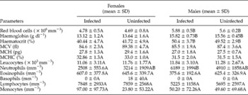
The haematological results (table 1) indicated that the number of red blood cells, the haematocrit and the haemoglobin levels were influenced by the sex, as the males evidenced values significantly higher than the females (P < 0.05), irrespective of parasitism. As for the mean corpuscular volume (MCV), mean corpuscular haemoglobin (MCH) and mean corpuscular haemoglobin concentration (MCHC), no significant differences were observed.
Haematological analyses of Aotus azarai infulatus (n=20) infected or uninfected by Trypanoxyuris sp. (CENP, Ananindeua/Pará/Brazil, 2007).

MCV, mean corpuscular volume; MCH, mean corpuscular haemoglobin; MCHC, mean corpuscular haemoglobin concentration. Different letters in the same row indicate a significant difference (P < 0.05).
The white blood cell analysis did not reveal significant changes in the number of eosinophils, basophils, lymphocytes and monocytes from infected and uninfected animals. However, there was a significant difference in the segmented neutrophils when comparing infected males and females.
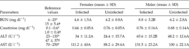
Table 2 lists the blood chemistry values (BUN, creatinine, ALT and AST) in male and female A. azarai infulatus parasitized or not by Trypanoxyuris sp. Infected males had BUN levels significantly higher when compared to other groups (P < 0.05). The same occurred with the ALT values.
Values of blood chemistry analyses for Aotus azarai infulatus (n=20) infected and uninfected by Trypanoxyuris sp. (CENP, Ananindeua/Pará/Brazil, 2007).
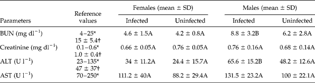
BUN, blood urea nitrogen; ALT, alanine aminotransferase; AST, aspartate aminotransferase. Different letters in the same row indicate a significant difference (P < 0.05).
* Reference interval for Aotus spp. using the products Vitros System® according to Ortho-Clinical Diagnostics.
Discussion
Coproparasitological examinations were not performed with the specific objective of investigating oxyurids. Therefore, the exams were sporadic in the CENP's Aotus colony. Repeating the examinations utilizing a perianal tape test was not possible due to the fact that therapeutic and prophylactic protocols were initiated soon after the results were confirmed.
The identification of parasitism by Trypanoxyuris sp. is supported by data from Tantaleán & Gozalo (Reference Tantaleán, Gozalo, Baer, Weller and Kakoma1994) who reported this to be the most frequently identified pinworm species in Aotus sp. Research on New World non-human primates in the wild indicated infection rates of 54.7% in the species Aotus nancymaae and lower (8.7%) in Aotus vociferans (Michaud et al., Reference Michaud, Tantalean, Ique, Montoya and Gozalo2003). Prevalence determination enables the comprehension of whether a disease is common or rare in a given population. Therefore, comparing prevalence data of free-ranging animals to our study indicates that these parasites can also be identified in captive populations.
The considerable number of infected animals observed in this study (50%) may be related to repeated self-infection events or typical oxiurid cross-infection (Flynn, Reference Flynn1973). It was also observed that all parasitized animals were previously established couples, suggesting that infection may have occurred among cagemates. This is compatible with the results from Felt & White (Reference Felt and White2005), as they determined that the risk of infection by T. microon in A. nancymaae was 14 times greater when animals were housed with an infected cagemate.
Data presented in table 1 indicated significant differences for red blood cells, haemoglobin and haematocrit values, despite the fact that the same did not occur in the MCV, MCH and MCHC. Sex-related differences observed in the haematological examinations were reported in the literature (Malaga et al., Reference Malaga, Weller, Buschbom and Ragan1990) and confirmed by our results. This may be explained by physiological variation in erythrocyte parameters, as testosterone stimulates erythropoiesis, while oestrogen is an inhibitor (Jain, Reference Jain1993). Malaga et al. (Reference Malaga, Weller, Buschbom and Ragan1990) described significant differences in the number of red blood cells, haemoglobin, MCV and platelet values between female and male owl monkeys. In the present study, platelet information was not obtained. Therefore, the differences in the relative values of erythrocytes are due to sexual differences rather than infection.
According to Jain (Reference Jain1993), evaluation of erythrocytic values can reveal disorders such as haemorrhagic syndromes, anaemia and decreased vascular fluid due to haemoconcentration, dehydration and hypovolaemia. Leucocyte values can also indicate infectious, inflammatory, toxic and stress conditions. Eosinophils correspond to 1–3% of the total leucocytes in the peripheral bloodstream, varying, in absolute terms, from 0 to 0.45 × 10³ μl− 1 in most NHP. The number of these cells increases in the tissues and in the peripheral blood as a response to parasitic infections and hypersensitivity reactions (Albert et al., Reference Albert, Brodie, Sasseville and Ringler1996). The same authors observed that in Aotus azarai boliviensis the number of eosinophils was significantly higher than the number observed in A. nancymaae, suggesting that there is variation in the count within the genus Aotus. In the present study no significant alterations were observed in the eosinophil count for infected and uninfected animals, probably due to the low infection level identified in animals. The absence of this alteration corroborates the findings by Felt & White (Reference Felt and White2005) when diagnosing parasitism by Trypanoxyuris microon in A. nancymaae.
When infected male and female data were compared, the significant difference identified in the segmented neutrophils was within the normal values when compared to clinically healthy individuals, as described by Baer (Reference Baer, Baer, Weller and Kakoma1994). Therefore, we believe that this result was not biologically meaningful. However, an increase in the catecholamine numbers in animals submitted to acute stress promotes the transfer of neutrophils from the marginal pool to the circulating pool, resulting in increased number of these cells in the leucogram (Thrall, Reference Thrall and Thrall2004; Cunha et al., Reference Cunha, Lopes and Sousa2005).
Laboratory tests may serve many functions, including prognosis, response to therapy and diagnosis. In some cases it may be necessary to run blood chemistry tests to assess different organ-systems, including the kidney and liver (Porter, Reference Porter2005). In this study the blood chemistry (BUN, creatinine, ALT and AST) results were not clinically relevant, since the values were identified as normal when compared to reference values reported by Linn et al. (Reference Linn, Duran-Struuck, Trivedi, Zajic, Wrobleski, Hawley, Myers, Reuter and Suckow2006) and the Vitros System® as a pattern for the species (Aotus spp.).
According to the literature, differences may exist in the haematology and serum chemistry data from different sources for owl monkeys, but it is not known whether these are related to the methods utilized for data collection, examination techniques, karyotype, husbandry or other environmental factors (Baer, Reference Baer, Baer, Weller and Kakoma1994).
Therefore, in the circumstances of the present study, it was possible to infer that the infection by Trypanoxyuris sp. did not influence the clinical and laboratory examination results. This also explains some differences identified in the haemogram and serum chemistry of animals utilized in this study, when compared to the literature (Malaga et al., Reference Malaga, Weller, Buschbom and Ragan1990; Baer, Reference Baer, Baer, Weller and Kakoma1994; Brieva et al., Reference Brieva, Romero, Umaña, Herrera and Barreiro2001; Linn et al., Reference Linn, Duran-Struuck, Trivedi, Zajic, Wrobleski, Hawley, Myers, Reuter and Suckow2006). Therefore, a detailed clinical examination is very important to ensure an effective evaluation.
The level of parasite infection did not appear to adversely effect the clinical condition of animals. This is emphasized by the combined analysis of the clinical and laboratory examinations, confirming the literature reports about Trypanoxyuris sp. as a parasite well tolerated by its host (Tantaleán & Gozalo, Reference Tantaleán, Gozalo, Baer, Weller and Kakoma1994). However, it is a fact that some facilities experienced difficulty when attempting to eradicate the parasite (Felt & White, Reference Felt and White2005). Therefore, control measures and therapeutic protocols must be routinely implemented in order to avoid transmission and dissemination of the parasite in the colony.
Acknowledgements
We would like to thank the National Primate Center (CENP) keepers and technicians for their support during the laboratory work and handling of animals utilized in this study, and Dr Christina Wippich Whiteman, DVM, PhD, for translating this paper. We also would like to thank Dr James Else, DVM, MPVM (Yerkes National Primate Research Center, Emory University, Atlanta, Georgia, USA) for suggestions and a technical English review. This study was approved by the Animal Research and Ethics Committee from the Evandro Chagas Institute (Health Vigilance Secretary of the Health Ministry of Brazil).