Introduction
Nuclear transfer (NT) technique has important implications in the studies on cell totipotency, plasticity and inter-relationship between karyoplast and cytoplasm. In 1983, McGrath & Solter first reported successful NT in mice, employing a new NT approach, in which penetration of the plasma membrane was avoided and a small portion of ovum plasma membrane and the surrounding cytoplasm was sucked into the enucleation pipette followed by the nucleus. Thereafter, normal pups have been obtained from 4- to 8-cell embryos (Cheong et al., Reference Cheong, Takahashi and Kanagawa1993), morulae (Tsunoda & Kato, Reference Tsunoda and Kato1997), inner cell mass (Tsunoda & Kato, Reference Tsunoda and Kato1998) and somatic cells. Thus far, mice have been cloned from many kinds of somatic cells, such as cumulus cells (Wakayama et al., Reference Wakayama, Perry, Zuccotti, Johnson and Yanagimachi1998), embryonic stem cells (Wakayama et al., Reference Wakayama, Rodriguez, Perry, Yanagimachi and Mombaerts1999a), male mouse tail-tip cells (Wakayama et al., Reference Wakayama and Yanagimachi1999b), immature Sertoli cells (Ogura et al., Reference Ogura, Inoue, Ogonuki, Noguchi, Takano, Nagano, Suzuki, Lee, Ishino and Matsuda2000a), fetal fibroblast cells (Ono et al., Reference Ono, Shimozawa, Ito and Kono2001), mature T and B cells (Hochedlinger & Jaenisch, Reference Hochedlinger and Jaenisch2002) and olfactory sensory neurons (Eggan et al., Li et al., Reference Li, Ishii, Feinstein and Mombaerts2004), respectively.
Different cloning strategies were adopted by investigators depending on the different donor cell types. When the karyoplasts of early embryo blastomeres served as donor nuclei in mouse NT, the donor–recipient fusions were generally mediated by inactivated Sendai virus and activated via 7% ethanol treatment or/and electrical pulses (Kono et al., Reference Kono, Kwon and Nakahara1991; Tsunoda & Kato, Reference Tsunoda and Kato1997). Whereas, when somatic cells were used as donor nuclei, direct injection method was mainly adopted. In addition, fusion method could also be used in some circumstances and the reconstructed embryos were activated by SrCl2 treatment (Wakayama et al., Reference Wakayama, Perry, Zuccotti, Johnson and Yanagimachi1998; Ogura et al., Reference Ogura, Inoue, Takano, Wakayama and Yanagimachi2000b; Ono et al., Reference Ono, Shimozawa, Ito and Kono2001).
Enucleation of the recipient oocytes is one of the key steps in nuclear transfer. However, the nucleus of a MII mouse oocyte cannot be distinguished clearly from the cytoplasm under phase contrast or interference microscopy (Tsunoda et al., Reference Tsunoda, Shioda, Onodera, Nakamura and Uchida1988). In general, MII oocytes are enucleated blindly by aspirating the first polar body (PB1) and the adjacent cytoplasm, which presumably contains nuclear material (Mohamed Nour & Takahashi, Reference Mohamed Nour and Takahashi1999). The position of the PB1 in mouse oocytes migrates randomly with time after the injection of human chorionic gonadotropin (hCG). Only 10% of MII nuclei are beneath the PB1 (Kono et al., Reference Kono, Kwon and Nakahara1991). Enucleation of mouse metaphase oocytes can be performed precisely under an inverted microscope equipped with Normarski or Hoffmann optics (Ogura et al., Reference Ogura, Inoue, Takano, Wakayama and Yanagimachi2000b). In addition, Wang et al. (Reference Wang, Liu, Li, Lian and Chen2001b) found that the mouse MII oocytes would show a swelling around the chromosomes and a transparent spindle area, shaped like ‘∞’ and ‘0’ in the manipulation medium containing 3% sucrose. It is easy, therefore, to remove the well distinguished spindle and chromosomes and consequently the enucleation rate can reach 100%. Furthermore, after treatment with 3% sucrose, the reconstructed oocytes, from transfer of MII chromosomes, could support full-term development of fertilized embryos (Wang et al., Reference Wang, Chen, Liu, Li and Sun2001a). Nevertheless, to date, there is no evidence to show whether the mouse oocytes treated with 3% sucrose can support the development of NT embryos.
In the present study, we investigated the effects of different sucrose concentrations on the manipulation time for NT, electrofusion and in vitro and in vivo development of NT embryos. For the first time, to our knowledge, we have demonstrated that mouse oocytes enucleated using sucrose treatment still have the potential to support NT embryos to develop to term in vivo.
Materials and Methods
Collection of MII oocytes
Female Kunming mice (Institute of Genetics, Chinese Academy of Science), 8–12 weeks old, were superovulated with 7.5–10 IU of equine chorionic gonadotropin (eCG, Tianjin Experimental Animal Center, Tianjin, China) followed by 7.5–10 IU hCG (Institute of Zoology, Chinese Academy of Sciences, Beijing, China) 48 h later. Matured oocytes were collected from the ampullae of oviducts 13–15 h after hCG injection and placed in 200 μl of CZB medium containing 300 IU/ml hyaluronidase (Sigma). After complete removal of cumulus cells from the oocytes, they were washed three times in CZB medium containing 10 mM HEPES, and then transferred for manipulation.
Preparation of compacted morula blastomeres
Female C57Bl/6 mice, 8–12 weeks old, were superovulated with 7.5–10 IU eCG and 7.5–10 IU hCG at 48 h intervals and mated with male C57Bl/6 mice. The eggs with cumulus cells were collected 17–20 h after hCG injection and treated with hyaluronidase (300 IU/ml) in CZB medium to remove the cumulus cells. Then the eggs were washed several times and cultured in CZB medium in a 5% CO2 and 95% air atmosphere at 37 °C. After 3 days, the eggs developed to morulae. The morulae were treated with 0.5% pronase (Gibco) at 37 °C for 3–5 min to remove the zona pellucida. Morulae were then transferred into Ca2+- and Mg2+-free Dulbecco's PBS (Gibco) droplets containing 0.5% FCS for 15–20 min in order to separate the blastomeres used for NT.
Nuclear transfer
The collected MII oocytes were pretreated in CZB medium containing 1, 2 and 3% sucrose, respectively, 5 μg/ml cytochalasin B (CB, Sigma) and 10 mM HEPES for 10 min. The transparent area was used as the position for enucleation (Wang et al., Reference Wang, Liu, Li, Lian and Chen2001b). The zona pellucida was slit with a glass needle along one-fifth to one-quarter of its circumference (Tsunoda et al., Reference Tsunoda, Yasui, Nakamura, Uchida and Sugie1986), close to the position of the transparent region or spindle. For all recipients, the MII chromosomes were removed with an enucleating pipette (inner diameter about 18–20 μm) with a non-bevel tip. A morula blastomere was inserted into the perivitelline space of the enucleated oocyte. Each manipulation group contained 10 oocytes and the time for manipulation of each group was recorded.
Electrofusion
The manipulated donor–recipient pairs were incubated for 15–30 min in CZB medium in an atmosphere of 5% CO2 and 95% air at 37 °C to recover normal osmotic pressure. The fusion of donor–recipient pairs was induced by a DC pulse of 1.6 kv/cm for 10 μs using an ECM2001 (BTX) in 300 mM mannitol containing 0.1 mM MgSO4, 0.1 mg/ml polyvinyl alcohol and 3 mg/ml bovine serum albumin (Ogura et al., Reference Ogura, Inoue, Takano, Wakayama and Yanagimachi2000b).
Activation
The fused pairs were cultured for 1–2 h and then were activated by treatment with 10 mM SrCl2 in Ca2+-free CZB medium for 6 h. The activation medium did not contain cytochalasin B to permit the extrusion of a pseudo-second polar body to re-establish the normal 2C DNA complement of the reconstituted embryos (Wakayama et al., Reference Wakayama, Rodriguez, Perry, Yanagimachi and Mombaerts1999a), because most of the blastomeres used in the experiment were at the metaphase stage.
In vitro culture
Activated nuclear transfer embryos (pseudo-pronuclear formation) were cultured in CZB medium in 5% CO2, 95% air at 37 °C.
Embryo transplantation
A total of 59 NT embryos of pseudo-pronuclear and 2-cell stages were transplanted into the oviducts of two pseudopregnant Kunming females at day 1, each recipient received about 30 embryos.
Data statistics
Data were analysed by mean ± SD and chi-squared test.
Results
In vitro development of fertilized C57BL/6 mouse embryos
To obtain blastomeres used as nucleus donors, fertilized C57Bl/6 pronuclear stage embryos were cultured in glucose-free CZB medium for 24 h, at this time point, most of the embryos were at the late 2-cell or 4-cell stages. Then the embryos were transferred into glucose-containing CZB medium and cultured further for about 40 h to form compacted morulae. The developmental rates of 2-cell, 4-cell, 8-cell and morula stage embryos were 97.8, 94.7, 92.9 and 92.2%, respectively (Table 1).
In vitro development of fertilized embryos

Effects of different sucrose concentrations on the manipulation time of NT
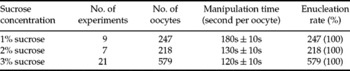
When 1, 2 or 3% sucrose concentrations were used to treat the MII oocytes for enucleation, respectively, the enucleation rates of MII oocytes could reach 100%. Nevertheless, when the micromanipulation was performed with medium containing 1% sucrose, the MII oocyte spindles could not be seen clearly and more time was needed to rotate the oocytes and to look for the nuclei and, therefore, more time was needed to complete the enucleation procedure for each oocyte. Whereas, the mean time of each NT performance was significantly reduced when using manipulation media containing 2 or 3% sucrose (Table 2).
Effects of different sucrose concentrations on the time of nuclear transfer manipulation

Effects of different sucrose concentrations on the fusion and in vitro development of NT embryos
The reconstructed embryos were put in CZB manipulation medium containing 1, 2 or 3% sucrose and then induced to fuse with an electric DC pulse and activated by treatment with SrCl2 in Ca2+-free CZB medium for 6 h, and finally, cultured in CZB medium to morula/blastocyst stages. Our results showed that the different concentrations of sucrose had no significant effect on fusion and in vitro development of the NT embryos (p > 0.05, Table 3).
Effects of different sucrose concentration on the fusion rate and in vitro development of reconstructed embryos

Fusion rate = No. of fused oocytes/No. of oocytes; Development rate of embryos at different stages = No. of embryos at different stages/No. of fused oocytes.
aValues in the same column indicate no significant difference (p > 0.05).
M, morula; B, blastocyst.
In vivo development of NT embryos
A total of 59 NT embryos at the pseudo-pronuclear or 2-cell stages were transferred into the oviducts of two pseudopregnant Kunming females at day 1. One of the two recipient mothers became pregnant, but this pregnant mother became sick and was very weak from day 17 and three dead fetuses were obtained at 21 day by Caesarean section.
Discussion
Enucleation of the recipient oocytes is one of the key steps in NT. The mouse MII oocyte nuclei could be observed clearly under an inverted microscope equipped with Nomarski or Hoffmann optics, so that enucleation could be performed accurately (Ogura et al., Reference Ogura, Inoue, Takano, Wakayama and Yanagimachi2000b). Wang et al. (2001a, b) reported that the enucleation rate of mouse MII oocytes could reach to 100%, when they were treated with 3% sucrose and resulted in live mouse offspring if reconstructed by MII chromosome exchanged and combined with in vitro fertilization. Kong & Zhang et al. (Reference Kong, Zhang, Zhong, Li, Sun and Chen2005) also demonstrated that 3% sucrose-treated mouse MII oocytes could support full-term development of pronuclear transplanted embryos and result in normal offspring. These results demonstrated that sucrose treatment for enucleation might be a safe method. In our present study, we used 1, 2 and 3% sucrose concentrations to treat mouse MII oocytes and the enucleation rate was the same as found by the Wang group. In addition, because of the high osmotic pressure generated by sucrose, the perivitelline spaces of mouse oocytes were enlarged. This necessitated slitting the zona pellucida, therefore, to avoid injury to the oocyte membrane.
To investigate whether or not sucrose treatment was a safe way to generate enucleated oocytes for mouse NT, we used three different sucrose concentrations to treat oocytes and compared the effects of concentration on the fusion and in vitro development of NT embryos. The results demonstrated that higher sucrose concentration (3%) had no more negative effects than lower concentrations (1 and 2%) on the embryos (Table 3). On the other hand, 3% sucrose could facilitate the enucleating operation and reduce the total time needed for nuclear transfer (120 ± 10s versus 180 ± 10s for each NT operation). Therefore, the use of 3% sucrose treatment was the preferred method.
In mouse cloning, when the blastomeres from early embryos served as nuclear donors, fusion of NT embryos was mediated in general by inactivated Sendai virus (McGrath & Solter, Reference McGrath and Solter1983; Kono et al., Reference Kono, Kwon and Nakahara1991; Tsunoda & Kato, Reference Tsunoda and Kato1997). In some cases, the electrofusion method was also employed by researchers (Cheong et al., Reference Cheong, Takahashi and Kanagawa1993; Tsunoda & Kato, Reference Tsunoda and Kato1997). Kono et al. (Reference Kono, Kwon and Nakahara1991) reported that the fusion rate was above 90% when the nuclei of 2-cell and 8-cell embryos were used as donors, which was significantly higher than our results (around 60%, Table 3). However, there was also report that when mouse fetal fibroblast cells were used as donors, the fusion rate was around 58–69% (Ono et al., Reference Ono, Shimozawa, Ito and Kono2001). This result was similar to our findings. Furthermore, electrofusion times could affect the developmental capacity of NT embryos; when the embryos were electrofused twice, the developmental competence decreased drastically and the fragmentation of embryos improved significantly (unpublished data).
Electrical stimulation and 7% ethanol treatment were generally adopted to activate the NT embryos when using early embryonic blastomeres as nuclear donors (Kono et al., Reference Kono, Kwon and Nakahara1991; Tsunoda & Kato, Reference Tsunoda and Kato1997); whereas, SrCl2 treatment was commonly used for the activation of NT embryos from somatic cell donors (Wakayama et al., Reference Wakayama, Perry, Zuccotti, Johnson and Yanagimachi1998, Reference Ono, Shimozawa, Ito and Kono1999; Ogura et al., Reference Ogura, Inoue, Takano, Wakayama and Yanagimachi2000b; Ono et al., Reference Ono, Shimozawa, Ito and Kono2001). When the nuclei of 2-cell, 8-cell and inner cell mass cells were used as donors, the activation rates by ethanol treatment were 86, 90 and 72% (Kono et al., Reference Kono, Kwon and Nakahara1991). There was no significant difference compared with our result (data not shown). Therefore, SrCl2 activation is a practical approach for NT embryos derived from nuclei of early embryos.
To demonstrate the in vivo developmental potential of NT embryos with compacted morula blastomeres as nuclear donors, we transplanted 59 pseudopronuclear and 2-cell NT embryos into the oviducts of two pseudopregnant Kunming mouse recipients and three full-term developed fetuses were obtained. These results indicated that sucrose treatment, electrofusion and SrCl2 activation protocols were applicable for mouse embryonic cloning. Although the cloning efficiency might be relatively low (three out of 59), this method could be an alternative approach for mouse cloning, especially when Normarski or Hoffmann optics was not available in the laboratory.
Acknowledgements
Grant sponsor: the National Natural Science Foundation of China (39830280); Major State Basic Research Project (G20000161).