The prevalence of patent foramen ovale in the general population ranges from 20 to 30%, Reference Horlick, Kavinsky and Amin1,Reference Kuramoto, Kawamura and Dembo2 and PFO can lead to various conditions, such as cryptogenic stroke, ischaemic stroke, and migraine. Researchers have investigated whether patients with cryptogenic stroke would benefit from patent foramen ovale closure with interventional treatment compared with antiplatelet treatment. Reference Mas, Derumeaux and Guillon3 The Amplatzer® Occluder (St. Jude Medical, Inc.; St Paul, MN) is widely used for most types of patent foramen ovale closure. However, after patent foramen ovale interventional closure, puncture of the interatrial septum is difficult across an existing patent foramen ovale occluder device, but is often needed for interventional therapy of atrial fibrillation and valvular heart disease. These complications exhibit an increasing incidence and are urgent problems that must be solved in the clinic. To facilitate atrial septal puncture and minimally invasive interventional therapy after patent foramen ovale transcatheter closure, we designed and manufactured a nitinol occluding device (Shanghai Push Medical Device Co., Ltd) to reserve the atrial septal puncture area. Here, we explore the feasibility of transseptal puncture after patent foramen ovale closure with the occluder in a canine animal model.
Materials and methods
Device and laboratory animals
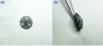
This paper presents the results of an in vitro study of a reserved atrial septal puncture area patent foramen ovale occluder. The reserved atrial septal puncture area was made of superelastic nitinol wire. The occlude device’s left atrial and right atrial side plates were reserved for puncture atrial septal access, where only the polyester fibre membrane was set for blocking blood flow. The right and left sides of the plate through the diaphragm path were symmetrical, and the centre was riveted with stainless steel. Three layers of polyester fibre membrane lined the middle of the device to prevent diversion (Fig 1), and the occluder was delivered using 8F to 12F delivery sheaths.
The shape of RASPAO; (a): Right ventricular surface-view of the occluder; (b): Sideview of the occlude.

Ten experimental canines were used to establish patent foramen ovale models, and all of them were prepared 3 days before the operation. The average weight of the experimental canines was 29.14 ± 2.25 kg. Canines were subject to 12 h/12 h alternating lighting per day, fed twice per day, given free access to water daily, and adapted to constant temperature and humidity in the animal room. All canines were pre-operatively prepared before operation.
Pre-operative preparation
Anaesthesia (tiletamine hydrochloride and zolazepam hydrochloride) was administered by intramuscular injection. Then, the trachea was intubated to establish respiratory access. Sevoflurane was inhaled to maintain anaesthesia. The jugular vein was punctured to establish venous access. The femoral artery was punctured to monitor ambulatory blood pressure, and the femoral vein was punctured to establish transcatheter access. The canines were placed supine and fixed on the operating table with a special fixator for vital sign monitoring.
Establishing patent foramen ovale animal models
After routine disinfection, sterile tissue sheets were laid. The right femoral vein was punctured, and a 7Fr vascular sheath was inserted. Right atrial, right ventricular, and pulmonary artery pressure were examined by right cardiac catheterisation after intravenous infusion of 2000U heparin. Simultaneously, the atrial septum and superior vena cava locations of the bilateral atria were determined using transoesophageal ultrasound. An 8F septal sheath was guided from the femoral vein to the superior vena cava with a 0.032-inch guidewire under fluoroscopy, and a Swartz catheter was used to perform fossa punctures based on a three-point positioning method: (1) The top of the tube core was slowly lowered to the oval socket at 4° ‘clock and fixed. (2) The tube was turned to the left at 90° until the top direction of the tube core was parallel to the spine and was located midpoint to lower-middle 1/3 of the line between the heart shadow trailing edge and aorta. Then, the sliding sheath catheter was inserted into the ovale, and the tube core was pushed until a breakthrough was achieved. Left atrial pressure was shown in the monitoring device, and a contrast agent was injected into the left atrium to guide the sheath catheter through the valve. (3) Then, the sliding sheath catheter is turned to the right, moved forward 30°, and secured to the core. Then, the Swartz catheter was gently delivered. The internal catheter was secured before approaching the ascending aorta. The external sheath was pushed to bend the distal end of the outer sheath under the aortic valve. The tube core was withdrawn and sent into the left atrial steel wire via the Swartz catheter. A model of PFO with a specific diameter was formed after the high-pressure balloon (DK MedTech, Inc; Suzhou Jiangsu) was sent into the ovale through the left atrial wire. The stiffened guidewire was exchanged and fed after the diameter and shape of the artificial patent foramen ovale were achieved.
Closure of artificial patent foramen ovale with reserved atrial septal puncture area
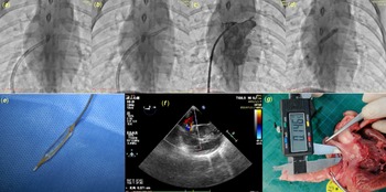
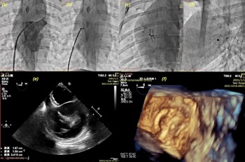
Finally, the patent foramen ovale was successfully set up. ECG and transoesophageal ultrasound were used to observe the arrhythmia and shunt after interventional closure. An intramuscular injection of 100,000 U/kg of penicillin was administered to prevent infection during the operation, and penicillin was administered 3 days after the procedure (Figs 2 and 3).
PFO animal model; ( a ): Atrial septal puncture site; ( b ): Atrial septal puncture; ( c ): Left atrial angiography; ( d ): Balloon expansion; ( e ): Dkonquer high-pressure balloon; ( f ): Morphology and diameter of PFO; ( g ): Anatomical view of the measurement of PFO diameter.

Model of PFO closure; ( a ): Left atrial angiography after an atrial septal puncture; ( b ), ( c ), and ( d ): Interventional closure using a RASPAO; ( e ): Transoesophageal ultrasonography after closure; ( f ): Reconstruction of the left atrial surface of the reserved atrial septal puncture area occlusion using transoesophageal ultrasonography.

Transarterial septal puncture through reserved atrial septal puncture area device
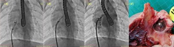
The patent foramen ovale canine models undergoing patent foramen ovale interventional closure using reserved atrial septal puncture area were fed for 3 months. Then, the trans-septal puncture was performed through the novel occluder using a puncture catheter. First, the puncture needle was pointed to the reserved area on the occluder, and the sheath catheter was moved to the reserved puncture area. Then, the puncture needle was pushed forward to achieve a sense of breakthrough. Pressure monitoring showed a left atrial pressure. Contrast agents were injected into the left atrium after the puncture and left atrial pressure monitoring was performed (Fig 4).
Transatrial septal puncture after interventional closure using a new occluder; ( a ): Locating the reserved puncture area; ( b ): Transatrial septal puncture based on the reserved puncture area; ( c ): Left atrial angiography; ( d ): Anatomical view of the left atrial surface of the new occluder after transeptal puncture.

The morphology and properties
The new occluders were removed immediately after a trans-septal puncture, and the gross shape of the occlusion device was observed. The tissue around the occlusion device was obtained for pathological examination, and then the occlusion device was embedded in methyl methacrylate. The occlusion device was sectioned with a diamond knife and stained. The occlusion device’s gross shape and histological changes were observed with a light microscope and scanning electron microscope.
Results
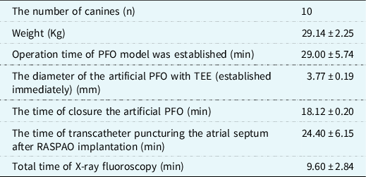
Patent foramen ovale models were successfully established in all canines, and the average diameter of the patent foramen ovale was 3.77 ± 0.19 mm. In the animal model of patent foramen ovale, interventional closure without a residual shunt was successfully achieved as assessed using transoesophageal echocardiography. Moreover, no deaths or complications, including residual shunt, occluder emboliation, or device-related thrombosis were seen. All reserved atrial septal puncture areas implanted in canines remained stable and maintained an optimal shape. The novel occluders did not affect the left ventricular ejection fraction and cardiac structure (Table 1).
Experimental data.

The structural integrity of new reserved atrial septal puncture areas was observed in the pathological specimens after an atrial septal puncture. The occluders maintained their structure without wire breakage or overall damage. The bilateral plates were in close contact with both sides of the atrial septum, and thrombosis was not noted on the surface of the occluders. A layer of grey, smooth translucent tissue covered the device at 3 months of follow-up. Additional puncture holes were observed in the reserved puncture area, and no further damage or tamponade was noted in the heart cavity.
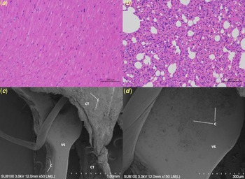
The thickness of endothelial cells increased with the time after implantation, as shown by optical microscopy, and an intact layer of endothelial tissue was formed 3 months after implantation. In addition, the surface of the RASPAOs was covered with endothelial cells, as demonstrated by scanning electron microscopy (Fig 5).
Assessment of new occluders based on pathology and electron microscopy. (a): Myocardial cells and interstitial structures were normal; (b): Epithelial cells in airway tissues were normal without apparent inflammatory cell infiltration; (c) and (d): A small number of flat cells attached to the surface on the new occluder and connective tissue surrounding it.

Discussion
The prevalence of patent foramen ovale is 20–30% in the general population Reference Horlick, Kavinsky and Amin1–Reference Kuramoto, Kawamura and Dembo2 and 27% based on autopsy. Reference Ning, Lo and Ning4 Currently, interventional closure is used for the treatment of most patent foramen ovales. Still, puncture of the interatrial septum is challenging to achieve given the influence of occluder for further interventional treatments, such as atrial septostomy for therapy of atrial fibrillation, treatment of valvular heart disease, and heart failure, which have an increasing incidence. Reference Schnabel, Yin and Gona5–Reference Fordyce, Roe and Ahmad7 For example, the incidence and prevalence of atrial fibrillation are increasing worldwide, Reference Benjamin, Muntner and Alonso8 and the treatment of atrial fibrillation is percutaneous catheter ablation. Reference Haissaguerre, Jais and Shah9–Reference Calkins, Hindricks and Cappato10 Furthermore, transcatheter valve replacement or repair is widely used to treat mitral regurgitation to reduce heart failure and death in the clinic. Reference Nishimura, Vahanian and Eleid11–Reference Whitlow, Feldman and Pedersen12 Atrial septostomy is the basis for these interventional procedures; therefore, it is imperative to develop a reserved atrial septal puncture area that does not prevent atrial septostomy. In theory, occluders made from biodegradable material do not prevent atrial septostomy. However, these occluders have had many problems preventing application. For example, the occluders have lacked sufficient strength and elasticity and require a larger delivery sheath, and it was not easy to expand the sheath. Reference Lin, Liu and Liu13
A residual right-to-left shunt is rarely noted after patent foramen ovale closure using a traditional occluder. Rigatelli et al. suggested that such strategy driven from identification and measurements of the right atrium and inter-atrial septum components resulted in low complications and high-success rates, mandatory conditions when facing with otherwise healthy subjects, such as the patients with patent oval foramen. Reference Rigatelli, Dell’Avvocata and Ronco14 Rubine et al. analysed six kinds of patent foramen ovale occluders and found that the Cardioform occluder had a higher rate of effective closure. However, the incidence of atrial fibrillation- and device-related thrombotic events was greater than that of the Amplatzer occluder after the procedure, Reference Regmi, Lara Garcia and Parajuli15,Reference Hardt, Eicken and Berger16 and the incidence of atrial fibrillation was 13%. Reference Gevorgyan Fleming, Kumar and West17 In addition, more risks of a forward residual shunt and wireframe fracture upon closure were noted. Reference Sommer, Love and Paolillo18 RESPECT suggested that the incidence of atrial fibrillation was 4% after the closure of patent foramen ovale with an Amplatzer occluder Reference Carroll, Saver and Thaler19 in a study of 1000 patients undergoing patent foramen ovale closure; however, no deaths occurred. Reference Krumsdorf, Ostermayer and Billinger20 Therefore, in this study, the reserved atrial septal puncture area was modelled based on the structure of a traditional occluder. It has a double-disc shape with some holes in the unique weaving way. Thus, the performance of the reserved atrial septal puncture area was not inferior to that of the Amplatzer occluder, and the incidence of atrial fibrillation and thrombus events was low when using the novel occluder. The reserved atrial septal puncture area could completely block off the area without residual shunt in this experiment. The atrial septostomy area was preserved in the novel occlude to facilitate subsequent puncture operations.
Moreover, none of the experimental canines with patent foramen ovale occluders had atrial fibrillation, suggesting that RASPAO positively influenced reducing the incidence of atrial fibrillation after the intervention. A retrospective study found that patent foramen ovale with traditional occluders might increase the risk of new arrhythmias, either temporarily or persistently, compared with medication, and the risk of atrial fibrillation increased 5.3-fold. Reference Chen and Thijs21 The mechanism might be related to atrial stimulation caused by compression of the atrial septum, an inflammatory response, and the formation of return circuits in atria from traditional occluders. Reference Aladdin, Feghali and Jenkins22–Reference Chubb, Whitaker and Williams23 The authors noted that the metal part of the plates is significantly reduced on both sides of the reserved atrial septal puncture area. As a result, the coverage area of the nitinol wire on the atrial septum decreased. In addition, there was only a polyester-fibre membrane to obstruct the shunt in the reserved atrial septostomy area. This design significantly reduced pressure on the atrial septum to minimise tissue oedema, inflammation, and fibrosis, thus reducing the risk of atrial fibrillation.
The occluder with biodegradable material retained the function of atrial septostomy in theory; however, this occluder was associated with many problems that made its application in the clinic complex. These problems include insufficient strength and elasticity, a larger delivery sheath tube, and difficulty expanding the sheath tube after being sent out. Reference Lin, Liu and Liu13 These results showed that the reserved atrial septal puncture area had a positive efficiency for closure in the canine model of patent foramen ovale, and residual shunt and displacement of the reserved atrial septal puncture area were not observed. We found that the novel occluder was covered entirely with endothelial cells without thrombi, exhibited a new structure, and had an intact nitinol wire based on biological tissue samples from experimental canines.
Atrial septostomy was performed successfully three months after patent foramen ovale interventional closure with reserved atrial septal puncture area, but many problems were found. First, the site of atrial septostomy below the reserved atrial septostomy area had a higher rate of success than the area above it. Second, the reserved atrial septal puncture area still has some influence on atrial septostomy. Third, it was challenging to maintain coaxial puncture holes on both sides in the reserved atrial septostomy area. These defects will be improved in future studies.
This study had some limitations, such as the small animal numbers and short follow-up time. The study did not include a regular PFO control group, and the canine model of patent foramen ovale was established using balloon dilatation. Therefore, morphological differences were noted compared with patent foramen ovale patients. A more extensive cohort study will be performed to provide sufficient data for potential clinical application in the future.
Acknowledgements
This work was funded by the Translational Medicine Project of the Wuxi Municipal Health Commission [ZH202107].
Conflicts of interest
None.
Ethical approval
All applicable institutional and/or national guidelines for the care and use of animals were followed.