Introduction
Obsessive-compulsive disorder (OCD) has been discussed as a disorder with potential neurodevelopmental risk factors (Rosenberg & Keshavan, Reference Rosenberg and Keshavan1998; Huyser et al. Reference Huyser, Veltman, De Haan and Boer2009), but surprisingly few studies investigated the potential neural indicators for this assumption.
One useful marker to assess early defects in neurodevelopment in the brain is cortical folding or gyrification. Cortical folding is known to develop during prenatal life and to be terminated to a very large degree before the age of 2 years (Armstrong et al. Reference Armstrong, Schleicher, Omran, Curtis and Zilles1995; Magnotta et al. Reference Magnotta, Andreasen, Schultz, Harris, Cizadlo, Heckel, Nopoulos and Flaum1999), before it starts to slightly decrease between childhood and young adulthood [according to a review by Mills & Tamnes (Reference Mills and Tamnes2014) by up to 7%]. Therefore, cortical folding seems to be a reliable marker for early neurodevelopmental alterations in the brain.
Answering the question if early developmental alterations already occur in patients would help to better understand the nature and mechanisms behind the existing structural alterations in OCD which – according to a recent review (Piras et al. Reference Piras, Piras, Chiapponi, Girardi, Caltagirone and Spalletta2015) – provide a rather heterogeneous picture. To the best of our knowledge cortical folding patterns in OCD have been investigated in only four studies up to now (Shim et al. Reference Shim, Jung, Choi, Jung, Jang, Park, Choi, Kang and Kwon2009; Wobrock et al. Reference Wobrock, Gruber, Mcintosh, Kraft, Klinghardt, Scherk, Reith, Schneider-Axmann, Lawrie, Falkai and Moorhead2010; Venkatasubramanian et al. Reference Venkatasubramanian, Zutshi, Jindal, Srikanth, Kovoor, Kumar and Janardhan Reddy2012; Fan et al. Reference Fan, Palaniyappan, Tan, Wang, Wang, Li, Zhang, Jiang, Xiao and Liddle2013), which show discrepant results overall.
Two of them found hypogyrification in the OCD sample compared to healthy controls using a classification approach of cortical folding patterns in regions of interest (ROIs). This hypogyrification was detectable in the left anterior cingulate cortex (ACC; Shim et al. Reference Shim, Jung, Choi, Jung, Jang, Park, Choi, Kang and Kwon2009) and the left prefrontal cortex (PFC; Wobrock et al. Reference Wobrock, Gruber, Mcintosh, Kraft, Klinghardt, Scherk, Reith, Schneider-Axmann, Lawrie, Falkai and Moorhead2010). The other two studies used a different methodological approach by calculating a local gyrification index (lGI). While Venkatasubramanian et al. (Reference Venkatasubramanian, Zutshi, Jindal, Srikanth, Kovoor, Kumar and Janardhan Reddy2012) did not find any differences in lGI between OCD patients and healthy subjects, Fan et al. (Reference Fan, Palaniyappan, Tan, Wang, Wang, Li, Zhang, Jiang, Xiao and Liddle2013) found a hypergyrification in OCD patients in the left insula, the left middle frontal and left lateral occipital regions extending to precuneus as well as in the right supramarginal gyrus.
Little is known about the association between clinical characteristics and altered cortical folding in OCD although there is first evidence indicating that medication, symptom severity and even disorder insight might be related to folding abnormalities in OCD. Comparing severely and mildly affected patients with healthy controls Wobrock et al. (Reference Wobrock, Gruber, Mcintosh, Kraft, Klinghardt, Scherk, Reith, Schneider-Axmann, Lawrie, Falkai and Moorhead2010) reported a stronger hypogyrification in patients with severe symptoms. Venkatasubramanian et al. (Reference Venkatasubramanian, Zutshi, Jindal, Srikanth, Kovoor, Kumar and Janardhan Reddy2012) found a negative association between lGI of right lateral orbitofrontral cortex (OFC) and compulsion score and between lGI left medial OFC and disorder insight. Opposite to these findings Fan et al. (Reference Fan, Palaniyappan, Tan, Wang, Wang, Li, Zhang, Jiang, Xiao and Liddle2013) reported a positive association between left insula lGI and symptom severity and an effect of medication on lGI values in the left insula in medication naive compared to previously medicated patients that, however, did not reach statistical significance.
Gyrification has also been investigated in several other psychiatric disorders (i.e. schizophrenia, autism, depression, panic disorder). In patients with psychotic disorders and their first-degree relatives, Nanda et al. (Reference Nanda, Tandon, Mathew, Giakoumatos, Abhishekh, Clementz, Pearlson, Sweeney, Tamminga and Keshavan2014) found a hypogyrification of the cingulate cortex compared to healthy participants suggesting that hypogyrification may mark a certain familial risk for psychotic disorders. In patients with panic disorder Yoon et al. (Reference Yoon, Jun, Jeong, Lee, Lim, Ma, Ko, Cho, Yeum and Lyoo2013) showed a hypogyrification in lateral brain areas extending from fronto-parietal areas (including precuneus) to the temporal pole, as well as an association between hypergyrification in posterior-medial areas and alleviation of symptoms, suggesting that an increased gyrification could constitute a compensational mechanism for hypogyrification affecting other, partly adjacent, areas. Similar findings were reported in major depressive disorder (MDD), where Zhang et al. (Reference Zhang, Yu, Zhou, Li, Li and Jiang2009) were the first to show a decreased gyrification in precuneus and posterior cingulate cortex (PCC), insula and OFC. Nixon et al. (Reference Nixon, Liddle, Nixon, Worwood, Liotti and Palaniyappan2014) replicated this finding of decreased precuneus gyrification bilaterally in patients recovered from MDD, and furthermore showed this hypogyrification to be associated with a hyperconnectivity between precuneus and dorsolateral prefrontal cortex (DLPFC).
Overall, these cortical folding studies in psychiatric disorders point towards an altered gyrification in areas responsible for emotional processing but also cognitive control. Considering that these disorders share – to some degree – some of their symptomatology and often co-occur or precede each other, it may be meaningful that they exhibit structural alterations in partly the same anatomical regions. Hence, these findings seem to support the hypothesis that neurodevelopmental deficits, partly mirrored in cortical folding deficits, may represent possible risk factors for the development of psychiatric disorders.
Against this background, the question whether structural alterations in OCD occur with or because of disorder progression or whether they have an early, neurodevelopmental origin needs to be further elucidated. Studying cortical folding in OCD may thus lead to a better understanding of possible neurodevelopmental risk factors that could constitute an early cause for characteristic alterations in OCD, such as disruptions within the cortico-striato-thalamo-cortical circuit (Saxena & Rauch, Reference Saxena and Rauch2000). The heterogeneity of results from the existing studies (Shim et al. Reference Shim, Jung, Choi, Jung, Jang, Park, Choi, Kang and Kwon2009; Wobrock et al. Reference Wobrock, Gruber, Mcintosh, Kraft, Klinghardt, Scherk, Reith, Schneider-Axmann, Lawrie, Falkai and Moorhead2010; Venkatasubramanian et al. Reference Venkatasubramanian, Zutshi, Jindal, Srikanth, Kovoor, Kumar and Janardhan Reddy2012; Fan et al. Reference Fan, Palaniyappan, Tan, Wang, Wang, Li, Zhang, Jiang, Xiao and Liddle2013) as well as methodological differences illustrate the need for further research in this field.
On this account we intended to examine cortical folding differences in a large sample of OCD patients and healthy controls (i.e. 75 OCD patients, 75 healthy subjects) and to investigate how potential structural alterations might relate to age of onset and symptom type (i.e. obsessions v. compulsions) or severity. We used the approach described by Schaer et al. (Reference Schaer, Cuadra, Schmansky, Fischl, Thiran and Eliez2012) to quantify local gyrification by computing the lGI which represents the amount of cortex buried within the sulcal folds as compared with the amount of visible cortex in circular ROIs.
Method
Participants
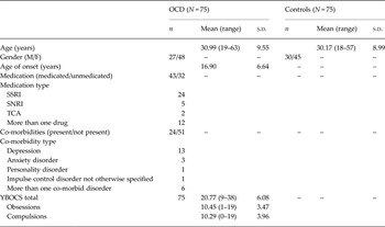
The study sample comprised 75 right-handed patients meeting the DSM-IV criteria for OCD and 75 right-handed healthy controls matched for age (t 148 = 0.54, p = 0.58) and gender (χ2 1 = 0.1, p = 0.73).
Forty-two patients were recruited from the Windach Institute and Hospital of Neurobehavioral Research and Therapy (WINTR), Germany. Thirty-three patients were recruited from the University Hospital for Psychiatry and Psychotherapy Jena, Germany. All 75 were in-house patients in wards specialized on OCD with a standardized admission process, standardized psychopathological screenings and standardized assessment of disorder history performed by an experienced psychiatrist. 57% of all patients were medicated and 32% suffered from one or more co-morbid psychiatric disorder (see Table 1).
Demographic and clinical characteristics of the sample

SSRI, selective serotonin reuptake inhibitor; SNRI, serotonin-norepinephrine reuptake inhibitor; TCA, tricyclic antidepressant; YBOCS, Yale–Brown Obsessive Compulsive Scale.
Exclusion criteria for both groups were a history of clinically important head injuries, seizures or neurological diseases. Healthy controls with a history of psychiatric illness were excluded. Exclusion criteria for patients were schizophrenia, autism, substance and alcohol abuse/dependency, mental retardation, pregnancy, and severe medical conditions.
After complete description of the study aims, written informed consent was obtained from the subjects. The study protocol was in compliance with the Declaration of Helsinki and approved by the Ethics Committees of the Klinikum rechts der Isar and the University of Jena. Prior to the scanning session we assessed demographic characteristics and symptom severity using the Yale–Brown Obsessive Compulsive Scale (YBOCS; Goodman et al. Reference Goodman, Price, Rasmussen, Mazure, Fleischmann, Hill, Heninger and Charney1989).
Image acquisition
Controls and patients recruited from WINTR were scanned at the Department of Neuroradiology, Klinikum rechts der Isar, Technische Universität München, Germany. Patients from Jena were scanned at the University Hospital Jena.
High-resolution anatomical T1-weighted scans from Jena were acquired in a 3-T whole body system equipped with a 12-element receive-only head matrix coil (MAGNETOM TIM Trio, Siemens Medical Solutions, Germany). High-resolution anatomical T1-weighted volume scans (MP-RAGE) were obtained in sagittal orientation [TR = 2300, TE = 3.03, TI = 900 ms, flip angle = 9°, FOV = 256 × 256 mm2, matrix = 256 × 256 mm, number of sagittal slices = 192, acceleration factor (PAT) = 2] with an isotropic resolution of 1 × 1 × 1 mm3.
Data from Munich were collected on a 3-T whole-body system equipped with a 12-element receive-only head matrix coil (INGENIA, Philips Healthcare, The Netherlands). High-resolution anatomical T1-weighted volume scans (MP-RAGE) were obtained in sagittal orientation (TR = 9, TE = 4, TI = 900 ms, flip angle = 8°, FOV = 240 × 240 mm2, matrix = 240 × 240 mm, number of sagittal slices = 170) with an isotropic resolution of 1 × 1 × 1 mm3.
Image processing and computation of lGI
We used the FreeSurfer software package (version 5.3.0, http://surfer.nmr.harvard.edu) to process the T1 images according to the standard and automatic processing stream (Fischl & Dale, Reference Fischl and Dale2000). This processing stream includes removal of non-brain tissue, transformation to Talairach-like space, and segmentation of gray/white matter tissue (GM, WM) resulting in two meshes (a white mesh = the WM/GM boundary and a pial mesh = GM/cerebrospinal fluid boundary). The meshes are composed of about 150 000 points (vertices) for each hemisphere. As a next step we computed the lGI at each vertex. This 3D approach of analyzing gyrification takes into account buried sulcis, is not restricted by sulcal walls and takes into consideration the significant variations of lGIs across all sulco-gyral regions of the cortex (Schaer et al. Reference Schaer, Cuadra, Tamarit, Lazeyras, Eliez and Thiran2008). The automatic lGI computation (Schaer et al. Reference Schaer, Cuadra, Schmansky, Fischl, Thiran and Eliez2012) involves: the creation of an outer surface (the smoothed pial surface), creation of 800 ROIs on this surface and their corresponding ROIs on the pial surface. This results in calculation of individual maps, which contain one lGI value for each vertex on the cortical surface. The lGI can have a value ranging between 1 (flat) and 5 (i.e. there is five times more cortical surface buried in sulci than visible cortex in the surrounding area). The individual lGIs were projected on a sample specific template (average subject) and smoothed using a Gaussian kernel of 10 mm.
Statistical analysis
Using the framework of the general linear modeling (GLM) we assessed the regional differences of lGIs between patients and controls at the level of each vertex for each hemisphere separately and included age, gender and scanner type (i.e. Siemens v. Philips) as covariates to correct for potential confounding effects. To correct for multiple comparisons across the whole brain, Monte Carlo simulations (Hagler et al. Reference Hagler, Saygin and Sereno2006) with 10 000 iterations were performed in order to identify significant contiguous clusters of vertex-wise group differences (p < 0.05).
Further statistical analyses were performed using the Statistical Package for Social Sciences [SPSS Inc. (2002), version 11.5.1, USA]. Differences in age and gender were assessed using the χ2 test. The potential association between lGI alterations and clinical parameters (symptom severity: YBOCS total, obsessions and compulsion scores; age of onset) was assessed using multiple linear regression with symptom severity and age of onset as predictors and mean lGI values of the brain regions that were identified as significantly different between the groups as criterion and age, gender and scanner as covariates. The regression analyses were done separately for each hemisphere. In case of a significant relationship the corresponding partial correlation coefficient was reported.
To control for the potential confounding effect of the two scanner types (Siemens v. Phillips) and their different sequences, besides taking scanner type as a covariate in the GLM analysis, we also performed a whole-brain lGI comparison between the two scanner groups.
In addition, to control for the effect of medication and co-morbidity we first performed a whole-brain lGI analysis (GLM) comparing medicated v. unmedicated patients as well as co-morbidity-free v. co-morbid patients with age, gender and scanner type as covariates.
Moreover, to evaluate if medication or co-morbidity affected lGI differences between patients and healthy controls we furthermore performed two MANCOVAs with average lGI values (extracted from the clusters found to be different between patients and controls) as dependent variables, medication or co-morbidity status as independent variables, and age, gender and scanner as covariates.
Results
Differences in gyrification
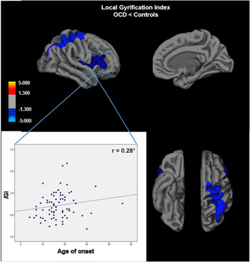
Whole-brain lGI analysis revealed two clusters in the right hemisphere consistently showing a decreased gyrification (hypogyria) in OCD patients compared to controls. The first cluster (cluster 1 in Table 2) showing a significantly reduced lGI (p < 0.01) extended from inferior parietal, superior parietal to supramarginal, post-and precentral and superior frontal areas. The second cluster (cluster 2 in Table 2) showing a significantly reduced lGI (p < 0.05) contained insula, superior- and transverse- temporal areas, pars opercularis, pars triangularis, rostral middle frontal and lateral orbitofrontal regions (for details see Fig. 1 and Table 2). The left hemisphere showed no differences in lGI between the groups. No clusters with increased gyrification were noted in patients. Annotation of clusters is according to the Desikan–Killiany Freesurfer atlas (aparc.annot).
Group difference in local Gyrification Index (lGI). Shown are clusters with significantly decreased lGI of the right hemisphere in OCD patients. The clusters are displayed on the pial surface of the participants’ average brain (lateral, medial, inferior and superior view). The color bar indicates the t value after clusterwise correction for multiple comparisons using Monte Carlo simulations (p < 0.05). The scatterplot represents the significant positive association between age of onset and mean lGI of cluster 2.

Brain regions with significant group differences in local Gyrification Index (lGI). Clusters with a significantly decreased lGI in OCD patients compared to healthy subjects in the right hemisphere after clusterwise correction for multiple comparisons using Monte Carlo simulation (p < 0.05)

VtxMax, Number of peak vertex of the significant cluster; CWP, cluster-wise probability and the nominal p value; CWPLow, CWPHi – the 90% confidence intervals of the p value; NVtx, number of vertices in cluster; *p < 0.05, **p < 0.01.
Effects of medication, co-morbidity and scanner sequence
The whole-brain lGI GLM analysis revealed no significant differences between medicated and unmedicated patients or between co-morbid and co-morbidity-free patients. Furthermore, the results of the MANCOVA showed that the lGI alterations were not influenced by medication status (medicated v. unmedicated patients, cluster 1: F = 0.238, p = 0.788, cluster 2: F = 0.878, p = 0.418) or presence of co-morbidity (co-morbid v. co-morbidity-free patients, cluster 1: F = 0.243, p = 0.785, cluster 2: F = 0.859, p = 0.426). Moreover, no significant differences in lGI between the data from the different scanner types of the two centers could be found.
Gyrification and clinical variables
The regression analysis revealed that in OCD patients altered gyrification (lGI values extracted from the clusters showing a significant alteration in patients v. controls) was not associated with any of the symptom severity scores (YBOCS total, obsessions or compulsions). The second regression analysis revealed that age of onset was positively associated (β = 0.27, T = 2.35, p = 0.02; partial correlation r = 0.28, p = 0.02) with the average lGI in the second cluster (see Table 2 and Fig. 1) in the right hemisphere including insula, superior- and transverse- temporal areas, pars opercularis, pars triangularis, rostral middle frontal and lateral orbitofrontal regions. This association remained significant also after Bonferroni correction.
Discussion
The present study which investigated whole-brain lGI in patients with OCD revealed that, compared to healthy controls, OCD patients showed a decreased gyrification in several cortical areas. These morphological alterations were not associated with symptom severity, medication status or co-morbidity although there was a positive association with age of onset.
Hypogyrification in OCD
Our results partly confirm previous findings which reported also hypogyrification in OCD (Shim et al. Reference Shim, Jung, Choi, Jung, Jang, Park, Choi, Kang and Kwon2009; Wobrock et al. Reference Wobrock, Gruber, Mcintosh, Kraft, Klinghardt, Scherk, Reith, Schneider-Axmann, Lawrie, Falkai and Moorhead2010), although they used a ROI-based approach and employed slightly different methods in calculating gyrification by either using the automated-gyrification index (Moorhead et al. Reference Moorhead, Harris, Stanfield, Job, Best, Johnstone and Lawrie2006) or employing a manual 2D segmentation (Van Essen & Drury, Reference Van Essen and Drury1997) instead of an automatic 3D approach.
When relating our findings to the existing OCD studies that are methodologically more comparable to ours [i.e. studies assessing the lGI as proposed by Schaer et al. (Reference Schaer, Cuadra, Tamarit, Lazeyras, Eliez and Thiran2008)] such as a study by Venkatasubramanian et al. (Reference Venkatasubramanian, Zutshi, Jindal, Srikanth, Kovoor, Kumar and Janardhan Reddy2012) one notices rather divergent results. Thus, Venkatasubramanian and colleagues found no overall gyrification differences but an association between reduced lGI and increased symptom severity. It should be noted, however, that they employed a ROI based lGI approach and studied medication naive patients whereas we employed a whole-brain lGI analysis and our sample was partly medicated.
Our results moreover contradict the results by Fan et al. (Reference Fan, Palaniyappan, Tan, Wang, Wang, Li, Zhang, Jiang, Xiao and Liddle2013) who studied, however, unmedicated and co-morbidity-free patients. Fan and colleagues reported altered gyrification in similar regions but in the opposite direction (i.e. they found an increased gyrification in OCD patients compared to controls). As mentioned above, neither medication nor co-morbidity which affected a certain percentage of our patients had a statistically significant influence. Nevertheless, a certain influence of medication and co-morbidity cannot be excluded which might explain the contrary findings.
Overall, comparability between the current study and the existing literature must be regarded as rather limited, mainly due to differences in methodology and clinical characteristics of the sample. From this perspective, a generalization of the results seems premature and, likewise, one can only speculate about the processes and mechanisms underlying altered cortical folding in OCD.
There are several theories regarding the driving force of cortical folding which are discussed in a review paper of Zilles et al. (Reference Zilles, Palomero-Gallagher and Amunts2013), but recent research on the mechanistic processes underlying cortical folding leave a number of open questions (Stahl et al. Reference Stahl, Walcher, De Juan Romero, Pilz, Cappello, Irmler, Sanz-Aquela, Beckers, Blum, Borrell and GotZ2013; Tallinen et al. Reference Tallinen, Chung, Biggins and Mahadevan2014, Reference Tallinen, Chung, Rousseau, Girard, Lefevre and Mahadevan2016).
One established theory about the driving force of cortical folding assumes that cortical folding is the consequence of too many neurons restricted in a confident space, causing tension along WM axons (Van Essen, Reference Van Essen1997). Somewhat in accordance with this theory, previous OCD studies showed widespread alterations in WM fiber bundles (Piras et al. Reference Piras, Piras, Caltagirone and Spalletta2013; Koch et al. Reference Koch, Reess, Rus, Zimmer and Zaudig2014), with alterations in pathways targeting the orbitofrontal areas, but also consistent anatomical connectivity alterations between intra-hemispheric lateral frontal and parietal regions. Considering that the present study showed hypogyrification in proximal areas of the right hemisphere, one could speculate that a disrupted fiber integrity going along with a lower tension along these fiber bundles may result in a lower gyrification of connected brain regions.
Structural alterations linked to gyrification
On the one hand, it is known that cortical folding relates to and depends on several other structural measures, such as GM volume, surface area or cortical thickness (Raznahan et al. Reference Raznahan, Shaw, Lalonde, Stockman, Wallace, Greenstein, Clasen, Gogtay and Giedd2011). These measures have been repeatedly shown to be altered in OCD with cortical volume reductions affecting mainly OFC, ACC and temporo-limbic areas (Piras et al. Reference Piras, Piras, Chiapponi, Girardi, Caltagirone and Spalletta2015). Moreover, a recent mega-analysis also showed a reduction of both GM and WM in various frontal regions, the ACC and the insula (De Wit et al. Reference De Wit, Alonso, Schweren, Mataix-Cols, Lochner, Menchon, Stein, Fouche, Soriano-Mas, Sato, Hoexter, Denys, Nakamae, Nishida, Kwon, Jang, Busatto, Cardoner, Cath, Fukui, Jung, Kim, Miguel, Narumoto, Phillips, Pujol, Remijnse, Sakai, Shin, Yamada, Veltman and Van den Heuvel2014). Results also showed that patients lose more volume in temporal cortex with age compared to healthy subjects. In the present study parts of these areas, which were already known to show structural alterations in OCD were also affected by a decreased lGI. One could speculate that the lGI is altered already during early development and this, in turn, may favor structural alterations found in patients at a later age.
Alterations in the temporal course of development
On the other hand it has been shown that all these structural measures develop and change at their own pace, reaching their morphological maturation at different ages (Raznahan et al. Reference Raznahan, Shaw, Lalonde, Stockman, Wallace, Greenstein, Clasen, Gogtay and Giedd2011). One could assume that a certain delay in the maturational process of one morphological measure will lead to a delay in others, given that the single measures are not completely independent from each other. Furthermore, structural morphology studies in pediatric OCD patients showed structural alterations in subcortical regions in children/adolescents while alterations in more cortical regions could be observed in adult OCD patients (Huyser et al. Reference Huyser, Veltman, De Haan and Boer2009). This review article suggested that structural alterations in adulthood may be related to alterations already present in early childhood and discussed the possibility of a ‘migration of pathology’ during the course of the disorder. Our results reveal cortical alterations in adult patients with OCD and confirm partly previous results, although more longitudinal studies are needed to confirm this pathology migration hypothesis.
Moreover, studies in healthy individuals show that the lGI is slightly decreasing during adolescence in precentral, temporal and frontal areas (Klein et al. Reference Klein, Rotarska-Jagiela, Genc, Sritharan, Mohr, Roux, Han, Kaiser, Singer and Uhlhaas2014). After correcting for age, we could also find similar regions to be significantly reduced in OCD. This can lead to the assumption that, if alterations occurred, they could also be related to an altered developmental pace leading to a decreased overall gyrification in OCD patients compared to healthy subjects. Unfortunately, most results are based on cross-sectional studies up to know, and longitudinal or pediatric studies on brain gyrification in OCD to test this hypothesis are still missing.
General hypogyrification in psychiatric disorders
Interestingly, hypogyrification has been found also in several other psychiatric disorders, some of them known to have a neurodevelopmental predisposition (Zhang et al. Reference Zhang, Yu, Zhou, Li, Li and Jiang2009; Yoon et al. Reference Yoon, Jun, Jeong, Lee, Lim, Ma, Ko, Cho, Yeum and Lyoo2013; Nanda et al. Reference Nanda, Tandon, Mathew, Giakoumatos, Abhishekh, Clementz, Pearlson, Sweeney, Tamminga and Keshavan2014; Nixon et al. Reference Nixon, Liddle, Nixon, Worwood, Liotti and Palaniyappan2014). Furthermore, it is known that there are similarities between OCD and other psychiatric disorders in terms of circuitries and systems which are presumed to be psychopathologically relevant (e.g. alterations within fronto-striatal circuits in schizophrenia and OCD, altered serotonin/dopamine system in schizophrenia and OCD) as well as a high co-occurrence rate (Tibbo & Warneke, Reference Tibbo and Warneke1999; Bradshaw & Sheppard, Reference Bradshaw and Sheppard2000). It seems plausible to assume that similar patterns in neurobiology underlie these clinical commonalities between the disorders with altered cortical folding constituting an important common feature characterizing different psychiatric disorders.
Gyrification and clinical characteristics
There was no association between altered lGI and clinical scores (i.e. symptom severity) although such associations (i.e. symptom severity, disorder insight) had been previously reported by other OCD gyrification studies (Wobrock et al. Reference Wobrock, Gruber, Mcintosh, Kraft, Klinghardt, Scherk, Reith, Schneider-Axmann, Lawrie, Falkai and Moorhead2010; Venkatasubramanian et al. Reference Venkatasubramanian, Zutshi, Jindal, Srikanth, Kovoor, Kumar and Janardhan Reddy2012; Fan et al. Reference Fan, Palaniyappan, Tan, Wang, Wang, Li, Zhang, Jiang, Xiao and Liddle2013).
Our results go more in line with neurodevelopmental theories by supporting the assumption that lGI could be considered as a rather stable marker of early neurodevelopment, whereas symptoms are known to dynamically change over time. This lets us speculate that, whereas symptomatology may represent a state marker of the disorder, hypogyrification may indeed constitute a trait characteristic of OCD.
This speculation is also supported by the positive association between age of onset and extent of gyrification alterations in the second cluster containing mainly insular – lateral frontal areas indicating that stronger alteration in gyrification goes along with an earlier age of onset. This finding underlines once more the clinical relevance of these structural alterations and indicates that the degree of folding alterations may represent a neurodevelopmental marker predisposing for an earlier manifestation of the disorder, if we consider cortical folding a measure which remains stable after early childhood.
On the other hand, the above mentioned review by Piras et al. (Reference Piras, Piras, Chiapponi, Girardi, Caltagirone and Spalletta2015) suggests that potential relationships between clinical variables and observed morphological alterations in OCD are rather heterogeneous. Moreover, this review underlines the fact that previous findings which do find significant associations could be triggered by multiple other factors as co-morbid illness or medication use and may even be driven by progressive changes evolving in dynamic trajectories during illness course or merely by the phenotypic heterogeneity of OCD.
Therefore, it is crucial to keep in mind that our knowledge about the influence of disorder onset or progression as well as treatment on brain morphology is still very limited. Thereby, the possibility that cortical alterations occur later in life, potentially as a result of these influencing factors, cannot be ruled out. Hence, further studies, ideally longitudinal designs based on large samples allowing for a stratification of clinical symptoms, are strongly needed to increase our understanding of these mechanisms and relationships regarding the cortical folding and its changes over time.
The current results need also to be viewed in light of certain limitations, i.e. co-morbidities and medication may have influenced our results and their generalizability to some extent. On the other hand, the rather large sample size of 150 participants in total ensures reasonable power and generalizability. Our attempts to control for these confounding effects revealed no significant influence of these variables on our results. It should be noted that these findings need to be interpreted with caution as other OCD studies indicate that medication (e.g. SSRIs) seems to reduce structural brain differences (in terms of GM volume) between healthy controls and patients (Hoexter et al. Reference Hoexter, De Souza Duran, D'alcante, Dougherty, Shavitt, Lopes, Diniz, Deckersbach, Batistuzzo, Bressan, Miguel and Busatto2012). However, there are no systematic studies on the effects of SSRI treatment on cortical folding and there is little understanding of how medication influences brain morphology in OCD (Atmaca, Reference Atmaca2013). But keeping in mind that morphological characteristics (i.e. volume, thickness, surface area, cortical folding) are linked in their development (Raznahan et al. Reference Raznahan, Shaw, Lalonde, Stockman, Wallace, Greenstein, Clasen, Gogtay and Giedd2011; Mills & Tamnes, Reference Mills and Tamnes2014) and alterations in one characteristic might also affect the other parameters, an indirect effect on cortical folding seems plausible. Moreover, animal research demonstrated that modulations in serotonergic neurotransmission by SSRIs mediate neuroplasticity (neurogenesis and gliogenesis) in various cortical and subcortical structures involved in OCD (Kodama et al. Reference Kodama, Fujioka and Duman2004). Hence, more systematic longitudinal studies are needed to clarify potential effects of medication on cortical folding in OCD.
As a final remark it should be mentioned that the hypogyrification found in the present study might not by itself be the core characteristic of the disorder. Altered cortical folding may be at most one aspect of a complex conglomerate with potential alterations at a functional, structural and cellular level.
Conclusion
In summary, our results of hypogyrified areas in OCD point towards disturbances in cortical brain surface and partly confirm previous findings in OCD patients as well as findings in related psychiatric disorders. Longitudinal studies are needed to reveal if such alterations occur due to different developmental pace or variations in time-point of cortical maturation.
Acknowledgements
We thank the Windach Institute and Hospital of Neurobehavioural Research and Therapy, Windach, Germany, for giving us the opportunity to recruit our patient sample at their institution. This study was supported by a Deutsche Forschungsgemeinschaft (DFG) grant to K.K. (KO 3744/2-1) and to G.W. (WA 3001/3-1). We also thank the Graduate School of Systemic Neurosciences (GSN) for making it possible to share the results with the neuroscientific community at various occasions such as conferences, retreats and symposia.
Declaration of Interest
None.