Introduction
The order Siluriformes is a monophyletic group popularly known as ‘catfish’, with about 35 families and over 3000 valid living species distributed in freshwater, brackish and marine environments in every continent of the world (Nelson, Reference Nelson2006; Sullivan et al., Reference Sullivan, Lundberg and Hardman2006; Eschmeyer et al., Reference Eschmeyer, Fricke and van der Laan2016). Most of these species are widely distributed in Neotropical regions (around 50% of the total number of species), although a large minority (about 72 species) are endemic and have restricted geographic ranges, being known only from the original site and basins in which they were discovered (Burguess, Reference Burguess1989; Eschmeyer et al., Reference Eschmeyer, Fricke and van der Laan2016; Froese & Pauly, Reference Froese and Pauly2016). Loricariidae is one of the most diverse families of this order in numbers of genera and species. This family includes seven subfamilies (Chiachio et al., Reference Chiachio, Oliveira and Montoya-Burgos2008) and 900 valid species, making this taxon the second most species-rich family of Neotropical freshwater fishes after Characidae (Roxo et al., Reference Roxo, Albert, Silva, Zawadzki, Foresti and Oliveira2014). Many species are of considerable economic importance, being sold for use in aquariums or for human consumption, mainly by riverside populations.
In general, loricariids present high intraspecific phenotypic plasticity, resulting in a great variability of shapes and colour throughout their ontogenetic development. Our limited knowledge of their biological characteristics, together with the biogeographic patterns of distribution and high morphological variability of the loricariids, make studies on the group difficult (Reis et al., Reference Reis, Weber and Malabarba1990; Weber, Reference Weber, Reis, Kullander and Ferraris2003; Armbruster, Reference Armbruster2004). Catfish are suitable hosts for a rich and diverse fauna of gill monogeneans, representing an interesting model for phylogenetic studies of monogeneans from the Neotropical region (Mendoza-Palmero et al., Reference Mendoza-Palmero, Scholz, Mendoza-Franco and Kuchta2012).
Monogeneans represent a diverse group of fish parasites, in terms of number of species, morphology and ecology (Poulin, Reference Poulin2002; Thatcher, Reference Thatcher2006). These parasites are known to be highly host specific, parasitizing only one host species or a narrow group of closely related fishes (Thatcher, Reference Thatcher2006). The host specificity shown by most monogeneans makes it easier to relate the ecological characteristics of the hosts and the diversity of their monogenean parasites, and to control the phylogenetic history of their associations (Poulin, Reference Poulin2002). Dactylogyridae is one of the most important and largest families among these parasites, occurring in different species of fishes from continental waters of South America (Thatcher, Reference Thatcher2006). Approximately 379 nominal species belonging to 31 genera have been described in siluriform fishes, with an increasing number of new descriptions in recent years (Mendoza-Palmero et al., Reference Mendoza-Palmero, Scholz, Mendoza-Franco and Kuchta2012, Reference Mendoza-Palmero, Blasco-Costa and Scholz2015; Cohen et al., Reference Cohen, Justo and Kohn2013). Most of the descriptions are based on morphological data, and do not provide the molecular characterization of the species described. Molecular techniques have been used to support taxonomic and morphological descriptions of monogeneans, and provide ways of performing phylogenetic analysis based on different markers (e.g. 18S, internal transcribed spacer (ITS) and 28S rDNA regions of the rDNA gene, and cytochrome c oxidase (COI) and 16S mitochondrial genes) (Plaisance et al., Reference Plaisance, Littlewood, Olson and Morand2005; Simková et al., Reference Simková, Matejusová and Cunningham2006; Wu et al., Reference Wu, Zhu, Xie and Li2006, Reference Wu, Zhu, Xie and Li2007, Reference Wu, Zhu, Xie, Wang and Li2008; Fehlauer-Ale & Littlewood, Reference Fehlauer-Ale and Littlewood2011; Hahn et al., Reference Hahn, Bakke, Bachmann, Weiss and Harris2011; Gilmore et al., Reference Gilmore, Cone, Lowe, King, Jones and Abbott2012; Sarabeev & Desdevises, Reference Sarabeev and Desdevises2014; Mendoza-Palmero et al., Reference Mendoza-Palmero, Blasco-Costa and Scholz2015), and a means of elucidating the relationship between parasite species, their hosts and the relationship between them (Poulin, Reference Poulin2002; Braga et al., Reference Braga, Araújo and Boeger2014).
Demidospermus was erected to include Demidospermus anus Suriano, 1983 as the type species, collected from the gills of Loricariichthys anus (Valenciennes, 1835) (= Loricaria anus) from Argentina. Currently, Demidospermus represents one of the most specious genera among dactylogyrids of freshwater siluriforms (Mendoza-Palmero et al., Reference Mendoza-Palmero, Scholz, Mendoza-Franco and Kuchta2012), with 29 valid species (including two new species described here) reported in a great variety of catfish species from South America (Mendoza-Palmero et al., Reference Mendoza-Palmero, Scholz, Mendoza-Franco and Kuchta2012). Mendoza-Palmero et al. (Reference Mendoza-Palmero, Blasco-Costa and Scholz2015), in a study on the phylogeny of monogenean parasites on Neotropical catfishes, presented the first molecular data of Demidosmermus species (D. mortenthaleri Mendoza-Palmero, Scholz, Mendoza-Franco & Kuchta, 2012 and two other undescribed species, Demidospermus sp. 11 and Demidospermus sp. 23), parasites of Brachyplatystoma juruense (Boulenger, 1898) and Brachyplatystoma vaillantii (Valenciennes, 1840) (Pimelodidae), respectively, from Iquitos, Peru. The results of their analyses reveal that dactylogyrids from Neotropical catfishes do not represent a monophyletic group, and suggest that the genus Demidospermus is not natural, requiring revision of the species composition and phylogeny.
The aim of the present study was to describe two new species of Demidospermus, supported by morphological and molecular data, both gill parasites of Loricaria prolixa Isbrücker & Nijssen, 1978 from the Sapucaí-Mirim River, in the state of São Paulo, Brazil. Additionally, the molecular characterization of D. anus parasites of the gills of Loricariichthys platymetopon Isbrücker & Nijssen, 1979, from the floodplain of the Upper Paraná River, Brazil, and their phylogenetic relationship with other parasites of siluriform fishes are presented.
Materials and methods
Collection and examination of samples
Fifty specimens of L. prolixa were collected between March 2012 and December 2013 in the reservoirs of three small hydroelectric power plants (SHPP) (ANEEL, 2008): Palmeiras (20°32′57.33″S, 47°48′47.26″W), Anhanguera (20°29′38.38″S, 47°51′33.11″W) and Retiro (20°26′12.5″S, 47°53′18.59″W), all in the Sapucaí-Mirim River, near the municipalities of Guará and São Joaquim da Barra, in the state of São Paulo, Brazil. Additionally, five specimens of L. platymetopon were collected in June 2016 in the Upper Paraná River floodplain, near the municipality of Porto Rico, Brazil (20°43′S, 53°10′W) during the sampling of the Long-Term Ecological Research Program (Pesquisas Ecológicas de Longa Duração – PELD) of the Ichthyology and Aquaculture Research Center (Nupélia) of the State University of Maringá.
Fish were collected from 17.00 to 07.00 hours using a nylon monofilament gill net. The fish specimens were stored individually in plastic bags and placed in a Styrofoam box with ice for immediate transportation to the laboratory where they were measured (cm), weighed (g) and necropsied.
The gills were removed and analysed fresh or placed in vials containing hot water (about 60°C). They were then shaken and absolute ethanol was added (Thatcher, Reference Thatcher2006). The monogeneans were collected using a stereomicroscope and some specimens were mounted in Gray and Wess, Hoyer or GAP (glycerin and picric acid) medium to highlight the sclerotized structures, while others were stained with Gömöri's trichrome and mounted in Canada balsam for analysis of the internal organs (Kritsky et al., Reference Kristsky, Thatcher and Boeger1986). Measurements (in micrometres) are presented as the mean, followed by the range and number of specimens measured (n) in parentheses.
Morphological and morphometrical analyses were performed using a computerized image analysis system with differential interference contrast (DIC) (Leica Application Suite, V3; Leica Microsystems, Wetzlar, Germany). The illustrations of the sclerotized structures were produced with the aid of a camera lucida mounted on a Leica DMLS microscope, with phase contrast optics.
Voucher specimens of the fish host were deposited in the Ichthyological Collection of the Limnology, Ichthyology and Aquaculture Research Center) (NUP) of the State University of Maringá, state of Paraná, Brazil (NUP 15002). The holotype and paratypes of the proposed new species were deposited in the Helminthological Collection of the Oswaldo Cruz Institute (CHIOC), Rio de Janeiro, Brazil, and voucher specimens were deposited in the zoological collection of the Amazonas National Research Institute, state of Amazonas, Brazil (INPA) and the Helminthological Collection of the Department of Parasitology, Institute of Biosciences, São Paulo State University – UNESP (CHIBB), in the municipality of Botucatu, São Paulo, Brazil.
Cohen & Kohn (Reference Cohen and Kohn2008) reported the occurrence of D. anus parasitizing L. platymetopon from the Itaipu Reservoir, in the state of Paraná, Brazil, and lent their material mounted on permanent slides for the analysis of the present study. Four paratypes of Demidospermus paranaensis Ferrari-Hoeinghaus, Bellay, Takemoto & Pavanelli, 2010 deposited at the Helminthological Collection of the Oswaldo Cruz Institute (CHIOC 37255) were also examined. The scientific names of the hosts follow Froese & Pauly (Reference Froese and Pauly2016).
Molecular analysis
To ensure that species separation was performed correctly and to confirm identification, each parasite specimen subjected to molecular analysis was mounted on a slide with glycerin and photographed. The same specimens were then used for molecular characterization. The total genomic DNA was extracted using the Qiagen Dneasy® Blood and Tissue Kit (Qiagen, California, USA), 30 μl for each sample, according to the manufacturer's protocol. Conventional polymerase chain reaction (PCR) amplifications were performed in 25-μl reaction mixes, containing 5 μl of DNA extract, 0.5 μl of each PCR primer using Ready-to-Go PCR beads (Pure Taq™ Ready-to-Go™ beads, GE Healthcare, Chicago, USA), a solution consisting of the stabilizer bovine serum albumin (BSA), deoxynucleoside triphosphates (dNTP: dATP, dCTP, dGTP and dTTP), ~2.5 units of puReTaq DNA polymerase and reaction buffer. The beads were reconstituted to a final volume of 25 μl and the concentration of each dNTP was 200 μm in 10 mm Tris–HCl (pH 9.0 at room temperature), 50 mm KCl and 1.5 mm MgCl2. The thermocycling profile employed was: initial denaturation of DNA at 94°C for 3 min; followed by 34 cycles of amplification at 94°C for 30 s, 56°C for 30 s and 72°C for 1.5 min; and a final extension at 72°C for 7 min (Mendoza-Palmero et al., Reference Mendoza-Palmero, Blasco-Costa and Scholz2015). The primers used for amplification and sequencing were partial 28S rDNA (large subunit, LSU) fragments U178 (5′-GCA CCC GCT GAA YTT AAG-3′) and L1642 (5′-CCA GCG CCA TCC ATT TTC A-3′) (Lockyer et al., Reference Lockyer, Olson and Littlewood2003), and L1200R (5′-GCA TAG TTC ACC ATC TTT CGG-3′) for sequencing (Littlewood et al., Reference Littlewood, Curini-Galletti and Herniou2000).
PCR products were run on agarose gels using GelRed and loading buffer, and purified using the QIAquick PCR Purification Kit (Qiagen). Automated sequencing was performed directly on the purified PCR products from specimens using BigDye v.3.1 Terminator Cycle Sequencing Ready Reaction kit (Applied Biosystems, Foster City, California, USA) for cycle sequencing. Sequences were run on an Applied Biosystems ABI 3500 DNA genetic analyser.
Phylogenetic analysis
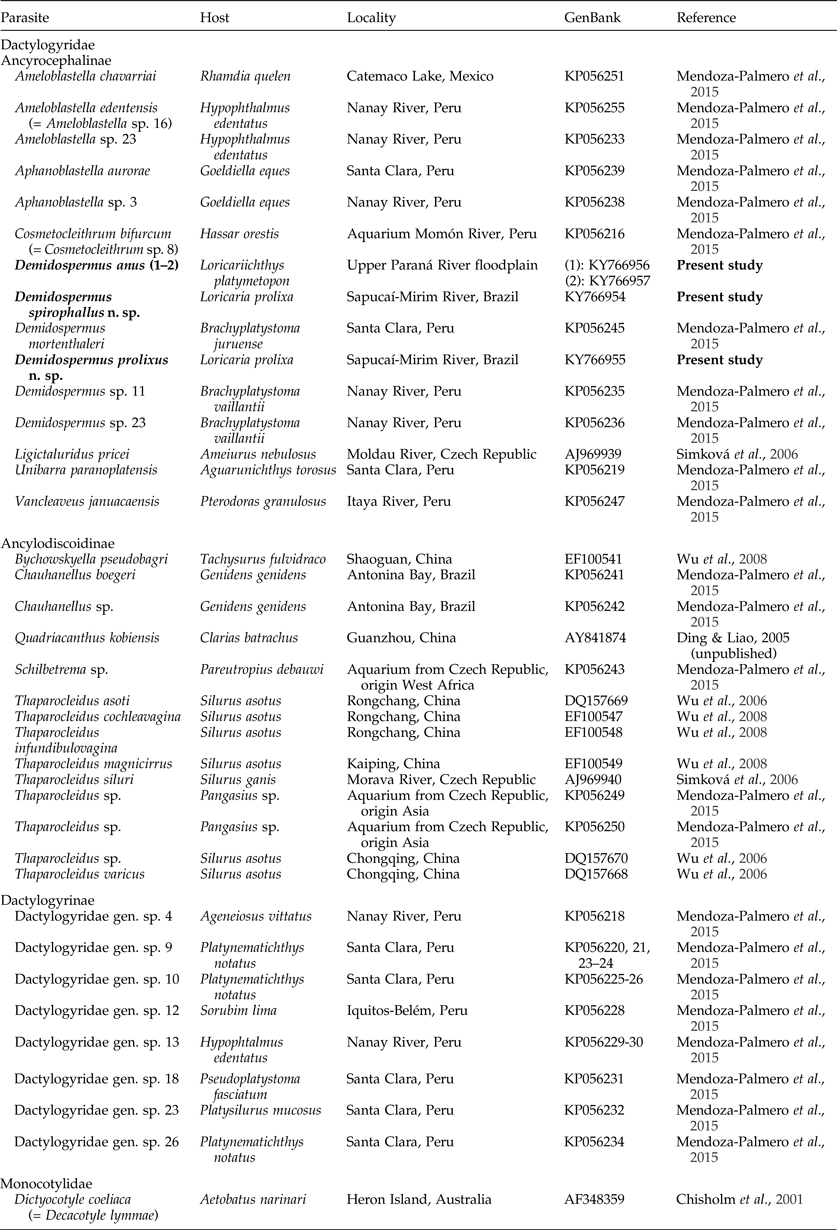
For phylogenetic analysis, contiguous sequences were edited in Sequencher™ v.5.2.4 (Gene Codes, Ann Arbor, Mitchigan, USA) and were subjected to BLAST analysis (http://blast.ncbi.nlm.nih.gov) to confirm identity sequences. Newly generated sequences of partial 28S rDNA were aligned with related sequences obtained previously from dactylogyrids of catfishes retrieved from GenBank (table 1). Dictyocotyle coeliaca Nybelin, 1941 (AF348359) was used as an outgroup. The species, hosts and accession numbers used in this study are described in table 1.
Species of the Dactylogyridae of siluriforms used in the present study, with details of locality, host and GenBank accession numbers.

Nucleic acid sequences were aligned using the Muscle program implemented in Geneious version 7.1.3 (Kearse et al., Reference Kearse, Moir, Wilson, Stones-Havas, Cheung, Sturrock, Buxton, Cooper, Markowitz, Duran, Thierer, Ashton, Mentjies and Drummond2012). All analyses were performed using only positions that were unambiguously alignable across all taxa. The phylogenetic analyses were performed including species of the Dactylogyridae, which were polarized using Monocotylidae as an outgroup.
The JModelTest 2.1.1 program (Posada, Reference Posada2008) was used to select the most appropriate evolutionary model for Maximum Likelihood (ML) and Bayesian inference (BI). Prior to ML analysis the best-fitting models of nucleotide substitution were determined based on Akaike Information Criteria (AIC) using jModelTest 2.1.1 (Posada, Reference Posada2008), with GTR+I+G for the partial LSU gene. The supports for ML were determined by performing 100 bootstrap replicates.
Bayesian inference (BI) was performed using the BEAST program (Drummond et al., Reference Drummond, Suchard, Xie and Rambaut2012) and run with a GTR+I+G nucleotide substitution model. Markov Chain Monte Carlo (MCMC) chains were run for 50 million generations and the log-likelihood scores plotted. Only the final 75% of trees were used to produce the consensus trees by setting the burn-in parameter at 5 million generations. Phylogenetic trees were generated and edited in FigTree v.1.3.1 (Rambaut, Reference Rambaut2009).
Results
Demidospermus spirophallus n. sp.
Dactylogyridae Bychowsky, 1933; Demidospermus Suriano, 1983.
Description
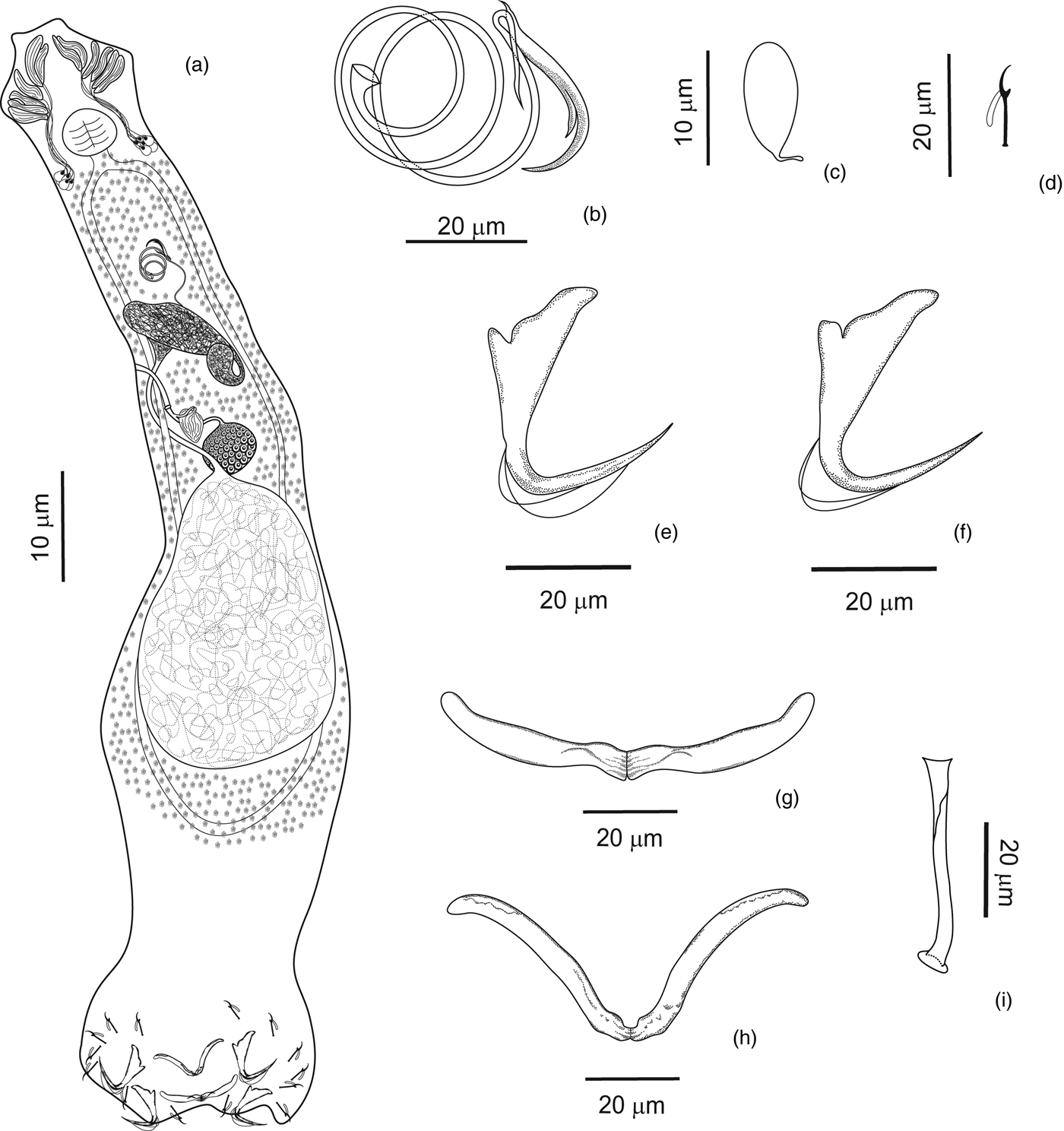
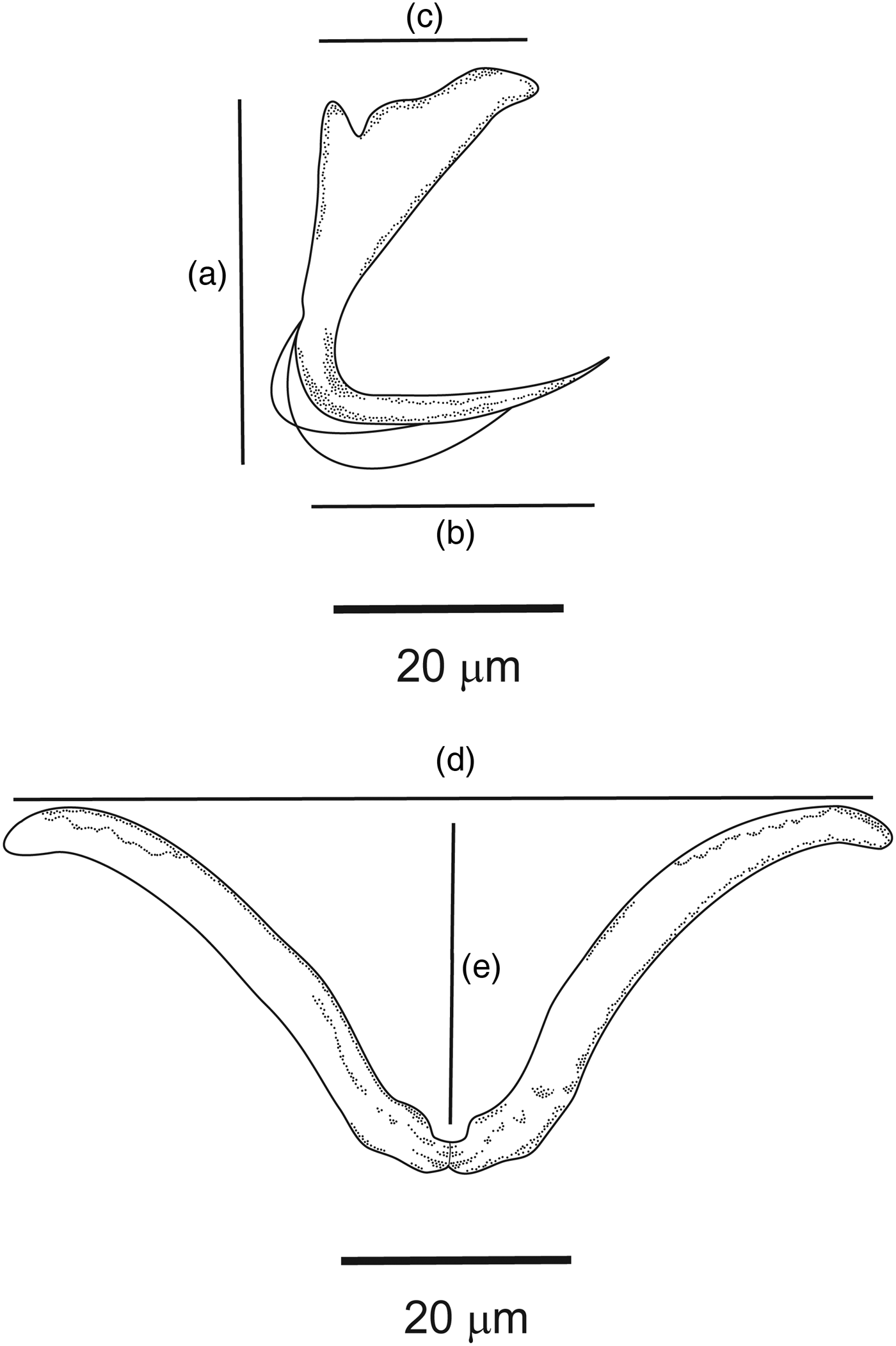
Based on 13 specimens mounted in Gray and Wess and 10 specimens stained with Gömöri trichrome and mounted in Canada balsam (fig. 1a–i). Body elongated, fusiform, 880 (670–1225; n = 8) long, divisible into cephalic region, trunk, short peduncle and haptor. Greatest width 125 (89–177; n = 12) at level of testis. Two terminal and two bilateral cephalic lobes, moderately developed. Three bilateral pairs of head organs; cephalic glands in the pharyngeal region. Eyes and accessory eyespot granules absent. Pharynx subspherical, 43 (33–53; n = 12) long, 45 (24–60; n = 12) wide. Oesophagus short, intestinal caecum confluent posterior to gonads. Counterclockwise-coiled male copulatory organ (MCO), 212 (193–230; n = 11) long, with 2½ rings, proximal ring 21 (18–22; n = 10) in diameter. Sheath-like accessory piece serving as guide for the MCO, with one of the ends bifurcated, 53 (49–57; n = 5) long. Gonads in tandem, ovary pretesticular. Testis elongated 254 (n = 1) long and 150 (n = 1) wide; between the intestinal caeca. Vas deferens conspicuous, looping around left intestinal caecum. Seminal vesicle a distal dilatation of vas deferens, with great variation of forms. Oviduct, ootype, uterus not observed. Ovary 52 (40–65; n = 4) long, 59 (21–86; n = 4) wide. Egg oval with a small posterior filament. Seminal receptacle anterior to ovary. Sinistral vagina aperture weakly sclerotized, curved and tubular, opening into subspherical seminal receptacle through a weakly sclerotized duct. Vitelline follicles densely scattered throughout trunk, absent in region of reproductive organs. Haptor 106 (63–135; n = 10) long and 136 (84–206; n = 10) wide, with two pairs of anchors and two bars. Anchors with well-developed superficial root, short deep root, short and straight shaft, and elongated straight point. Dorsal anchor 32 (29–37; n = 11) long (fig. 2a), base 26 (24–28; n = 11) wide (fig. 2b) and root 16 (13–19; n = 11) wide (fig. 2c). Ventral anchor 28 (25–31; n = 12) long; short, deep root; root 18 (20–25; n = 12) wide and base 24 (20–26; n = 12) wide. Bars with medial narrowing in the base and variable aperture and distance between ends. Dorsal bar ‘V’ wide open-shaped, articulate, with variable aperture of angulation, distance between ends 58 (23–76; n = 11) (fig. 2d), 81 (73–99; n = 11) total length (fig. 2e) and 24 (14–38; n = 11) height. Ventral bar ‘V’ shaped with ends laterally directed, with an evident narrowing in the middle and varying aperture of angulation; 89 (78–102; n = 12) total length, distance between ends 61 (24–97; n = 11) and 29 (14–37; n = 11) height. Seven pairs of hooks, similar in form and size, with distribution as proposed by Mizelle (Reference Mizelle1936), each with a recurved point, delicate throughout; protruding thumb, and filamentous hooklet loop approaching three-quarters of shank length. Shanks of hooks were 8 (6–9; n = 11) long, and hooklets 4 (3–5; n = 11) long.
Morphology of Demidospermus spirophallus n. sp., showing: (a) entire body, dorsal view (composite); (b) copulatory complex, ventral view; (c) egg; (d) hook; (e) dorsal anchor; (f) ventral anchor; (g) dorsal bar; (h) ventral bar; and (i) vagina, dorsal view.

Anchor and bar of Demidospermus spirophallus n. sp., showing: (a) length of superficial root to base; (b) width of base; (c) root width; (d) distance between ends; and (e) bar length.

Taxonomic summary
Type host
Loricaria prolixa Isbrücker & Nijssen, 1978 (Siluriformes, Loricariidae).
Type locality
Sapucaí-Mirim River (20°29′38.38″S, 47°51′33.11″W), municipality of São Joaquim da Barra, state of São Paulo, Brazil.
Site of infection
Gills.
Infection rate
Prevalence: 98.0%; mean intensity of infection: 92.5 ± 10.5 (9.0–400.0); mean abundance: 90.7 ± 10.5 (0–400.0).
Specimens deposited
Holotype CHIOC (38611a); paratypes CHIOC (38611b–d); vouchers INPA (705 a–d) and CHIBB (226L–232L).
Etymology
The specific epithet is from the Latin and is derived from the coiled disposition of the male copulatory organ (MCO) (spiro = anything wound up or coiled + phallus = penis).
Remarks
Demidospermus spirophallus n. sp. was included in the genus proposed by Suriano (Reference Suriano1983) based on agreement with the amended generic characteristics of the diagnosis presented by Kritsky & Gutiérrez (Reference Kritsky and Gutiérrez1998), such as the tubular MCO with a sheath-like accessory piece, sinistral vagina, the absence of eyes or accessory granules, gonads in tandem, and ventral and dorsal bars shaped in either ‘U’ or ‘V’ forms. In D. spirophallus n. sp., the bars showed varied opening angles, depending on the mounting of the permanent slides.
This new species differs from its congeners mainly due to the morphology, diameter and number of rings of the counterclockwise-coiled male copulatory organ (MCO); the accessory piece, the shape of the vagina and the presence of a weakly sclerotized duct that composes the seminal receptacle. The sinistral vagina is tubular, curved, delicate, weakly sclerotized, opening into a small seminal receptacle with a pouch-like shape, throughout a weakly sclerotized duct, versus the non-sclerotized vagina described in other species in this genus, such as D. luckyi (Kritsky, Thatcher & Boeger, 1987) (= Omothecium luckyi), D. pinirampi (Kritsky, Thatcher & Boeger, 1987) (= Omothecium pinirampi), D. ceccarellii Cepeda & Luque, 2010 and D. brachyplatystomae Cepeda & Luque, 2010. The presence of the sclerotized duct as part of the seminal receptacle has never been described before in Demidospermus specimens.
The MCO of this new species has 2½ rings and the accessory piece is tubular, with one bifurcated end, while the accessory piece of the type species D. anus is composed of only one sclerotized piece, the number of the cirrus rings is lower (1½) and the diameter of the rings are greater than in D. spirophallus n. sp. The species D. paranaensis, described parasitizing a loricariid from Brazil, also exhibits a counterclockwise-coiled MCO, although the cirrus is composed of just 1½ rings, long and elliptical shaped, and its tegument presents conspicuous annulations throughout the trunk and peduncle (inconspicuous in D. spirophallus n. sp.). Although some species of the same genus may present the cirrus as a sigmoid tube (D. centromochli Mendoza-Franco & Scholz, 2009), C-shaped (D. ichthyocercus Monteiro, Kritsky & Brasil-Sato, 2010) or a short and robust tube (D. brevicirrus Mendoza-Palmero, Scholz, Mendoza-Franco & Kuchta, 2012), the majority have a counterclockwise-coiled MCO.
Demidospermus spirophallus n. sp., as well as D. paranaensis, D. anus, D. ichthyocercus and D. centromochli, present no differences in terms of the morphology of the haptoral hooks, unlike some of its congeners (e.g. D. macropteri Mendoza-Franco & Scholz, 2009, D. peruvianus Mendoza-Palmero & Scholz, 2011, D. curvovaginatus Mendoza-Palmero & Scholz, 2011, D. striatus Mendoza-Palmero & Scholz, 2011, D. lebedevi (Kritsky & Thatcher, 1976), D. brevicirrus, D. mortenthaleri and D. pinirampi).
Although D. lebedevi also shows a cirrus composed of circular rings, it has 2–3 rings with a greater diameter (72 (56–83)) than those of D. spirophallus n. sp. Furthermore, D. lebedevi has eyes and an accessory eyespot, which are absent in the new species.
Demidospermus prolixus n. sp.
Description
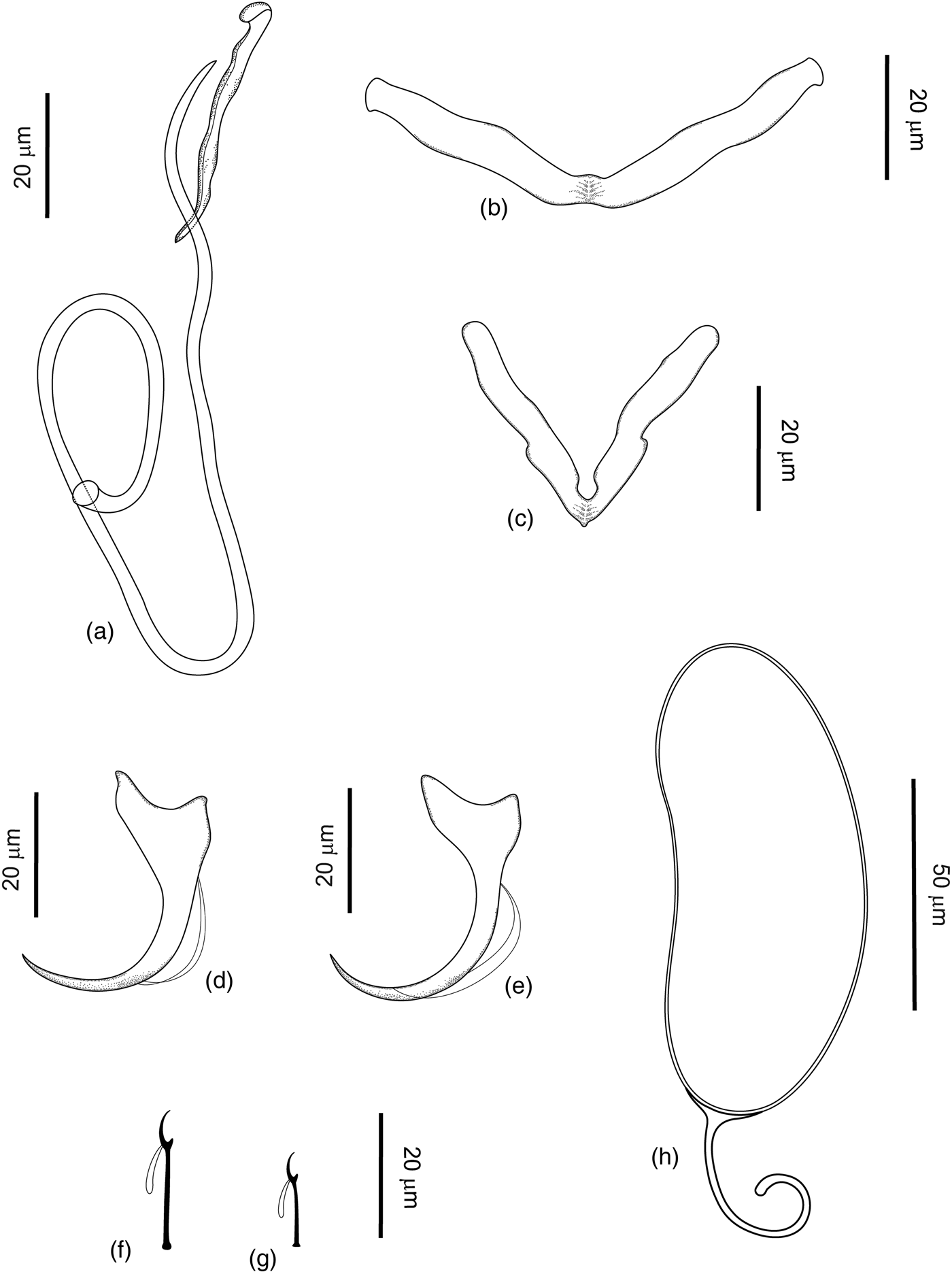
Based on two specimens mounted in Gray and Wess, five in Hoyer and one stained with Gömöri trichrome and mounted in Canada balsam (fig. 3a–h). Body elongated, fusiform, 747 (416–987; n = 7) long, divisible into cephalic region, trunk, short peduncle and haptor. Greatest width 119 (109–130; n = 7) at level of testis. Two terminal and two bilateral cephalic lobes, moderately developed. Three bilateral pairs of head organs; cephalic glands in the pharyngeal region. Eyes and accessory eyespot granules absent. Pharynx subspherical, 37 (34–48; n = 7) long; 31 (25–58; n = 7) wide. Oesophagus short, intestinal caecum confluent posterior to gonads. Counterclockwise-coiled male copulatory organ (MCO), 223 (210–234; n = 7) long, and 1½ rings, 66 (60–74; n = 7) long and 27 (20–31; n = 7) wide. Accessory piece straight and strongly sclerotized, groove-like, 36 (32–38, n = 7) long. Gonads in tandem, ovary pretesticular. Testis elongated 138 (113–164; n = 2) long and 64 (54–74; n = 2) wide; between the intestinal caeca. Vas deferens conspicuous, looping around left intestinal caecum. Seminal vesicle a distal dilatation of vas deferens, with great variation in forms. Oviduct, ootype, uterus not observed. Egg oval, 106 (100–112; n = 2) long and 52 (49–54; n = 2) wide, with a posterior filament, 52 (45–58; n = 2) long. Seminal receptacle not observed. Sinistral vagina aperture non-sclerotized. Vitelline follicles densely scattered throughout trunk, below the pharynx and anteriorly to the peduncle. Haptor 78 (60–103; n = 7) long and 170 (152–205; n = 7) wide, slightly subtrapezoidal, with two pairs of anchors and two bars. Anchors with well-developed superficial root; short, deep root; short and curved shaft. Ventral anchor 36 (34–38; n = 5) long; root 15 (13–16; n = 7) wide and base 19 (16–22; n = 7) wide. Dorsal anchor 36 (34–39; n = 5) long; root 15 (13–16; n = 5) wide, and base 21 (20–24; n = 6) wide. Bars with medial narrowing in the base and variable aperture and distance between ends. Ventral bar ‘V’ wide open-shaped, articulate, with variable aperture of angulation (conspicuously more open than the dorsal bar), total length 85 (83–86; n = 5), distance between ends 73 (71–80; n = 5) and 19 (11–26; n = 5) height. Dorsal bar ‘V’ shaped, with an evident narrowing in the middle and varying aperture of angulation; 72 (70–73; n = 5) total length, distance between ends 39 (31–47; n = 5) and height 30 (28–33; n = 5). Seven pairs of hooks, dissimilar in terms of the shank size: hook pairs 1–6 are similar (shanks 9 (8–10; n = 15) long and hooklets 5 (4–6; n = 15) long), but hook pair 7 presents a longer shank (shanks 13 (10–14; n = 15) long and hooklets 6 (5–7; n = 15) long). The hook distribution is as proposed by Mizelle (Reference Mizelle1936), each with a recurved point, delicate throughout; protruding thumb and filamentous hooklet loop approaching half the shank length.
Morphology of Demidospermus prolixus n. sp., showing: (a) copulatory complex, ventral view; (b) ventral bar; (c) dorsal bar; (d) dorsal anchor; (e) ventral anchor; (f) hook pair 7; (g) shape of hook pairs 1–6; (h) egg.

Taxonomic summary
Type host
Loricaria prolixa Isbrücker & Nijssen, 1978 (Siluriformes, Loricariidae).
Type locality
Sapucaí-Mirim River (20°29′38.38″S, 47°51′33.11″W), municipality of São Joaquim da Barra, state of São Paulo, Brazil.
Site of infection
Gills.
Infection rate
Prevalence: 98.0%; mean intensity of infection: 169.4 ± 19.3 (16.0–732.0); and mean abundance: 166.0 ± 19.2 (0–732.0).
Specimens deposited
Holotype CHIOC (38609a); paratype CHIOC (38609b–c; 38610); vouchers INPA (704 a–e) and CHIBB (233L–236L).
Etymology
The specific epithet is derived from the name of the type host (Loricaria prolixa).
Remarks
Demidospermus prolixus n. sp., as well as D. spirophallus n. sp., was included in this genus based on agreement with the amended generic characteristics of the diagnosis presented by Kritsky & Gutiérrez (Reference Kritsky and Gutiérrez1998), previously cited. The morphology of the copulatory complex of this new species is very similar to that of D. paranaensis described by Ferrari-Hoeinghaus et al. (Reference Ferrari-Hoeinghaus, Bellay, Takemoto and Pavanelli2010) from L. platymetopon, as the presence of a longer, counterclockwise-coiled MCO, with an ovate base and non-articulated groove-like accessory piece serving as a MCO guide. However, the MCO and the accessory piece are more robust and larger than those observed in D. paranaensis.
This new species differs from other congeners mainly due to the morphology of the bars (dorsal bar with conspicuous protuberances), two different hook shapes (fig. 3a–g), and inconspicuous tegumental annulations (conspicuous in D. paranaensis). Demidospermus prolixus n. sp. shares the presence of a non-sclerotized vagina with other congeners, such as D. pinirampi, D. luckyi, D. ceccarellii and D. brachyplatystomae, and has no eyes or accessory eyespots, like D. anus, D. paranaensis, D. macropteri and D, spirophallus n. sp. However, other congeners have subspherical eyespots and/or scattered granules.
Demidospermus anus Suriano, 1983
Redescription
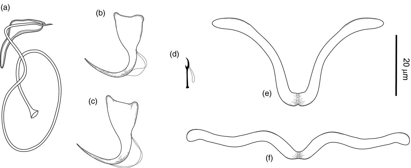
Characteristics based on two specimens mounted in Gray and Wess, and six specimens mounted in Hoyer (fig. 4a–f). Body elongated, fusiform, 356 (319–392; n = 3) long, divisible into cephalic region, trunk, short peduncle and haptor. Greatest width 65 (55–75; n = 3) at level of testis. Two terminal and two bilateral cephalic lobes, moderately developed. Three bilateral pairs of head organs; cephalic glands in the pharyngeal region. Eyes and accessory eyespot granules absent. Pharynx subspherical, 28 (24–32; n = 4) long. Oesophagus short, intestinal caecum confluent posterior to gonads. Counterclockwise-coiled male copulatory organ (MCO), 152 (143–156; n = 5) long, curved tube, with variable diameter and loop number (1–1½ rings), depending on the mounting slide. Sheath-like accessory piece serving as guide for the MCO, 31 (30–33; n = 5) long. Gonads intercaecal, tandem; ovary pretesticular. Vas deferens conspicuous, looping around left intestinal caecum; seminal vesicle a distal dilatation of vas deferens. Oviduct, ootype, uterus not observed. Sinistral vagina aperture weakly sclerotized, curved and tubular. Vitelline follicles densely scattered throughout trunk, absent in region of reproductive organs. Haptor 48 (46–52; n = 3) long and 71 (69–73; n = 3) wide, with two pairs of anchors and two bars. Anchors with short superficial root, deep root, short and straight shaft. Ventral and dorsal anchors similar, 20 (19–21; n = 4) long; base 15 (14–16; n = 4) wide; and root 11 (10–12; n = 4) wide. Dorsal bar ‘V’ shaped, wide, open-shaped, articulate, with medial narrowing in the base and variable aperture and distance between ends, 60 (50–70; n = 3) total length. Ventral bar ‘V’ shaped, more open than the dorsal bar, with an evident narrowing in the middle; 65 (60–70; n = 3) total length. Seven pairs of hooks, similar in form and size, 11 (10–12; n = 8) total length, with distribution as proposed by Mizelle (Reference Mizelle1936), each with a recurved point, delicate throughout; protruding thumb and filamentous hooklet loop approaching three-quarters of shank length.
Morphology of Demidospermus anus, dorsal view: (a) copulatory complex; (b) ventral anchor; (c) dorsal anchor; (d) hook; (e) dorsal bar; and (f) ventral bar.

Taxonomic summary
Type host
Loricariichthys anus (Valenciennes, 1835) (= Loricaria anus) (Siluriformes, Loricariidae).
Type locality
Laguna de Chascomús (35°37′S, 58°04′W), Province of Buenos Aires, Argentina.
Site of infection
Gills.
Material studied (present study)
Eight specimens from Loricariichthys platymetopon Isbrücker & Nijssen, 1979 (Siluriformes, Loricariidae), collected in June 2016 in the Upper Paraná River floodplain, near the municipality of Porto Rico, Paraná, Brazil (20°43′S, 53°10′W).
Specimens deposited
Vouchers CHIOC (38608 a–e), INPA (703 a–d) and CHIBB (237L–243L).
Additional hosts
Loricariichthys platymetopon from the reservoir of Itaipu Hydroelectric Power Station, Paraná, Brazil (Cohen & Kohn, Reference Cohen and Kohn2008).
Remarks
Demidospermus was proposed by Suriano (Reference Suriano1983) with D. anus as the type species. However, as described by the authors, the holotype, paratypes or vouchers of the type species were never deposited at the Helminthological Section of the Museo de La Plata, Argentina. The scarcity of information about this material, its incomplete description and the absence of sequences of the type species make studies of this monogenean genus difficult, and represent a problem in its circumscription.
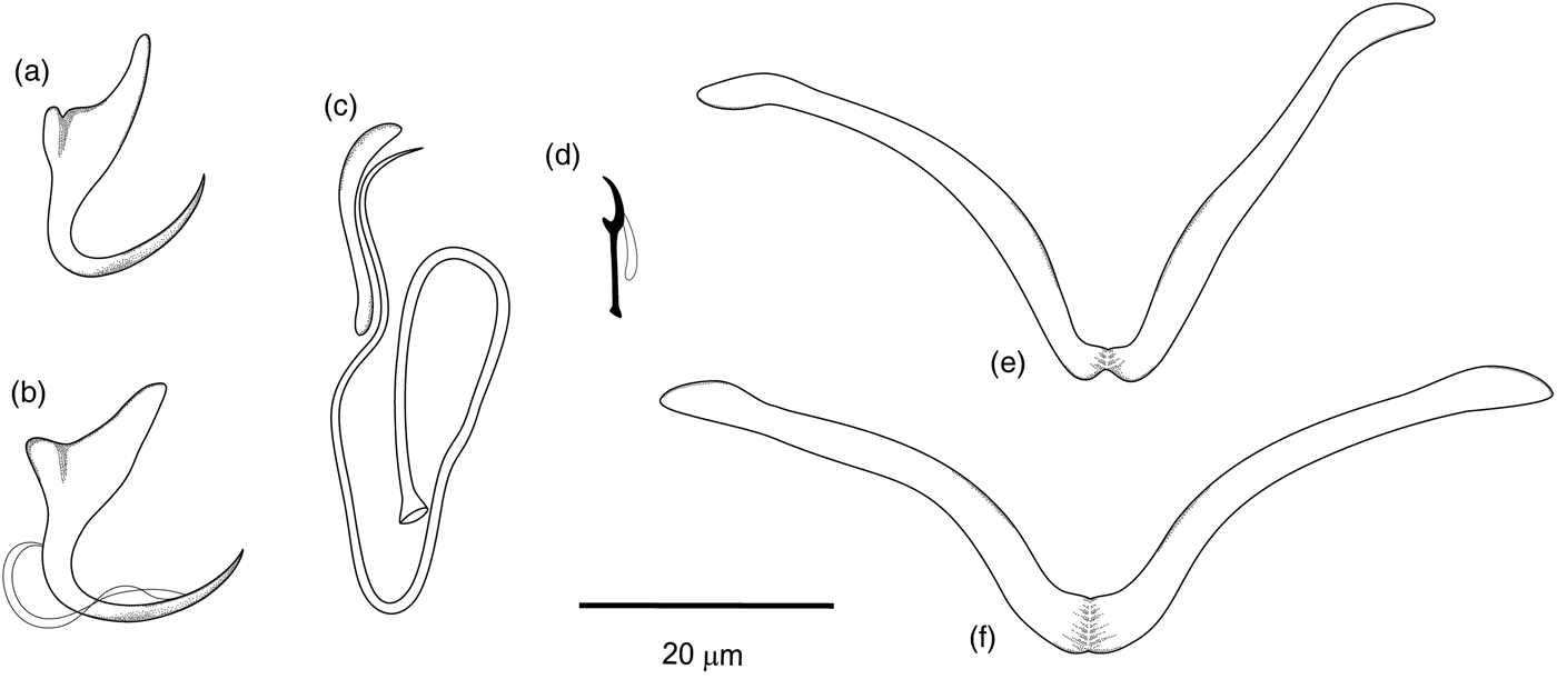
Ferrari-Hoeinghaus et al. (Reference Ferrari-Hoeinghaus, Bellay, Takemoto and Pavanelli2010) described the species D. paranaensis from L. platymetopon collected in the Upper Paraná River floodplain, which is the same reservoir and the same host species from which the specimens of D. anus analysed in the present study were collected. In another study, Cohen & Kohn (Reference Cohen and Kohn2008) reported, for the first time, the occurrence of D. anus in L. platymetopon from the reservoir of the Itaipu Hydroelectric Power Station, Brazil. Ferrari-Hoeinghaus et al. (Reference Ferrari-Hoeinghaus, Bellay, Takemoto and Pavanelli2010) considered that D. paranaensis differed from D. anus mainly based on the presence of annulations in the tegument (present in D. paranaensis and not described in D. anus), the size of the haptoral pieces and the morphology of the MCO. However, analysing the paratypes of D. paranaensis deposited in the CHIOC and the specimens analysed by Cohen & Kohn (Reference Cohen and Kohn2008) described as D. anus, no differences were found between the morphology and measurements of the specimens. In both cases, all the specimens had annulations in the tegument, an absence of eyespots, and similar morphologies and measurements of the cirrus (124 (123–125; n = 10) total length), accessory piece (18 (17–19); n = 5) and haptoral pieces (fig. 4a–f and fig. 5a–f). Moreover, the diameter of the cirrus ring varies greatly, depending on the mounting and compression. Considering that in both cases the gills were removed and fixed in 1:4000 formalin solution, the annulations may be a consequence of the technique used for fixation prior to analysis.
Morphology of Demidospermus paranaensis, dorsal view: (a) ventral anchor; (b) dorsal anchor; (c) copulatory complex; (d) hook; (e) dorsal bar; and (f) ventral bar.

These annulations were not evident in the specimens of D. anus analysed in the present study or those analysed by Suriano (Reference Suriano1983) and, in both cases, the material was analysed from frozen fish and in vivo, respectively. In general, the morphologies of D. paranaensis and D. anus analysed in the present study are very similar (figs 4 and 5), including the presence of partially encapsulated sperm, as described by Suriano (Reference Suriano1983). However, differences in the morphology of the anchors, and the measurements of the cirrus and accessory piece (greater in D. anus than D. paranaensis) were observed. Careful future morphological and molecular studies are required on these species.
Molecular characterization
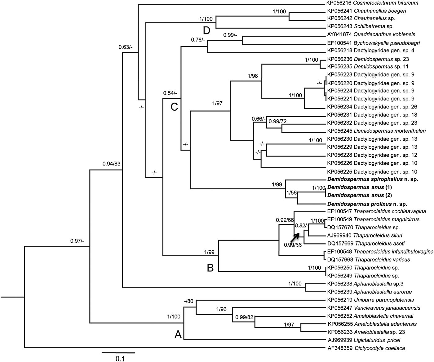
The new partial LSU sequence length varied from 1483 bp for D. spirophallus n. sp., to 1281 bp for D. prolixus n. sp., 684 bp for D. anus (1) and 918 bp for D. anus (2). An unambiguous alignment including Dactylogyridae from the Siluriformes available in GenBank spanned 480 positions. A total of 39 monogenean specimens of siluriforms from the Neotropical, Palaearctic, Ethiopian and Oriental regions were used in the analyses (table 1). The ML and BI analyses yielded similar branching topology, with supported values in the most basal and terminal nodes. Tree main clades, A, B, C and D, were observed and corroborated the findings of Mendoza-Palmero et al. (Reference Mendoza-Palmero, Blasco-Costa and Scholz2015) (fig. 6), with a few changes. Clade A comprises parasites of siluriforms from the Neotropical region and Ligictaluridus pricei (Mueller, 1936) a parasite of ictalurid fish from the Czech Republic, introduced to Europe with its host. Clade B consists of all Thaparocleidus spp. and Aphanoblastella spp., forming a polytomic clade related to clades A and B, with strong branching support. Clade C consists of all Dactylogyridae gen. spp. and Demidospermus spp. from the Neotropical region, and Bychowskyella pseudobagri Achmerow, 1952, and Quadriacanthus kobiensis Ha Ky, 1968 from Oriental regions such as China. Demidospermus spirophallus n. sp. and D. prolixus n. sp. parasites of L. prolixa, and specimens of D. anus parasites of L. platymetopon clustered together as sister taxa in a separate clade within the main clade C, with high supporting values. Finally, clade D consisted of dactylogyrids from the Neotropical and Ethiopian regions (Chauhanellus spp. from Brazil and Schilbetrema sp. from West Africa). The position of clade D with the species Chauhanellus sp., Chauhanellus boegeri Domingues and Fehlauer, 2006, and Schilbetrema sp. was not supported, as observed by Mendoza-Palmero et al. (Reference Mendoza-Palmero, Blasco-Costa and Scholz2015). The non-identified Dactylogyridae gen. sp. 4 is allocated differently from that in the study by Mendoza-Palmero et al. (Reference Mendoza-Palmero, Blasco-Costa and Scholz2015), within the Bychowskyella pseudobagri and Quadriacanthus kobiensis clade, with high Bayesian support and low bootstrap. Cosmetocleithrum bifurcum Mendoza-Franco, Mendoza-Palmero & Scholz, 2016 (= Cosmethocleithrum sp. 8) was not resolved, forming a polytomy.
Bayesian topology based on partial 28S ribosomal DNA sequences of Dactylogyridae from siluriforms. GenBank accession numbers precede species names. Support values are above nodes as follows: posterior probabilities for Bayesian analyses and bootstrap for maximum likelihood analyses. Dashes represent branches not supported by analyses (posterior probabilities <0.50 and bootstrap scores <50). Arrow indicates support values above the node. Branch length scale bar indicates number of substitutions per site. The species Dictyocotyle coeliaca (AF348359) was used as an outgroup.

Discussion
In addition to the problems regarding the circumscription of the genus Demidospermus due to its incomplete description and the absence of deposited material of the type species, a number of morphological characteristics described in the description of specimens of this genus vary considerably, such as the position of the vaginal opening (sinistral versus dextral; anterior versus medial opening); sclerotized versus non-sclerotized vagina; presence versus absence of eyespots or scattered granules; hook shape (similar morphology versus variable morphology), counterclockwise-coiled versus clockwise MCO. This represents a problem for the accurate identification of the specimens based on morphology, as some characteristics may overlap with those observed in other genera, and perhaps reinforces the possibility that this group is not natural.
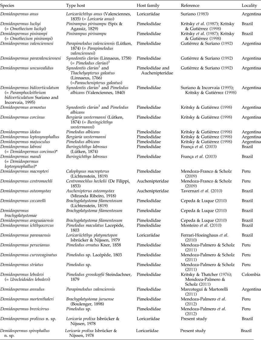
The closeness of the phylogenetic relationships of host species has a strong influence on the distribution of monogenean parasites, to the extent that the majority of hosts are susceptible to a specific group of these parasites (Thatcher, Reference Thatcher2006; Braga et al., Reference Braga, Araújo and Boeger2014). Neotropical siluriforms, specifically, do not represent a monophyletic assemblage, and include descendants of four independent monophyletic clades (Sullivan et al., Reference Sullivan, Lundberg and Hardman2006; Braga et al., Reference Braga, Araújo and Boeger2014). The origin of their monogenean fauna is unclear, reflecting the differences of the fish lineages that make up this order (Braga et al., Reference Braga, Araújo and Boeger2014). The reports of monogeneans of Demidospermus described until now (29 species) are limited to freshwater siluriforms from the Neotropics (table 2), belonging to the Loricariidae, Pimelodidae and Auchenipteridae families, distributed from the Peruvian Amazon to Argentina. These fish families are not phylogenetically closely related (see Sullivan et al., Reference Sullivan, Lundberg and Hardman2006, and references therein; Mendoza-Palmero & Scholz, Reference Mendoza-Palmero and Scholz2011) and the occurrence of some species of Demidospermus parasitizing these fishes, along with recently reported molecular data about these monogeneans (see Mendoza-Palmero et al., Reference Mendoza-Palmero, Blasco-Costa and Scholz2015), suggest the need to review the species composition of this genus and others that occur in catfishes, for the correct generic assignation of these species.
Species of the genus Demidospermus described in the present study with details of locality, host type and family.

*Junior synonyms of Demidospermus leptosynophallus and Demidospermus corcinus, respectively (invalid species).
1 According to Monteiro et al. (Reference Monteiro, Kritsky and Brasil-Sato2010), the fish identified as Synodontis clarias (= Pimelodus clarias) is in fact Pimelodus maculatus.
Ten species belonging to Demidospermus have been recorded in catfishes from Brazil, and other species have been described in fishes collected in Argentina (11 species), Peru (Peruvian Amazon) (7 species) and Colombia (1 species). Among these, only four species of Demidospermus were described specifically from loricariid fishes (table 2), including the type species D. anus. The loricariid recorded as hosts to Demidospermus spp. are L. anus (= Loricaria anus), which was reported as a host to the type species D. anus (Suriano, Reference Suriano1983); L. platymetopon, a host of D. paranaensis (Ferrari-Hoeinghaus et al., Reference Ferrari-Hoeinghaus, Bellay, Takemoto and Pavanelli2010) and D. anus (Cohen & Kohn, Reference Cohen and Kohn2008); Loricariichthys castaneus (Castelnau, 1855), parasitized with Demidospermus sp. (Azevedo et al., Reference Azevedo, Abdallah and Luque2010), and L. prolixa, a host of the two new species described in this study.
Mendoza-Palmero et al. (Reference Mendoza-Palmero, Blasco-Costa and Scholz2015), in a study on the phylogeny of Neotropical monogeneans from catfishes (pimelodids), suggested a possible polyphyly of the group, based on the subdivision of the monogeneans into three main clades (A, B and C) within Dactylogyridae. In addition, the authors presented the first molecular data of Demidospermus species (D. mortenthaleri and two other undescribed species – Demidospermus sp. 11 and Demidospermus sp. 23), parasites of the pimelodids B. juruense and B. vaillantii, respectively, from Iquitos, Peru. However, observing their phylogenic results and carefully analysing the morphology of D. mortenthaleri (see Mendoza-Palmero et al., Reference Mendoza-Palmero, Scholz, Mendoza-Franco and Kuchta2012), and other species of Demidospermus described parasitizing Brachyplatystoma filamentosum (Lichtenstein, 1819) from Brazil (Cepeda & Luque, Reference Cepeda and Luque2010) (D. ceccarelli, D. brachyplatystomae and D. araguaiaensis), we hypothesize that these species do not in fact belong to this genus, and so should be included in a new genus. Although these species resemble each other, they show conspicuous differences in terms of the diagnostic characteristics of the Demidospermus genus (e.g. bar shapes). The same may have occurred with the undescribed species, namely Demidospermus sp. 11 and Demidospermus sp. 23, but the absence of a morphological description for these species (Mendoza-Palmero et al., Reference Mendoza-Palmero, Blasco-Costa and Scholz2015) limits conclusions at the present time. No sequences of either Demidospermus specimens from Brazil or monogenean parasites from loricariid fishes have been performed until now. The present study therefore provides, for the first time, data regarding the molecular characterization of Demidospermus species parasitizing loricariid fishes, supporting future investigations and phylogenetic studies.
Demidospermus spirophallus n. sp., D. prolixus n. sp. and D. anus clustered together within the most diverse clade, the Neotropical region (clade C, see Mendoza-Palmero et al., Reference Mendoza-Palmero, Blasco-Costa and Scholz2015), and were closely related to the monogeneans previously described in the Ancylodiscoidinae subfamily. These three species of Demidospermus from Brazilian loricariids did not group together with other Demidospermus species parasites of pimelodids from Peru (Demidospermus sp. 23, Demidospermus sp. 11 and D. mortentahleri), exhibiting a distant relationship, as described previously by Mendoza-Palmero et al. (Reference Mendoza-Palmero, Blasco-Costa and Scholz2015), reinforcing the fact that this genus is not natural. Based on the results of the morphological and molecular data presented in this study, we suggest that, in fact, lineages of dactylogyrids from loricariids belong to the Demidospermus genus, and an erection of two new genera should be proposed to accommodate parasite species of pimelodids from Peru (one to accommodate the species Demidospermus sp. 23 and Demidospermus sp. 11, and another for D. mortentahleri). However, future morphological and phylogenetic analyses with the inclusion of new sequences of dactylogyrids identified as belonging to the Demidospermus genus from other siluriform fish may clarify the host associations, and confirm whether this dactylogyrid possesses specificity for parasitizing loricariid fishes or if, in fact, they can be distributed among siluriform fishes in general.
Acknowledgements
We are most grateful to Edmir Daniel Carvalho (in memoriam), professor and researcher at the Universidade Estadual Paulista, Botucatu Campus, São Paulo, Brazil, who dedicated his life to the study of ecology and environmental impacts on rivers and reservoirs caused by anthropic actions, and actively participated in the development of this project. We would like to thank Sandro Geraldo de Castro Britto and Diogo Freitas Souza for logistical support in the collection of hosts; Aline Gouveia de Souza Lins for help in the analysis of parasites; and Cláudio Henrique Zawadzki, Walter Antonio Pereira Boeger, Rodney Kozlowiski de Azevedo and Vanessa Doro Abdallah Kozlowiski for taxonomic suggestions. We are also most grateful to Simone Chinicz Cohen and Anna Kohn (Oswaldo Cruz Foundation – FIOCRUZ) for allowing us to examine some specimens of Demidospermus in their care.
Financial support
The authors would like to thank CAPES (Coordination for the Improvement of Higher Education Personnel) and FAPESP (São Paulo Research Foundation) (L.F. – Process: 2012/07850-7) for financial and scientific support, and the post-graduate scholarships granted to M.I.M. (grant number AUX-PE-PNPD 3005/2010); and the Young Researcher Program PROPE-UNESP 02/2016 and New Doctor Program PROPE-UNESP 03/2016 for the scholarships granted to M.I.M. and L.F., respectively. R.J.S. is supported by CNPq (307808/2014-9) and CNPq-PROTAX (440496/2015-2)/FAPESP 2016/50377-1. We would also like to thank FUNEP (Teaching and Extension Research Support Foundation) (Process: 1.01852/2011), Duke Energy and CELAN (Central Elétrica Anhanguera) for financial and logistical support.
Conflict of interest
None.
Ethical standards
Specimens of L. prolixa were collected under Permanent License for the Collection of Zoological Material – SISBio 13794-1.