Introduction
Meiotic maturation of mammalian oocytes is spontaneously blocked at the stage of metaphase II and further progress in meiosis beyond this block depends upon an activating stimulus introduced into the oocyte by sperm (Yanagimachi et al., Reference Yanagimachi, Knobil and Neil1988). Inside the fertilized oocyte, the sperm induces oscillation of intracellular free calcium (Ca2+) ion levels that triggers the activation process (Swann & Ozil, Reference Swann and Ozil1994). The parthenogenetic activation of oocytes in vitro is, therefore, generally induced by an artificial increase of intracellular Ca2+ levels using e.g. Ca2+ ionophore A23187 (Steinhardt et al., Reference Steinhardt, Epel, Carroll and Yanagima1974; Bos-Mikich et al., Reference Bos-Mikich, Swann and Whittingham1995; Sedmikova et al., Reference Sedmíková, Burdova, Petr, Etrych, Rozinek and Jílek2003). However, the progress of meiotic maturation may be also triggered by nitric oxide (NO) synthesized in the cells by nitric oxide synthase (NOS) from molecular oxygen and l-arginine (Kwon et al., Reference Kwon, Nathan, Gilker, Griffith, Matthews and Stuehr1990; Lamas et al., Reference Lamas, Marsden, Li, Tempst and Michel1992). As a cellular messenger the NO is involved in numerous physiological processes including those associated with reproduction (Biswas et al., Reference Biswas, Kabir and Pal1998). In sea urchins, a rapid increase of nitrosation preceding the calcium pulse was observed within eggs after insemination, and microinjection of NO donor or recombinant NOS was sufficient to initiate the events of egg activation (Kuo et al., Reference Kuo, Baxter, Thompson, Stricker, Patton, Bonaventura and Epel2000). It seems, however, that the activation of mammalian oocytes by NO donor is a more complicated process dependent, at least, upon the release of Ca2+ from extracellular spaces (Petr et al., Reference Petr, Rajmon, Rozinek, Sedmíková, Jeseta, Chmelíková, Svestková and Jílek2005a).
Signalling pathways in animal cells are frequently characterized by oscillation of cellular messenger levels (Dyachok et al., Reference Dyachok, Isakov, Sagetorp and Tengholm2006). Intracellular Ca2+ release essential for oocyte activation at fertilization (Kline & Kline, Reference Kline and Kline1992; Swann & Ozil, Reference Swann and Ozil1994; Wu et al., Reference Wu, He and Fissore1997; Jones, Reference Jones2005) represents a typical example of such mode of signalling (Rottingen & Iversen, Reference Rottingen and Iversen2000). Also NO signalling exhibits a similar pulsatile pattern (Fujie et al., Reference Fujie, Yamamoto, Murakami, Hatakeyama, Shiga, Suzuki and Ito2005) and our recent experiments suggested that the use of intermittent, instead of continuous, exposure of porcine oocytes to NO donor could significantly improve the effectivity of parthenogenetic activation (Petr et al., Reference Petr, Chmelíková, Krejčová, Rehak, Novotná and Jílek2008).
Among numerous prerequisites of successful development, the genomic integrity undoubtedly plays one of the key roles. Single-cell gel electrophoresis (comet assay) provides a suitable tool for analysis of DNA damage in any type of nucleated cell (McKelvey-Martin et al., Reference McKelvey-Martin, Green, Schmezer, Pool-Zobel, Meo and Collins1993; Collins, Reference Collins2004). As a simple, sensitive and rapid technique the comet assay has found applications in diverse fields of the biomedical research, including reproductive biology (e.g. Tatemoto et al., Reference Tatemoto, Sakurai and Muto2000; Marchesi et al., Reference Marchesi, Feng and Hershlag2007; Lewis & Agbaje, Reference Lewis and Agbaje2008). Using this approach Mattioli et al. (Reference Mattioli, Barboni, Gioia and Lio2003) demonstrated that cold-induced calcium rise may induce DNA fragmentation in immature porcine oocytes, probably through the disturbance of Ca homeostasis activating nuclear endonucleases. On the other hand, the results of mouse oocyte exposure to increasing concentrations of NO donor implied that NO may determine the oocyte quality and developmental potential through attenuating oocyte aging (Goud et al., Reference Goud, Goud, Diamond and Abu-Soud2005).
The development of optimal activation protocol is important for improvement of developmental competence of parthenogenetically activated porcine oocytes. An increased DNA instability could signal a possible failure of activated oocyte to pass through early stages of embryonic development. We, therefore, decided: (1) to examine the DNA integrity in porcine oocytes matured in vitro after intermittent or continuous exposure to NO donor S-nitrosoacetylpenicillamine (SNAP); (2) to compare the findings with the effect of standard Ca2+ ionophore A23187 activation; and (3) to investigate the relationship between the extent of DNA instability and the mode of activation protocol.
Materials and Methods
Isolation and culture of oocytes
Ovaries from gilts at an unknown stage of the estrous cycle obtained from a local slaughterhouse were placed into a saline solution (0.9% sodium chloride; 39 °C) and transported to the laboratory within 1 h. Fully grown oocytes were aspirated by a 20-gauge needle from follicles with a diameter of about 2–5 mm. Then, the oocytes with compact cumuli were selected, washed three times in a culture medium and cultured at 39 °C and in an atmosphere of 5% CO2 in air using a 3.5 cm diameter Petri dishes (Nunc) with 3 ml of culture medium. Modified M199 medium (GibcoBRL, Life Technologies) containing sodium bicarbonate (0.039 ml of a 7% solution/ml of medium), calcium lactate (0.6 mg/ml), sodium pyruvate (0.25 mg/ml), gentamycin (0.025 mg/ml), HEPES (1.5 mg/ml), gonadotropins 13.5 IU eCG:6.6 IU hCG/ml (P.G.600 Intervet, Boxmeer, Holland), and 10% fetal calf serum (GibcoBRL, Life Technologies) was used for 48 h cultivation. Then, the oocytes devoid of cumulus cells by repeated pipetting through a narrow glass pipette were subjected to further treatment. Based on our previous experiments (Petr et al., Reference Petr, Rajmon, Lánská, Sedmíková and Jílek2005b), 2 mM SNAP (Alexis Biochemicals, USA) in M199 medium without P.G. 600 was used for continuous or intermittent activation of oocytes. Continuous activation (NOc) consisted of a 6 h exposure to the SNAP. Intermittent activation (NOi) involved 10 min exposure to SNAP followed by 10 min culture in medium without SNAP. This cycle was repeated 12 times and terminated by 2 h culture in medium without SNAP. Positive control group of oocytes was exposed to 2 mM calcium ionophore A23187 (Sigma-Aldrich) for 5 min. that was followed by a 2 h treatment with 2 mM 6-dimethylaminopurine (6-DMAP) (Jilek et al., Reference Jílek, Hüttelova, Petr, Holubova and Rozinek2001) and terminated by 4 h culture in the culture medium only. Oocytes cultured in parallel for 6 h in culture medium only served as negative control.
An independent 48 h incubation of oocytes in the culture medium alone was performed to check the course of in vitro maturation. At the end of this period, 96% of oocytes reached metaphase II while remaining oocytes were at the stage of metaphase I, anaphase I or telophase I.
Evaluation of oocyte activation
At the end of the activation procedure, the oocytes were placed on microscopic slides and covered with coverslips affixed by vaseline. Subsequently, the oocytes were fixed with acetic acid:ethanol (1:3, v/v) for at least 24 h, stained with 1.0% orcein and examined under a phase-contrast microscope (Olympus BX40, magnification ×100). Oocytes that reached the pronuclear stage, but not those remaining at metaphase II or arrested at anaphase II or telophase II, were considered as activated.
Comet assay
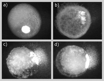
Activated oocytes were centrifuged (5 min, 14 000 g) to relocate the lipids to one pole of the oocytes, treated for 5 min with 0.1% (w/w) pronase solution (Sigma Aldrich) at 38 °C to remove the zona pellucida and washed quickly in culture medium. Then they were transferred into the eppendorf tubes with 30 μl of culture medium (around 30 oocytes per tube), and kept at 4 °C until the further processing. Slides were prepared according to the original method of Singh et al. (Reference Singh, McCoy, Tice and Schneider1988) with some modifications. Briefly, the medium was carefully removed from eppendorf tubes, the oocytes were mixed with 90 μl of 0.75% low melting point (LMP) agarose (Amresco) and 45 μl of this mixture was spread over the layer of 110 μl of 0.75% normal melting point (NMP) agarose attached to microscopic slide (SuperFrost Plus) precoated with 2% agarose. Two slides were prepared from each eppendorf tube. Following 5 min solidification on ice the layer with oocytes was overlaid with 75 μl of pure LMP agarose, covered with coverslip and the slides were again cooled on ice for 5 min. However, subsequent 1.5 h treatment in lysing solution (2.5 M NaCl, 100 mM EDTA, 10 mM Tris, 0.16 M DMSO, 0.016 mM Triton X-100, 1% lauryl sarcosinate, all Sigma) at pH 10 was not capable of removing the high content of lipids typical for porcine oocytes. This represented a serious obstacle preventing DNA migration during electrophoresis in the majority of oocytes (Fig. 1a). Therefore, we inserted an additional step – washing with acetone (2 × 5 min) – prior to the lysis which significantly improved the quality of final images. Despite this, some oocytes (up to 20–30%) still displayed the cytoplasmic material that disabled an objective measurement of DNA migration (Fig. 1b–d).
Porcine oocytes processed by the comet assay without (a) and with (b–d) acetone prior the lysis.

After lysis, the slides were equilibrated for 40 min in alkaline buffer (0.3 M NaOH, 1 mM EDTA, pH 13) to allow the DNA to unwind. Electrophoresis was performed in fresh alkaline buffer (20 min., 1.2 V/cm, 300 mA). Finally, the slides were neutralized in 0.4 M Tris (pH 7.5), fixed in methanol (15 min.), stained with 0.005% ethidium bromide (Sigma, Germany) for 7 min, washed in distilled water, dried at room temperature and stored. All steps until the lysis were performed under yellow light to prevent an artificial damage to DNA.
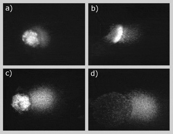
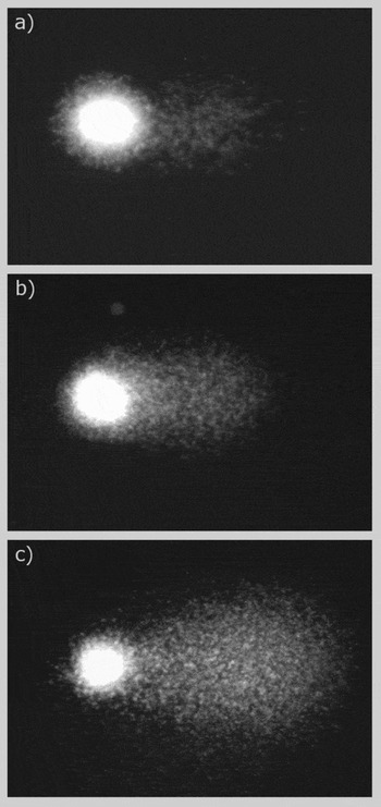
Before analysis, the slides were rehydrated in distilled water, covered with coverslip and images were captured with CCD camera (VDS) attached to a VANOX BHS fluorescence microscope (Olympus). Only the images showing no residue of oocyte cytoplasm were selected for comparative analysis (Fig. 2). In parallel, the images originating from polar bodies were captured. These were easily discernible on the basis of tightly coiled DNA in the ‘head of comet’ (Fig. 3). The extent of DNA migration was quantified using Lucia G 4.81 software (Laboratory Imaging) and the results were expressed as a percentage of DNA in the tail (tail DNA %).
Porcine oocytes processed by the comet assay with acetone prior to lysis: images with low (a, b), medium (c) and high (d) level of DNA instability.

Images from polar bodies of porcine oocytes with low (a), medium (b) and high (c) levels of DNA instability.

Statistical analysis
Activation experiments including negative control were performed four times. As the differences between experiments were not statistically significant, the data for comparative analysis were pooled from four replicate trials. The differences in the effectivity of various activation protocols were analysed by Pearson's chi-squared test. The differences in the level of DNA instability between the groups were evaluated using a non-parametric Mann–Whitney test. Oocytes and polar bodies were analysed separately. p-values lower than 0.05 were considered as statistically significant.
Results
Oocyte activation
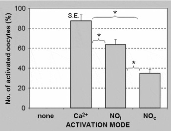
Figure 4 summarizes the effectivity of various experimental protocols in activating the oocytes. No parthenogenetic activation of oocytes was observed in negative control, i.e. after 6 h culture in the medium alone. This demonstrated that oocyte activation observed in other groups was not associated with aging of oocytes. Among three activation protocols, the 6 h treatment with calcium ionophore A23187 and 6-DMAP activating about 87% of oocytes appeared to be the most effective. In contrast, only 35.5% of oocytes started parthenogenetic development after continuous 6 h exposure to NO donor SNAP. However, the intermittent exposure to SNAP almost doubled the rate of activated oocytes (63.4%; p < 0.05).
The effect of different experimental protocols on parthenogenetic activation of porcine oocytes. The term ‘none’ means 6 h culture in free medium only, Ca2+ means activation with calcium ionophore A23187 and 6-dimethylaminopurine, NOi and NOc means intermittent and continuous activation with nitric oxide donor SNAP, respectively. Columns represent arithmetic means from four experiments; each group involves 120 oocytes, *p < 0.05.

Comet assay
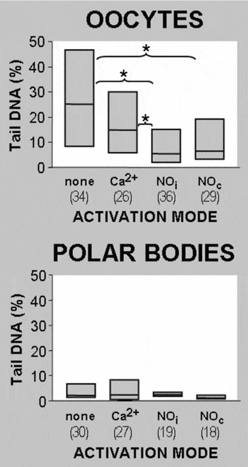
The untreated (negative control) and calcium ionophore A23187 activated oocytes (positive control) did not differ significantly in the degree of DNA migration (Tail DNA = 25.01% and 14.85%, respectively). On the other hand, both continuous and intermittent exposure to SNAP induced lower level of DNA damage than simple incubation in culture medium only (Tail DNA = 6.55% and 5.45%, respectively; p < 0.05). When compared to standard activation with Ca2+, however, only the group exposed repeatedly to SNAP displayed a significantly higher integrity of DNA (p < 0.05). The incidence of oocytes with high level of DNA instability (Tail DNA > 30%) within the particular groups decreased from 44% in negative control to 27% in positive control, 14% in oocytes continuously exposed to SNAP and 8% in oocytes repeatedly exposed to SNAP. The oocytes with completely destroyed nuclear DNA (Fig. 2d) appeared only within the negative control group.
Concerning the polar bodies, no significant differences in the level of DNA instability between the groups were detected. Nevertheless, lower variability of the results was observed within both SNAP-treated groups of oocytes in comparison with negative and positive control.
Discussion
Comet assay procedure
Two approaches have, so far, been applied to assess DNA fragmentation in porcine oocytes. The first approach consisted of visual qualitative evaluation under the microscope for the presence or absence of DNA fragments escaping the nucleus (Mattioli et al., Reference Mattioli, Barboni, Gioia and Lio2003). The second approach was based upon the measurement of migrated DNA length in individual cells using an eyepiece micrometer (Tatemoto et al., Reference Tatemoto, Sakurai and Muto2000; Yuan et al., Reference Yuan, Hao, Liu, Wu, Yang, Liu, Tian, Zhu and Zeng2008). However, the values of ‘tail length’, the parameter popular namely at the beginnings of the comet assay, may be influenced significantly by the electrophoresis setting such as adjusted voltage and the type of power supply (Vrzoc & Petras, Reference Vrzoc and Petras1996). Therefore, ‘% tail DNA’ is now considered as a more appropriate parameter reducing the variability of the multiple electrophoresis runs and facilitating inter-laboratory comparison of the results (Kumaravel et al., Reference Kumaravel, Vilhar, Faux and Jha2009). In addition, numerous commercially available specific image analysis software packages are recommended to quantify the extent of DNA damage more reliably and reproducibly (Kumaravel et al., Reference Kumaravel, Vilhar, Faux and Jha2009). Such measurements require high quality images with minimum background fluorescence. Unfortunately, the porcine oocytes are characterized by high content of lipidic components in the cytoplasm irremovable by the treatment with standard lysing solution, which may lead after the staining of slides to confounding fluorescence of remaining cytoplasmic material. An insertion of acetone pretreatment into the procedure dissolved the lipids (at least in part) and allowed us to measure exactly the proportion of migrated DNA on the basis of fluorescence intensity in a sufficient number of oocytes.
The effect of different activation modes on DNA integrity of porcine oocytes and polar bodies. The term ‘none’ means 6 h culture in free medium only, Ca2+ means activation with calcium ionophore A23187 and 6-dimethylaminopurine, NOi and NOc stand for intermittent and continuous activation with nitric oxide donor SNAP, respectively. The horizontal line in the box depicts the median value, the box 25–75 percentile, the number in the parentheses shows the number of analysed cells, *p < 0.05.

Activation protocols and DNA integrity
The results of the comet assay revealed a high level of DNA instability in the negative control, which undoubtedly reflected starting degeneration of mature oocytes in the absence of activation stimulus. Surprisingly, oocytes subjected to the consecutive treatment of Ca2+ ionophore A23187 and 6-DMAP, i.e. to the procedure with almost 90% activation efficiency, also displayed relatively high values of damaged DNA in comparison with those exposed to NO donor SNAP.
Defects in genetic material of mammalian oocytes are considered to be one of the main causes for the compromised developmental competence. Even a subtle DNA damage unidentifiable at the morphological level may lead to the failure of essential cell functions. For example, some cryopreserved bovine metaphase II oocytes exhibited an increased DNA fragmentation despite an apparently intact morphology (Men et al., Reference Men, Monson, Parrish and Rutledge2003a). Undoubtedly just this DNA damage triggered an apoptotic degeneration of oocytes during subsequent culture (Men et al., Reference Men, Monson, Parrish and Rutledge2003b) because damage to the haploid DNA cannot be readily repaired by endogenous mechanisms as in diploid cells (Long & Michod, Reference Long and Michod1995). Hence, although the Ca2+ elevation represents the primary intracellular signal necessary for parthenogenetic activation of porcine oocytes (Petr et al., Reference Petr, Rajmon, Rozinek, Sedmíková, Jeseta, Chmelíková, Svestková and Jílek2005a), our present data suggest that this mode of activation may be associated with an increased DNA instability in maturing oocytes that could compromise the further embryonic development.
Similar to Ca2+, NO also plays an important role in numerous physiological processes. During oocyte maturation, NO controls the meiotic resumption of oocytes, especially germinal vesicle breakdown and first polar body emission by regulating the anaphase–telophase transition (Huo et al., Reference Huo, Liang, Yu, Zhong, Yang, Fan, Chen and Sun2005). In addition, it delays oocyte aging. Exposure of mouse oocytes to increasing concentrations of NO diminished the rate of tubulin polymerization in the ooplasm, cortical granule exocytosis and zona pellucida dissolution timing, and improved spindle as well as chromosomal integrity (Goud et al., Reference Goud, Goud, Diamond and Abu-Soud2005). These protective effects of NO have been attributed to its ability to scavenge harmful reactive oxygen species contributing to the process of aging and decreased developmental competence of oocytes (Goud et al., Reference Goud, Goud, Diamond and Abu-Soud2005). We assume, therefore, that an increased oxidative stress resulting from an imbalance between the overproduction of unwanted free radicals under in vitro conditions and insufficient availability of endogenous NO could be responsible for a relatively high DNA instability in oocytes activated by Ca2+ ionophore A23187 and 6-DMAP.
On the other hand, continuous treatment of porcine oocytes with a NO donor is characterized by low activation efficiency such that parthenogenetic embryos only rarely develop beyond the 4-cell stage, probably due to an inadequate gene expression (Petr et al., Reference Petr, Rajmon, Lánská, Sedmíková and Jílek2005b, Reference Petr, Chmelíková, Krejčová, Rehak, Novotná and Jílek2008). It seems that intermittent exposure of oocytes to NO, resembling the oscillation of intracellular Ca2+ in fertilized mammalian oocytes, better corresponds to natural conditions because this experimental protocol preserves DNA integrity better (present results), increases the activation rate and improves the subsequent parthenogenetic development (Petr et al., Reference Petr, Chmelíková, Krejčová, Rehak, Novotná and Jílek2008).
In contrast to oocytes, the levels of DNA damage in the polar bodies showed no significant difference between the groups. As both polar bodies and oocytes were processed simultaneously on the same slides, we cannot exclude that, due to tight coiling, the DNA in polar bodies did not unwind sufficiently within a given time – which could mask possible differences between the groups. In any case, NO-mediated activation produced more homogeneous results than the standard activation with Ca2+ or simple incubation in culture medium.
In conclusion, the physiological nature of activating stimulus has not yet been fully clarified. This factor is one of the reasons why procedures such as in vitro fertilization or cloning of mammals using the nuclear transfer are still characterized by a relatively low efficiency. Our results suggest that the intermittent exposure to a NO donor could represent a promising approach to optimization of activation protocols.
Acknowledgements
The project was supported by grants GACR 523/06/0295, MSM 6046070901 and AVOZ 50390512.
Conflict of interest statement
The authors declare that there are no conflicts of interest.