Introduction
Cysts are frequently found on magnetic resonance imaging (MRI) and computed tomographic (CT) brain imaging. Because they possess a broad histopathologic spectrum, the ability to differentiate these cysts based solely on imaging findings can be difficult. Intracranial epidermoid cysts are congenital, comprising 0.2–1.8% of primary intracranial tumours, and are four to nine times as common as dermoid cysts.
Surgical intervention should be undertaken in patients presenting with hydrocephalus, neurologic symptoms or cyst enlargement. The endoscopic approach represents a minimally invasive and safe procedure in the treatment of symptomatic pineal cysts (1) In some cases clinically symptomatic glial cyst of the pineal gland, are presenting with cerebral deficits, sensory deficits and emotional disturbances (2).
We present a case with depressive symptomatology secondary to a pineal epidermoid cyst.
Case report
We here present the case of a 32-year-old man who reported abrupt onset of symptoms of severe headache, this accompanied by fatigue, depressed mood lasting most of the day, marked diminished interest or pleasure in all or nearly all activities, insomnia and reduced ability to think and concentrate. On the Beck Depression Inventory (BDI) he had a score of 24, a score higher than 15 representing clinical depression. His score on BDI was mainly shaped from his reported symptoms in the subscale that measures somatic–vegetative performance complaints (consisting of the last eight items of the BDI). The symptoms progressively increased and the patient visited a psychiatrist who recommended an MRI examination of which the result was the following:
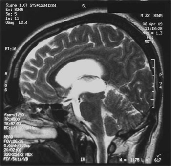
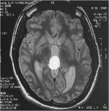
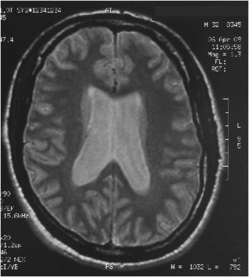
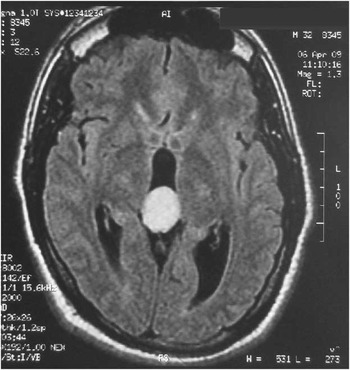
Brain MRI Examination techniqueT1W SE/FSE sequences in all three spatial planes before and after intravenous contrast administration. T2W SE/FSE sequences in all three spatial planes. Flair sequences in all three spatial planes MRI findings. A cystic lesion was detected in the posterior part of the third ventricle with a maximum diameter of ∼2.8 cm and obstructing the aqueduct of Sylvius (Figs 1 and 2), which resulted in obstructive hydrocephalus (Fig. 3) and, more centrally, dilatation of the third ventricle and the lateral ventricles. After the intravenous contrast administration, the lesion showed no enhancement (Fig. 4). No abnormal signal intensity was observed in the cerebral hemispheres, the brain stem or the cerebellum. No mass was visualised in the cerebral hemispheres, neither was any sign of intra-axial or extra-axial bleeding or haematoma identified.
Cystic lesion in the third ventricle.

Cystic lesion in aqueduct of Sylvius.

Cystic lesion and obstructive hydrocephalus.

Cystic lesion.

The images after the intravenous contrast administration showed no abnormal enhancement. Pituitary gland and paranasal sinuses appeared normal, as did orbital structures. Two weeks later he was operated upon: surgery was performed, the approach used being neuroendoscopic 3rd ventriculostomy via neuronavigator, and the cyst was removed. An MRI follow-up 8 months after surgery revealed a normal flow of cerebral spinal fluid through the Sylvius aqueduct. Histological examination revealed a cystic formation, the walls of which at the inner surface consisted of brain parenchyma with reactive gliosis lesions [confirmed by immunohistochemical staining for Glial Fibrillary Acidic Protein (GFAP) and protein S100] and haemosiderin deposition (revealed by histochemical staining for Perl's iron). The outer cyst wall consisted of pineal parenchyma with no evidence of malignancy. The patient made a full recovery and returned to his daily routine two months after the initial diagnosis.
Discussion
This is the case of a young man presenting depressive symptomatology after the appearance of hydrocephalus and a pineal epidermoid cyst. Patients with hydrocephalus present a variety of neuropsychiatric symptoms, these being mainly apathy, anxiety, agitation and depression. The frequently observed association of hydrocephalus and depressive symptomatology, which was described by Adams et al. (Reference Adams, Fisher, Hakim, Ojeman and Sweet3), is typically characterised in the early manifestations of illness by slowness of thought and action, lack of spontaneity or initiative as well as apathy and inertia. The above authors described the inner psychic life of the patient as impoverished and suggested that these symptoms might easily be mistaken for signs of depression.
The discovery of melatonin and its physiological effects stimulated interest in the relationship between the pineal gland and mental disorders, particularly those of an affective nature, as well as sleep alterations about which an extensive amount of documentation has been generated over the last few decades. The observation that melatonin was capable of increasing the levels of serotonin in the pineal gland gave rise to the hypothesis of its possible usefulness in the treatment of affective disorders, the serotoninergic theory of antidepressants being advanced at the end of the 1960s (Reference Lopez-Munoz, Molina, Rubio and Alamo4).
A rational protocol for the follow-up of asymptomatic pineal cysts detected by MRI depends upon the resolution of three problems. First, the imaging features of pineal cysts that should prompt follow-up imaging remain unclear. Although it has been recommended that particularly large pineal cysts (>1 cm or >1.4 cm in diameter) undergo follow-up imaging, no evidence has been presented to suggest that larger cysts are more likely to grow or become symptomatic. Second, the significance of small changes in pineal cyst size, seen on follow-up MRI imaging, is unknown. In part, this lack of knowledge is due to the fact that the natural history of incidentally discovered asymptomatic pineal cysts is incompletely understood. The finding that pineal cysts are relatively rare in young children and that, on average, these cysts are seen in adults <40 years old, suggests that pineal cysts may typically be formed in late childhood and later involute. Thus, an interval increase in cyst size could simply reflect the natural history of asymptomatic cyst development and may not presage the development of symptoms. Finally, appropriate intervals for imaging follow-up of atypical pineal cysts and criteria for cessation of cyst surveillance are as yet undetermined (Reference Kiely5).
Clinically speaking, the psychiatrist should bear in mind that many cases of depressive symptomatology could have an organic cause and he/she should therefore proceed to more laboratory exams, including a CT scan. Young people often manifest sudden psychological symptoms secondary to cerebral tumours or an epidermoid cyst, thus, surgical intervention should be the preferred treatment rather than rapid initiation of antidepressant pharmacotherapy.
Acknowledgement
The authors would like to thank the patient and his family for providing the medical information.