Introduction
Nasal dermoid sinus cysts were originally described as nasal lesions which included tissue of ectodermal and mesodermal origin.Reference Sessions1 They are congenital and usually present between the ages of 14 and 34 months,Reference Rahbar, Shah, Mulliken, Robson, Perez-Atayde and Proctor2 but many examples of adult presentation have been reported.Reference Vaghela and Bradley3, Reference Duz, Secer, Tosun and Gonul4 The incidence of intracranial extension of dermoids varies from 5 to 45 per cent.Reference Rahbar, Shah, Mulliken, Robson, Perez-Atayde and Proctor2, Reference Vaghela and Bradley3, Reference van Aalst, Luerssen, Whitehead and Havlik5 One retrospective study of surgical resection of nasal dermoids found that 27.5 per cent of adults who presented with a nasal dermoid cyst had intracranial extension.Reference Vaghela and Bradley3
A number of techniques have been described for the treatment of nasal dermoid tumours.Reference Zapata and Kearns6 Previously, a craniotomy was mandatory to achieve gross resection of dermoids with intracranial extension.Reference Blake, Chow, Holmes and Meara7 More recently, one case of successful endoscopic resection of a relatively small intracranial dermoid has been reported.Reference Duz, Secer, Tosun and Gonul4 In the cases reported below, we employed a different surgical technique to endoscopically remove two dermoids with massive intracranial frontal lobe involvement.
The goal of endoscopic removal is to eliminate the increased morbidity associated with a frontal craniotomy.
Case reports
Patient one
An otherwise healthy, 23-year-old man presented after having a seizure.
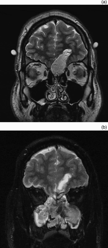
A magnetic resonance imaging (MRI) scan revealed a large skull base tumour extending 4 cm into the left frontal lobe (Figure 1a). This lesion had intracranial and extracranial components, with a fistulous tract extending from the skull base out to the nasal tip. A surgical navigation computed tomography (CT) scan and a neuronavigation MRI scan were also performed.
Magnetic resonance imaging (MRI) scans for patient one. (a) Pre-operative, coronal MRI scan showing the lesion extending 4 cm into the left frontal lobe. (b) Post-operative, coronal MRI scan showing total resection of the dermoid cyst, with packing supporting the skull base repair.

After discussing the surgical options and associated risks with the patient, he elected to undergo transnasal endoscopic resection of the intracranial dermoid cyst, rather than open bifrontal craniotomy.
The extracranial portion of the dermoid cyst was approached initially using an open rhinoplasty approach. When the nasal envelope was elevated, the fistulous tract was identified extending out through the nasal tip. The tract was tagged and the skin pit excised in an elliptical fashion, followed by closure of the incision. With the fistulous tract identified, the upper lateral cartilages were separated from a bifid nasal septum, which contained the extracranial portion of the cyst. The cartilage was removed on the left-hand side, the cyst was excised, and the nasal envelope was reattached to the columella. The remainder of the case was performed using nasal endoscopy.
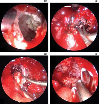
Full endoscopic sinus surgery was performed bilaterally, widely opening the maxillary, ethmoid, sphenoid and frontal sinuses. Opening the sinuses prevents iatrogenic sinus disease following the operation, increases working space for instrumentation, and helps define the limits of the intracranial portion of the tumour. With the skull base completely skeletonised bilaterally, middle turbinectomies were performed to expose the olfactory clefts. Next, a nasal septal flap pedicled on the septal branch of the sphenopalatine artery was raised from the nasal septum and placed into the nasopharynx for later use. A septectomy was performed to completely expose the anterior skull base, leaving 1 cm of anterior septum for nasal support. The frontal sinuses were then connected in one common cavity as a Draf III procedure. This was performed to ensure adequate opening and exposure of the anterior limit of the tumour (Figure 2).
Intra-operative views via a 70° nasal endoscope for patient one. (a) Sebaceous material can be seen as a component of the dermoid cyst. (b & c) The right frontal sinus is noted for perspective (arrow). The cyst wall is meticulously dissected off the surrounding dura and brain. (d) Approximately 2 cm of exposed brain is visualised through the skull defect following cyst removal.

The intradural component of the procedure was performed with complete exposure of the skull base. Due to the anterior location of the tumour, the skull base underlying the intracranial component was removed using a 70° diamond bur and visualised using a 70° nasal endoscope. With removal of the underlying bone, the cyst was entered, exposing sebaceous material and hair. The components were removed from the pocket of the dermoid cyst. The sebaceous material had tracked 4 cm into the left frontal lobe, at which point the dermoid contents had broken through the cyst wall. There was a visible brain tissue reaction in this area, with gliosis. With meticulous dissection and a four-handed technique, the cyst wall was removed from the surrounding brain in an inferior to superior and right to left direction, collapsing the cyst in on itself. The cyst was then removed, together with approximately 2 cm of exposed brain, through the defect.
The skull base defect was repaired with dural substitute laid in an underlay fashion beneath the remaining dura. Evicel fibrin sealant (Johnson & Johnson, Somerville, New Jersey, USA) was dripped onto the underlay graft to completely seal the breach and prevent CSF leakage. The nasal septal flap was removed from the nasopharynx and rotated onto the skull base defect for full closure. Frontal sinus stents, absorbable packing and two long finger cot dressings were inserted underneath the skull base for support.
Immediate post-operative MRI scanning demonstrated complete removal of the tumour (Figure 1b). The patient was discharged on post-operative day two in good condition. He suffered no recurrence over one year of clinical follow up.
Patient two
A 23-year-old woman was referred after a large skull base tumour was noted on a head CT scan performed as an investigation for headaches.
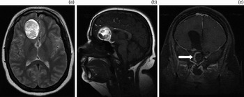
Subsequent CT and MRI scans revealed a large tumour extending 6 cm into the right frontal lobe, which had a small, tailed extension to the anterior suprasellar area (Figure 3). There was no extracranial component to the tumour.
Magnetic resonance imaging (MRI) scans for patient two. Pre-operative (a) axial and (b) sagittal views show a large dermoid extending 6 cm into the right frontal lobe. (c) Post-operative coronal MRI scan shows complete removal of the cyst, leaving a large, cerebrospinal fluid filled cavity. The nasal septal flap is draped along the skull base defect (arrow).

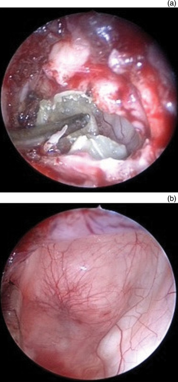
The skull base was exposed as described above. The skull base was then drilled down with 70° and 15° diamond burrs over the planum sphenoidale as well as the right anterior and posterior ethmoid and midline cribriform area. Once the skull base was removed down to the dura, the dura was entered and the dermoid cyst contents were encountered. The cyst contents extended 6 cm into the right frontal lobe, and were removed using a binostril, four-handed technique similar to that utilised in patient one (Figure 4).
Intra-operative views via a 70° nasal endoscope for patient two. The cyst and its contents are removed (a) leaving a large parenchymal defect (b).

The skull base was repaired with an underlay dural substitute and a right-sided nasal septal flap.
Immediate post-operative MRI scanning demonstrated complete cyst removal (Figure 3c).
The patient's discharge from hospital was delayed due to concerns about post-operative CSF leakage. She was returned to the operating room for an examination under anaesthesia, but no CSF leak was identified. She was subsequently discharged on post-operative day five in good condition.
At the time of writing, the patient had remained asymptomatic, with no recurrence at six months post-operatively.
Discussion
Nasal dermoid cysts cannot be cured without total excision of the tumour and any associated sinus tract.Reference Zapata and Kearns6 If dermal components remain after surgery, recurrence rates as high as 100 per cent have been reported.Reference Posnick, Bortoluzzi, Armstrong and Drake8
Traditionally, the extracranial component of dermoids has been resected via an open rhinoplasty or vertical midline incision. However, there are now several reports of endoscopic resection.Reference Zapata and Kearns6, Reference Weiss, Robson and Mulliken9 Despite this, numerous authors have stated that dermoid cysts that extend intracranially into or past the falx cerebri cannot be resected endoscopically.Reference Zapata and Kearns6, Reference Weiss, Robson and Mulliken9
Pollock has stated that any technique used to resect intracranial dermoid cysts successfully must meet four criteria: it must allow access to the cyst and permit osteotomies; it must enable repair of cribriform plate defects and prevention of CSF leakage; it must aid reconstruction of the nasal dorsum; and it must ensure a cosmetically acceptable scar.Reference Pollock10
Some surgeons have found that a brow incision and small window craniotomy is a superior technique with less morbidity than a frontal craniotomy. They have also claimed that the results are more cosmetically acceptable.Reference Heywood, Lyons, Cochrane, Hayward and Hartley11 Others have suggested a ‘keystone’ approach, requiring a bifrontal craniotomy, as being the most effective at achieving total resection.Reference van Aalst, Luerssen, Whitehead and Havlik5 Regardless of modifications, it has previously been thought that some form of frontal craniotomy was required to remove a dermoid with intracranial extension,Reference Blake, Chow, Holmes and Meara7 and numerous case reports have discussed various techniques.Reference Sanchez-Mejia, Limbo, Tihan, Galvez, Woodward and Gupta12, Reference Blythe, Revington and Nelson13
The use of a team-based approach, including an experienced endoscopic otolaryngologist and neurosurgeon who frequently operate together, is crucial to the success of the transnasal endoscopic technique. The above case reports demonstrate that it is possible to resect large intracranial dermoid cysts using this approach.
The non-invasive nature of dermoid cysts makes the plane of dissection between the cyst wall and the surrounding brain parenchyma or dura relatively smooth and easy to manipulate. The cyst contents are readily removed to enable decompression and to improve cyst wall mobility. The above-described transnasal endoscopic skull base approach meets all four of Pollack's criteriaReference Pollock10 for successful resection of intracranial dermoids, but also completely eliminates an external sur.
The morbidity associated with frontal craniotomy includes meningitis, hyposmia, CSF leakage, damage to the frontal and sagittal sinuses, and occasionally intracerebral haemorrhage, cerebral oedema, epilepsy, memory and concentration deficits, and osteomyelitis of the frontal bone flap.Reference Heywood, Lyons, Cochrane, Hayward and Hartley11 Many of these problems are minimised or eliminated when open excision is avoided. The transnasal approach does not require frontal lobe retraction, and patients have excellent post-operative recovery and a shorter hospital stay. Patient one was discharged on post-operative day two (although he felt well enough to leave the hospital on post-operative day). In addition, the endoscopic approach enables a substantial improvement in cosmetic result, as it leaves no scar.
• Nasal dermoids are composed of ectodermal and mesodermal tissues
• Up to 45 per cent of such tumours have intracranial extension
• Total excision of the tumour and sinus tract is necessary for cure
• Tumours with intracranial extension were previously thought to require frontal craniotomy for complete excision
• However, this paper reports successful endoscopic resection of two intracranial dermoids with large anterior frontal lobe extensions, using a 70° endoscope and a pedicled nasoseptal flap to close the skull base defect
• Such endoscopic removal has minimal morbidity and mortality, and better cosmesis, compared with open craniotomy
One other case of successful transnasal endoscopic resection of an intradural dermoid has been reported by Duz et al.,Reference Duz, Secer, Tosun and Gonul4 but this case had significant differences in tumour location and surgical technique. In this case, the cystic lesion was located in the anterior subfrontal region but more posteriorly than the present two cases, allowing resection using a 0° nasal endoscope.Reference Duz, Secer, Tosun and Gonul4 In the present two cases, the cysts were much larger. In addition, due to the large degree of parenchymal extension and the anterior location of the cysts, the use of 70° nasal endoscopy was absolutely crucial to enable intra-operative visualisation and complete removal.
Conclusion
The transnasal endoscopic skull base approach for large intracranial dermoid cysts represents a promising treatment option in experienced hands. Careful resection using an angled endoscope can prevent the undue morbidity associated with frontal craniotomy, while still achieving total resection of the tumour.