INTRODUCTION
The yellow-eyed penguin (Megadyptes antipodes), or Hoiho, is endemic to New Zealand and is one of the rarest species of penguin (McKinlay, Reference McKinlay2001). It is the only member of its genus and one of the most endangered of the 18 species of penguins. The yellow-eyed penguin (YEP) has been classified as endangered on the IUCN Red List since 2000 based on extreme population fluctuations, restricted breeding range and declines in quality and quantity of their natural habitat (Birdlife International, 2011). The population is estimated at between 6000 and 7000 birds (McKinlay, Reference McKinlay2001), with 630 pairs on the South Island southeast coast, 178 pairs on Stewart Island, 520–570 pairs on the Auckland Islands and around 405 pairs on Campbell Island (Moore, Reference Moore1992; McKinlay, Reference McKinlay2001). Unlike other penguin species, YEPs are not colonial but instead nest in sparse colonies and avoid visual contact between pairs at adjacent nest sites. Approximately two-thirds of the entire population of YEPs is found on the southern offshore and sub-Antarctic islands of New Zealand with 22% on Campbell Island (52°32′24″S, 169°8′42″E), 23% on the Auckland Island archipelago (50°42′0″S, 166°5′0″E) and 21% found on Stewart (47°00′0″S, 167°50′0″E) and Codfish (46°47′0″S, 167°38′0″E) Islands (Darby and Seddon, Reference Darby, Seddon, Davis and Darby1990; Moore, Reference Moore1992). The remainder of the population is found on the east coast of the South Island between South Otago and Banks Peninsula, with 3 main colonies located at Oamaru, the Otago Peninsula and the Catlins (Darby and Seddon, Reference Darby, Seddon, Davis and Darby1990).
Recent periodic mass mortality events or population declines have been documented in YEPs on the South and Stewart Islands since the 1980s, resulting in significant population declines (Moore et al. Reference Moore, Fletcher and Amey2001). These population declines have been attributed to non-infectious events such as unidentified phytotoxins (Gill and Darby, Reference Gill and Darby1993), starvation, poor nutrition (Vanheezik, 1990a; Vanheezik and Davis, Reference Vanheezik and Davis1990) and a possible relationship with climatic events such as El Nino or the Southern Oscillation (Moore and Wakelin, Reference Moore and Wakelin1997). Infectious causes of population decline have also been identified, in particular avian haemoparasites, such as Plasmodium and more recently Leucocytozoon (Graczyk et al. Reference Graczyk, Cranfield, Brossy, Cockrem, Jouventin and Seddon1995; Alley, Reference Alley2005; Hill et al. Reference Hill, Howe, Gartrell and Alley2010).
The significance of haemoparasites in mass mortality events is controversial. There is evidence that Leucocytozoon and Plasmodium are endemic parasites in YEP populations within their New Zealand range. Plasmodium spp. was first reported in YEPs in the 1940s–50s and, more recently, considered the cause of a mortality event on the Otago Peninsula during the 1989/90 breeding season, due to the pattern of mortality coupled with higher positive Plasmodium antibody titres in the mortality outbreak penguins compared with live penguins from the same geographical location (Gill and Darby, Reference Gill and Darby1993; Graczyk et al. Reference Graczyk, Cranfield, Brossy, Cockrem, Jouventin and Seddon1995). However, recent investigation into the prevalence of this disease failed to identify Plasmodium spp. in 143 YEPs from the Otago Peninsula (Sturrock and Tompkins, Reference Sturrock and Tompkins2007). During this same time period, Alley et al. (2005) described the first reported case of leucocytozoonosis, a disease caused by Leucocytozoon spp., in the YEP population on Codfish and Stewart Island (Alley, Reference Alley2005; Hill et al. Reference Hill, Howe, Gartrell and Alley2010). Leucocytozoon spp. has previously also been reported in New Zealand in wild Fiordland crested penguins (Eudyptes pachyrhynchus) (Fallis et al. Reference Fallis, Bisset and Allison1976).
Leucocytozoon spp. infections are usually benign; however, a few species are extremely pathogenic, for example L. simondi in young ducks and geese in the northern hemisphere, and L. smithi in wild and domestic turkeys (Meleagris gallopavo) throughout North America and Europe (Steele and Noblet, Reference Steele and Noblet1992; Remple, Reference Remple2004). The presence of Leucocytozoon can exert subclinical effects on the host compounding the effects from concurrent disease or other stressors. Infection with Leucocytozoon has also been shown to have negative effects on reproduction and body weight of the host (Merino et al. Reference Merino, Moreno, Sanz and Arriero2000; Remple, Reference Remple2004). Leucocytozoon toddi has been shown to have detrimental effects on juvenile great horned owls (Bubo virginianus) resulting in increased mortality during years of severe food shortage (Hunter et al. Reference Hunter, Rohner and Currie1997).
Leucocytozoon gametocytes develop in circulating leucocytes and erythrocytes, with schizogony occurring in fixed tissues. Two types of schizonts are produced following schizogony; those in hepatic cells form hepatic schizonts, while merozoites that develop in the cells of the reticulo-endothelial system form megaloschizonts. These larger schizonts can be found in a wide range of organs including the brain, liver, lungs, kidneys, intestines and lymphoid tissues. The development of these megaloschizonts in a variety of organs is probably the main mechanism contributing to pathogenicity of Leucocytozoon (Fallis et al. Reference Fallis, Desser and Khan1974; Steele and Noblet, Reference Steele and Noblet1992).
The prevalence and pathogenicity of Leucocytozoon spp. in YEP chicks has been reported on the Otago Coast of New Zealand and nearby Stewart Island (Hill et al. Reference Hill, Howe, Gartrell and Alley2010); however, little is known about the prevalence of this parasite in the sub-Antarctic population of YEPs. The aim of this study was to further investigate the prevalence of Leucocytozoon spp. and the possible role of this pathogen in a chick mortality event during the 2008/2009 breeding season of the endangered YEP sub-Antarctic Enderby Island population.
MATERIALS AND METHODS
Study sites
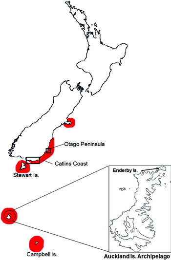
YEP samples were collected from the southeastern Otago and Catlins coast of the South Island of New Zealand (46°27′S 169°49′E), which supports a population of approximately 950 adult breeding penguins. Birds were also sampled from 2 sub-Antarctic islands. In the Auckland Islands archipelago (50°29′–50°59′S, 165°52′–166°20′E) which supports a YEP population of ∼1200 breeding adults, birds were sampled from Enderby Island which comprises 40% of the total breeding population of yellow-eyed penguins. Birds were also sampled on remote Campbell Island (52°33′S, 169° 09′E) which supports an estimated breeding population of 800 adult yellow-eyed penguins (IUCN Red List data) (Fig. 1).
Distribution and range of yellow-eyed penguins (Megadyptes antipodes) on the South Island Stewart Island and sub-Antarctic Islands of New Zealand. Shading indicates yellow-eyed penguin range. Inset map of Auckland Islands Archipelago (50°29′–50°59′S, 165°52′–166°20′E) indicates location of Enderby Island. The boxes indicate areas on the mainland where samples were collected. Samples were collected from all over Campbell and Enderby Islands.

Collection of blood samples and blood smear preparation
During the breeding season between December 2006 and January 2007, adult YEPs on Enderby Island (n = 19) and Campbell Island (n = 19) were randomly selected and blood samples collected to assess the presence of Leucocytozoon. In addition, 96 blood samples (27 chicks, 4 juveniles and 65 adults) were collected during the December 2008–January 2009 breeding season. It is unknown whether any penguins that were sampled on Enderby during the 2006–2007 season were re-sampled in 2008–2009 as these birds have no permanent method of identification.
All birds captured on Enderby Island in 2008/2009 were given a physical examination and either had a passive integrated transponder placed subcutaneously or were marked with non-permanent livestock marker to prevent re-sampling and were released after blood sample collection. A body condition score was subjectively assigned on a scale of 1–9 based on and modified from the American Animal Hospital Association nutritional assessment guidelines for cats and dogs (Baldwin et al. Reference Baldwin, Bartges, Buffington, Freeman, Grabow, Legred and Ostwald2010).
Between 0·5 and 2 mL of blood was drawn from either the brachial or medial metatarsal vein of each bird and placed into lithium heparin blood containers. Two fresh blood smears were prepared for each bird on glass slides. The blood films were air-dried and the slides stored in a sealed, dry, watertight container. Smears were fixed in 100% methanol and stained with modified Wright's solution (Diff Quik, Harleco, Gibbstown, NJ, USA). The entire field of each smear was initially examined for the presence of haemoparasites at low magnification (400×), and then at least 50 fields were studied at high magnification (1000×) for approximately 30 min.
The remaining blood was stored in liquid nitrogen or Queen's lysis buffer while on site and then transferred to a −80 °C freezer for long-term storage.
Post-mortem sample analysis and collection
Post-mortem examination was performed on 19 YEP chicks from Enderby Island during the 2008–2009 breeding season and tissues from a full range of organs were fixed in 10% buffered formalin for histopathology. In addition a sample of fresh liver from each chick was stored and frozen in liquid nitrogen and then transferred to a −80 °C freezer upon return to the laboratory for molecular studies. Post-mortem examination was also performed on 115 YEPs (84 chicks and 31 adults) from the Otago Peninsula/Catlin coast of the South Island that were submitted to the Wildlife Health Centre at Massey University during 2008. A full range of organs from these birds was fixed in 10% buffered formalin for histopathology and liver from 27 submitted chicks was frozen and stored at −10 °C for later molecular analysis. All fixed tissues were routinely processed, embedded in paraffin, cut at a thickness of 3 μm and stained with haematoxylin and eosin for subsequent histopathological examination.
Molecular studies
DNA was extracted from all collected blood (n = 134) and tissue samples (Enderby chicks n = 19, South Island yellow-eyed penguins n = 27) using a DNeasy blood and tissue kit (Qiagen, Victoria, Australia) following the manufacturer's instructions for blood or tissue respectively. All samples were screened for the presence of Leucocytozoon DNA using the nested PCR method to amplify the cytochrome b gene as described by Hill et al. (Reference Hill, Howe, Gartrell and Alley2010) in order to conform to the international database as recommended by Bensch et al. (2009) and Valkiunas et al. (Reference Valkiunas, Santiago-Alarcon, Levin, Iezhova and Parker2010). To confirm successful amplification 10 μL of the final PCR product was run on a 1·5% agarose gel containing ethidium bromide prior to purification and sequencing. A known Leucocytozoon spp. positive tissue sample, confirmed by sequencing, was used as a positive control and water blanks were included as negative controls.
When sufficient PCR product was amplified, Leucocytozoon spp. positive PCR amplicons were purified using a PureLink PCR purification kit (Invitrogen, Carlsbad, CA, USA) and subjected to automatic dye-terminator cycle sequencing with BigDyeTM Terminator Version 3.1 Ready Reaction Cycle Sequencing kit and the ABI3730 Genetic Analyzer (Applied Biosystems Inc, Foster City, CA, USA) to confirm genomic sequence using both the forward and reverse primers. The resulting sequences were submitted to the GenBank database (JX569268-70).
Phylogenetic analysis of Leucocytozoon isolates
Phylogenetic analysis of Leucocytozoon cytochrome b sequences (n = 21) obtained was compared by NCBI Blast to those other published cytochrome b sequences available from GenBank. Representative YEP isolates, 10 sequences obtained from the MalAvi database (Bensch et al. Reference Bensch, Hellgren and Perez-Tris2009) and known GenBank sequences, including 6 representatives of well-characterized Leucocytozoon species/lineages (L. majoris, GenBank FJ168563, L. macleani, GenBank DQ676825, L. schoutedeni, GenBank DQ676824, L. danilewskyi, GenBank EU627823, and L. fringillinarum, GenBank AY393796), a lineage from Tyto alba (GenBank EU627792) as suggested by Valkiunas et al. (Reference Valkiunas, Santiago-Alarcon, Levin, Iezhova and Parker2010) and 3 previously identified lineages from YEP (GenBank GU065716–18), were trimmed to the same length (411 base pairs) using Geneious™ (Biomatters, Auckland, New Zealand) and aligned using Clustal W (Higgins et al. Reference Higgins, Thompson, Gibson, Thompson, Higgins and Gibson1994) with gaps ignored. A Bayesian phylogenetic tree was generated in MrBayes version 3.1 (Ronquist and Huelsenbeck, Reference Ronquist and Huelsenbeck2003) using a general time-reversible model including invariable sites (GTR + I) was used. The Bayesian phylogeny was obtained using 1 cold and 3 hot Monte Carlo Markov chains, which were sampled every 1000 generations over 2 million generations. Of these trees, 25% were discarded as burn-in material. The remaining trees were used to construct a majority consensus tree. Bootstrap percentages from the Bayesian analysis were added to the tree at the appropriate nodes. The sequence divergence between and within the different lineages was calculated using a Jukes-Cantor model of substitution implemented in the program PAUP* 4.0 Beta version 10 (Swofford, Reference Swofford2002).
Statistical analysis
The 95% confidence interval for apparent prevalence was calculated using the Wilson binomial approximation from Brown et al. (Reference Brown, Cat and DasGupta2001). The prevalence was compared between age groups on Enderby Island, between prevalence in adults on Campbell and Enderby Islands in 2006/07, and between seasonal data on Enderby Island by chi-squared analysis. A Fisher's exact test was used to analyse the Otago Peninsula samples and to compare tissue sample results of chicks found on Enderby Island and the Otago Peninisula.
Ethics approval and permits
The research was carried out under the following permits: DOC banding permit: Enderby/Campbell 2006–2008 – SO-17933-FAU, Enderby 2008–09 –DOC AE permit # 175, Research permit for sub-Antarctic island – permissions database number SO-17658-RES (Invercargill permit # 0506-14); Massey University Animal Ethics permit MUAEC 08/91.
RESULTS
Clinical findings
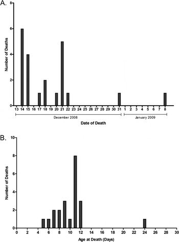
During the 2008/2009 breeding season, 48 nests were discovered and monitored on Enderby Island. Many eggs failed to hatch resulting in only 40 viable chicks. Between 14 and 23 December, 20 of 40 monitored chicks died and a further 2 chicks had died by mid-January (Fig. 2A). Most chicks were aged between 5 and 12 days old with 1 chick being found dead at an estimated 24 days after hatching (Fig. 2B). Eight carcasses were missing, and 19 carcasses were recovered in suitable condition for post-mortem examination. At the time of examination, the 40 live chicks were found to be underweight, lethargic, had poor to average feather growth and were in poor body condition. Chicks that survived the first few weeks showed significant improvements in demeanour and body condition; these improvements were most notable in chicks at nests where a sibling had died.
Distribution of number and date of death (A) and age of death (B) for yellow-eyed penguin chicks (Megadyptes antipodes) on Enderby Island from November 2008 to January 2009.

All 65 adult penguins that were sampled and examined during the 2008/09 breeding season on Enderby Island appeared healthy but were of moderate to poor body condition, with minimal subcutaneous fat, prominent keel and hips and lower body weight than expected.
Pathological findings
Post-mortem examination of the 19 chicks indicated they were in very poor body condition, with reduced pelvic and epaxial muscular mass, no subcutaneous, epicardial or abdominal fat reserves and the proventriculus was empty except for dark reddish brown/black mucus (melaena) and a few twigs and small pebbles. The most likely cause of death based on these findings was starvation. One of the 19 chicks that died had post-mortem findings inconsistent with this pattern. This chick had a full proventriculus and no notable gross abnormalities except for mild hepatomegaly. There were no significant histopathological findings in this chick.
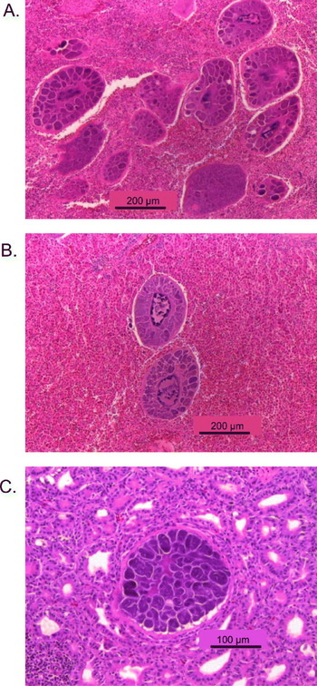
Another chick (YEP TL4B) of approximately 3·5 weeks age was found dead, trapped in a hole near to its nest. There were multiple gross abnormalities noted including widespread petechial and ecchymotic haemorrhages throughout most organs and hepato- and splenomegaly. There were 2–3 mL of serous fluid in the pericardial sac. The proventriculus was full and squid pieces were able to be identified. This chick had good fat reserves and was in good body condition. Histopathology results indicated severe disseminated leucocytozoonosis with megaloschizonts present in high numbers throughout most organs including the liver, spleen, kidneys, intestinal wall, thymus, heart and lungs (Fig. 3, Table 1). The most likely cause of death for this chick was severe disseminated leucocytozoonosis.
Haematoxylin and eosin-stained tissues from a yellow-eyed penguin chick (Megadyptes antipodes). Mature exo-erythrocytic meronts of Leucocytozoon spp. in the spleen (A), liver (B) and thyroid (C). Scale bars = 200 μm or 100 μm as indicated.

Prevalence of Leucocytozoon spp. by light microscopy, and PCR of blood and tissue samples

Eighty-four dead chicks from the Otago Peninsula/Catlin coast (South Island) were also examined. None of the mainland chicks showed any histological evidence of Leucocytozoon infection. The cause of death of these birds ranged from starvation, diphtheritic stomatitis, heat stress, predation, other diseases such as aspergillosis, or a combination of these factors.
Blood smears
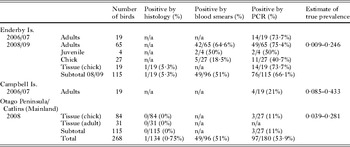
Examination of 96 blood smears taken from YEP adults, juveniles and chicks from Enderby Island during the 2008/09 breeding season found an overall prevalence of 51% of the smears containing intra-erythrocytic structures consistent with Leucocytozoon infection. This included 42/65 (64·6%) of adults, 2/4 (50%) juveniles and 5/27 (18·5%) chicks (Table 1).
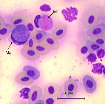
Morphological analysis of the gametocytes observed in blood smears (Fig. 4) of the 49 YEPs during the 2008–2009 breeding season on Enderby Island confirmed the presence of Leucocytozoon gametocytes that were structurally similar to L. tawaki (Valkiunas, personal communication, Reference Valkiunas, Santiago-Alarcon, Levin, Iezhova and Parker2010). There was no evidence of co-infection with other Leucocytozoon or Plasmodium spp. Blood smears were not available from other seasons or other study sites.
Gametocytes of Leucocytozoon spp. in host cells identified from Giemsa-stained blood smear of a yellow-eyed penguin (Megadyptes antipodes). Ma, macrogametocyte; Mi, microgametocyte. Scale bar = 20 μm.

Molecular analysis
During the 2006/07 season, the prevalence of Leucocytozoon, as confirmed by PCR, on Enderby and Campbell Islands was 73·7% and 21% respectively. The prevalence on Enderby Island during the 2008/09 season was 66·1% whereas a low prevalence (11%) was detected in YEPs on the mainland of New Zealand during this season. There was no significant difference (χ 2 = 0·023, d.f. = 1, P = 0·879) in the prevalence of Leucocytozoon DNA in peripheral blood samples from adult YEPs on Enderby Island between the breeding seasons 2006/2007 (73·7%) and 2008/2009 (75·4%). However, there was a significant difference (χ 2 = 19·8, d.f. = 1, P < 0·001) between the prevalence of Leucocytozoon DNA in peripheral blood samples from adult YEPs on Enderby Island during the 2006/2007 and 2008/2009 sessions when compared with those from Campbell Island in 2005/2006 (21·0%). The results of PCR analysis of yellow eyed penguin samples for the prevalence of Leucocytozoon DNA are presented in Table 1.
Additionally, there was a significant difference (χ 2 = 10·1, d.f. = 1, P < 0·001) between the prevalence of Leucocytozoon DNA in peripheral blood samples from adult (75·4%) and chick (40·7%) YEPs on Enderby Island in the 2008/2009 breeding season. There was also a significant difference (χ 2 = 4·88, d.f. = 1, P = 0·027) in the prevalence of Leucocytozoon DNA in peripheral blood samples from live chicks (40·7%) and the PCR analysis of post-mortem tissue samples from dead chicks (73·7%) on Enderby Island in the 2008/2009 breeding season. Four juvenile YEPs from Enderby Island were also sampled in 2008/2009 and 50% (2/4) were positive; however, given the low sample size of this group they have been excluded from further analysis.
A comparison of diagnostic methods for the detection of Leucocytozoon, showed that there was no significant difference between light microscopy and PCR analysis of peripheral blood samples in all adults (64·6% and 75·4% respectively) and all chicks (18·5% and 40·7% respectively). There was a significant difference (χ 2 = 14·6, d.f. = 1, P < 0·001) between detection of Leucocytozoon in chick post-mortem samples by histology (5·3%) and PCR analysis of tissue samples (73·7%). Additionally, there was a significant difference (P = 0·0132) between the prevalence of Leucocytozoon DNA in tissue histology (0/84) and PCR (3/27) for YEPs on the Otago Peninsula. There was also a significant difference (P < 0·0001) between the prevalence of Leucocytozoon DNA in tissue samples from YEP chicks on Enderby (14/19) during the 2008/2009 season compared with chicks from the Otago Peninisula (3/27) during the same season.
Phylogenetic analysis of Leucocytozoon isolates
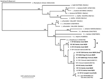
Eighty-one per cent (17/21) of isolates grouped into cluster B (Fig. 5) with 99% sequence homology to previously identified YEP lineages YEP-1 (GenBank GU065716) and YEP-2 (GenBank GU065717) and L. spp. BAOW5909 from a barn owl (Tyto alba, GenBank EU627792), SPOW44 from a spotted owl (Strix occidentalis, GenBank EU627793), and L-CIAE2 from a marsh harrier (Circus aerginosus, GenBank EF607287) (Fig. 5). Whereas previously identified lineage YEP-3 (GenBank GU065718) displayed a 98% sequence homology with the cluster B isolates (GenBank JX569268 and JX569270). The remaining 4 Enderby isolates (GenBank JX569269) displayed only 97% homology with L. spp. BAOW5909 and SPOW44 lineages and grouped into a separate cluster (cluster A, Fig. 5).
Phylogenetic analysis of Leucocytozoon spp. isolated from yellow-eyed penguins (Megadyptes antipodes). Bayesian analysis of mitochondrial cytochrome b gene from 1 lineage of Plasmodium spp., 9 Leucocytozoon sp. submitted to GenBank, and Leucocytozoon isolated in this study (bold font) from yellow-eyed penguins living on Enderby Island (n = 7), Campbell Island (n = 1) and the Otago Peninsula (n = 2). The tree is rooted on a lineage of Plasmodium relictum. Numbers above the branches indicate bootstrap support based on 1000 replicates. Names of the lineages (when available) and GenBank Accession numbers of the sequences are given after the species names of the parasites.

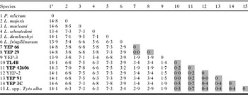
The isolates have no direct genetic relationship to a Plasmodium relictum sp. isolated from an African penguin (Spheniscus demersus, GenBank NC012426) with a sequence divergence of between 13·8 and 14·8% (Fig. 5, Table 2). Cluster A isolates clustered in their own group and included only isolates from YEPs residing on Enderby Island during the 2008/2009 breeding season (Fig. 5). However, cluster B comprises Leucocytozoon from YEPs on Enderby Island, including the chick (TLB4) (GenBank JX569268) that died due to disseminated leucocytozoonosis, Otago Peninsula, Campbell Is. (GenBank JX569270), previously described Leucocytozoon spp. from Stewart Island YEPs (YEP-1 and YEP-2) and L. spp. BAOW5909. Within this group, there was minor sequence divergence ranging between 0·0% and 0·7% (Table 2). Both clusters had 1·4–1·9% sequence divergence from cluster A with a 3·4–3·6% sequence divergence when compared with cluster B.
The sequence divergence (as a percentage) between 15 mitochondrial cytochrome b gene lineages of avian Leucocytozoon spp.
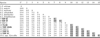
a The species are numbered as in Fig. 5 in which the GenBank Accession numbers of the lineages are given. Sequence divergence was calculated with the use of a Jukes-Cantor model of substitution. Names in bold identify isolates from this study. Shaded areas correspond to groups as shown in Fig. 5; Group A = column 7–8, Group B = columns 10–15.
DISCUSSION
The results of this study have identified significant differences in the prevalence of Leucocytozoon in YEPs between Enderby and Campbell Islands in the sub-Antarctic. The prevalence rates of Leucocytozoon in birds from these islands were also significantly higher than that found in birds from the southeast coast of the South Island of New Zealand (Hill et al. Reference Hill, Howe, Gartrell and Alley2010). However, in all sites the presence of the parasite in apparently healthy adult YEPs suggests that this is an endemic haemoparasite of YEPs in the sub-Antarctic islands. The high prevalence of Leucocytozoon in the sub-Antarctic, especially on Enderby Island, implies that these penguins may be the major reservoir of this haemoparasite in the larger YEP population.
The results of this study also suggest that infection with Leucocytozoon is a potential contributing factor to YEP chick mortality but epidemiological studies are required to further investigate the association between infection with Leucocytozoon and nestling disease or death. Based on our pathological findings that only 1 out of 19 chicks showed histological evidence of disease associated with the infection, it is unlikely that this strain of Leucocytozoon is of high pathogenicity. Nevertheless, the high prevalence of infection raises the possibility that subclinical effects of infection may play a role in chick mortality, especially in years when food supply is poor. No other common factor for the poor breeding success and high chick mortality observed on Enderby Island during the 2008–2009 breeding season could be identified. The pattern of mortality seen on Enderby Island is similar to the mortality observed by Hill et al. (Reference Hill, Howe, Gartrell and Alley2010) on Stewart Island in 2006/07 where it was found that younger chicks seemed to succumb to starvation first whereas older chicks developed leucocytozoonosis with megaloschizonts in many different tissues resulting in tissue damage and death. The chick (YEP TLB4) that died due to disseminated leucocytozoonosis, was infected with the same cluster B isolate as that affecting the majority of positive YEPs from Enderby (76·5%), Campbell and Stewart Island, as well as those on the Otago Peninsula. This finding is important as this isolate of Leucocytozoon has been shown to be capable of causing severe disease and death in this species.
The finding of 2 distinct phylogenetic clusters is also important and suggests that there may be a unique endemic isolate present only in Enderby Island YEP, while the cluster B isolates comprising Leucocytozoon from Enderby Island, Otago Peninsula and Campbell Island YEPs may be reflective of inter-island migration events. A study by Boessenkool (Reference Boessenkool, Star, Waters and Seddon2009) has demonstrated that migration events between sub-Antarctic and mainland YEPs, although rare, do occur. However, it could be possible that YEPs from within the sub-Antarctic islands may frequently move between islands particularly when hunting. Thus, based on prevalence data from this study and that of Hill et al. (Reference Hill, Howe, Gartrell and Alley2010), it is likely that Enderby and/or Stewart Island, and the Auckland Island archipelago are the main reservoirs for the disease, and that low levels of migration have resulted in spread of this parasite to Campbell Island as well as the South Island. The slightly higher prevalence noted on Campbell Island compared with the Otago Peninsula may indicate an increased migration rate from Enderby to Campbell as both islands are in the sub-Antarctic. It is currently unclear whether the cluster A isolate is truly endemic to Enderby Island and whether infection has an impact on reproductive success.
There are also differences in the nesting environment of YEPs between the sub-Antarctic and mainland and these could contribute to the exposure of birds to the pathogen. Although it is speculative, behavioural differences between the populations may contribute to the different prevalence rates seen and can be part of an animal's non-immunological defences. During the first 4–6 weeks after hatching, chicks are brooded continuously by either parent. A guard stage develops from true brooding at around 3 weeks of age as the chicks grow (Marchant and Higgins, Reference Marchant and Higgins1990). This behaviour could afford some protection from biting flies for at least the first 3 weeks. After guard-stage, chicks remain at or near the nest while both parents go out to feed during the day. The parents leave at dawn and return at night to feed the chicks (Marchant and Higgins, Reference Marchant and Higgins1990). During this post-guard stage the chicks may be more susceptible to bites from simuliids. It is also during this stage that chicks become more mobile and may seek shade under vegetation or cool, damp ground near streams (Marchant and Higgins, Reference Marchant and Higgins1990). This behaviour may increase the risk of exposure to simuliids as vectors of disease.
The likely vector of Leucocytozoon on the South Island and on Stewart Island is Austrosimilium ungulatum. This Simuliid has been shown (Desser and Allison, Reference Desser and Allison1979) to be the primary vector of Leucocytozoon tawaki that affects Fiordland crested penguins (Eudyptes pachyrhynchus). Simuliid blackflies are the usual vector for the majority of investigated Leucocytozoon spp. (Desser and Bennett, Reference Desser, Bennett and Kreier1993; Valkiunas, Reference Valkiunas2005). Austrosimulium campbellense is endemic to Campbell Island and A. vexans to the Auckland Islands. The latter is known from Enderby Island, Auckland Island and Adams Island (Dumbleton, Reference Dumbleton1963; Craig and Crosby, Reference Craig and Crosby2008; Craig, 2010, personal communication). Biological characteristics indicate that both those species are very closely related to A. ungulatum (Dumbleton, Reference Dumbleton1973). Based on this, A. vexans is highly likely to be capable of transmitting Leucocytozoon spp. to YEPs. Due to the very low numbers of Simuliids we observed (n = 3) on Enderby during the 2008–2009 breeding season, coupled with the very high prevalence of infection observed, it is possible that penguins become infected if landing on the main Auckland or Adams Islands where they are exposed to larger numbers of this vector. However, the presence of this parasite in young guard-stage penguin chicks lends strong support to the theory that the vector is present on Enderby Island. Although the prevalence of Leucocytozoon was significantly higher in the sub-Antarctic islands, it would seem logical for an arthropod vector to be more prevalent in warmer climates. The known invertebrate hosts of Leucocytozoon are simuliid flies with the exception of L. caulleryi whose vector is Culicoides arakawae (Hsu et al. Reference Hsu, Campbell and Levine1973). Detailed vector studies for Leucocytozoon spp. are required to determine whether Simuliids or another vector is capable of transmitting Leucocytozoon in YEPs on Enderby Island.
Despite the high prevalence of infection, only the older chicks examined post-mortem displayed pathology associated with Leucocytozoon infection. In birds, maternal antibodies and other immune factors are transmitted to the embryo via the egg yolk (Grindstaff et al. Reference Grindstaff, Brodie and Ketterson2003). These antibodies afford passive protection to the chick and may also affect the development of the juvenile's immune response (Staszewski and Siitari, Reference Staszewski and Siitari2010). This maternal transfer of immunity may explain the expression of leucocytozoonosis in the YEP chicks but needs further investigation. Alternatively, the Leucocytozoon that infects the YEPs may be well host-adapted and only cause disease in compromised hosts.
Subclinical effects of infection with Leucocytozoon are common in other species and it is possible that the haemoparasite contributes to mortality in less direct ways. One of the major impacts of infection with Leucocytozoon in other species of birds is a decrease in reproductive performance (Merino et al. Reference Merino, Moreno, Sanz and Arriero2000; Dunbar et al. Reference Dunbar, Torniquist and Giordano2003). There was no evidence to suggest that Leucocytozoon played a significant role during the 2008–2009 breeding season on the Otago Peninsula. The presence of Leucocytozoon spp. at any level of parasitaemia has been shown to exert subclinical effects on the host as well as influence or amplify effects from concurrent diseases or stressors resulting in increased mortality, or reduced reproductivity or body weight (Merino et al. Reference Merino, Moreno, Sanz and Arriero2000). Wild populations of penguins are experiencing increased pressure due to environmental and anthropogenic stressors including climate change, increased competition with fisheries, increased habitat destruction, increased tourism and human contact etc. (Jones and Shellam, Reference Jones and Shellam1999). Wild populations of birds that are infected with blood parasites are usually chronically infected with disease only occurring during stressful situations such as breeding and moulting, or due to increases in any of the above-mentioned stressors (Atkinson and van Riper, Reference Atkinson, van Riper, Loye and Zuk1991; Bennett et al. Reference Bennett, Peirce and Ashford1993). Infection with Leucocytozoon seems to follow this trend in the sub-Antarctic YEP population. Garvin et al. (Reference Garvin, Szell and Moore2006) showed that blood parasites do pose a physiological cost, at least in neotropical migrant passerines. Their research showed that migrants infected with blood parasites arrived on the northern coast of the Gulf of Mexico in poorer body condition than uninfected birds (Garvin et al. Reference Garvin, Szell and Moore2006). Concurrent with our study, there appeared to be a disruption of food supply during the 2008–2009 YEP breeding season, although there is no conclusive evidence to support this, with the results of most of the post-mortems on dead chicks indicating starvation, as well as low observed body condition of adult penguins attending nests. This stress may have amplified the subclinical level of parasitaemia resulting in increased mortalities during this season.
Valkiunas et al. (Reference Valkiunas, Iezhova, Krizanauskiene, Palinauskas, Sehgal and Bensch2008) found that both microscopic examination of blood films and nested PCR-based diagnostics showed a similar level of prevalence of infection of blood parasites in naturally infected birds. This is in contrast to studies conducted by Richards et al. (Reference Richards, Sehgal, Jones and Smith2002), Jarvi et al. (Reference Jarvi, Schultz and Atkinson2002) and Durrant et al. (Reference Durrant, Beadell, Ishtiaq, Graves and Olson2006), who all reported a much higher prevalence of haematozoa with PCR-based techniques compared with light microscopy. Studies by Valkiunas et al. (Reference Valkiunas, Iezhova, Krizanauskiene, Palinauskas, Sehgal and Bensch2008) did not support these conclusions as they found that the discrepancies were likely due to shortcomings in the microscopy methods used in those studies. Poor quality blood film preparation makes it very difficult to identify haemoparasites, thus resulting in a finding of lower prevalence as compared with PCR. Our study also found a similar prevalence of Leucocytozoon infection using light microscopy and PCR analysis of peripheral blood samples and, as such, supports the recommendation from Valkiunas et al. (Reference Valkiunas, Iezhova, Krizanauskiene, Palinauskas, Sehgal and Bensch2008) for continued use of optical microscopy in the research of haemosporidian parasites of vertebrates.
Further epidemiological studies are required to investigate the association between infection with Leucocytozoon and nestling disease and death, especially as the strain from cluster B has demonstrated a potential to cause severe disease and death in YEP chicks across their range. This could have implications for the population of this endangered penguin especially as, due to continually increasing environmental and anthropogenic stressors on penguins, more subclinical effects such as reduced reproductivity in adults and increased mortality in chicks could potentially result in further declines in the population.
ACKNOWLEDGEMENTS
We wish to thank the following people for their assistance during this study: Dr Trevor Crosby (Landcare Research), Dr Gediminas Valkiunas (Vilnius University, Lithuania), Professor Douglas Craig (University of Alberta, Canada), the Department of Conservation (NZ Sealion team 2008/09, Melanie Young, Dr Kate McInnes, Dr Louise Chilvers, Pete McLelland), and the members of the NZ Wildlife Health Centre. We would also like to acknowledge the Massey University Postgraduate Fund for their financial support.