Implications
Intestinal bacteria can actively metabolize amino acids (AA). This study showed that altering the gut microbiota with oral antibiotics changes the microbial fermentation of AA and proteins, with a parallel change in host nitrogen (N) metabolism, by reducing N utilization efficiency. These findings suggest a contribution by the gut microbiota to host N metabolism. These results also show that host N metabolism can be modulated by altering the gut microbiota.
Introduction
Microbiota in the gut, especially those in the large intestine, are actively involved in nutrient metabolism. Intestinal bacteria can metabolize AA that are closely linked to the health of the host (Dai et al., Reference Dai, Wu and Zhu2011). Our previous in vitro studies that investigated the variations in AA metabolism by examining bacteria from different gut niches, demonstrated that luminal bacteria tended to utilize free AA (Yang et al., Reference Yang, Dai and Zhu2014). Moreover, other studies have shown that 30% to 60% of dietary AA disappears through first-pass intestinal metabolism (Stoll et al., Reference Stoll, Henry, Reeds, Yu, Jahoor and Burrin1998), particularly glutamine, glutamate and aspartate, with a disappearance rate above 90% (Wu, Reference Wu2009). Our previous study showed that a considerable amount of AA could be incorporated into bacterial cells; for example, 16% lysine and 57% leucine were incorporated into bacterial cells derived from pig intestines during in vitro incubation (Dai et al., Reference Dai, Zhang, Wu and Zhu2012). In addition, a previous study has revealed that intravenously administered antibiotics increased whole body urea synthesis as indicated by the kinetics of (15N) threonine and (U-13C, 15N) threonine flux in neonatal pigs (Puiman et al., Reference Stein, Shipley and Easter2013). Therefore, intestinal bacteria can, to a certain extent, participate in the metabolism and utilization of nitrogen (N) nutrients (protein and AA) in the lumen of the intestine, in addition to their important roles in rapid passage, host enzymatic digestion, and absorption of nutrients by the host. However, the impact of the gut microbiota on in vivo host digestibility and metabolism, especially on N metabolism, remains unclear.
Antibiotics have been widely used for the control of infectious diseases and have also been used as growth promoters in production animals (Cromwell, Reference Cromwell2002). However, the wide spread and uncontrolled use of antibiotics in feed has led to the emergence of many resistant bacterial strains that have detrimental effects on both humans and animals. Thus, the use of antibiotics as growth promoters in animal feed has been banned since 2006 in Europe. In China, although the use of antibiotics in feed during the weaning period of the pig is still a common practice, it has increasingly led to safety concerns from consumers. Thus, to design better alternatives to antibiotics for pigs, it is important to understand the long-term effect and the possible mode of action of the antibiotics. The mode of antibiotic action on host physiology is considered to be due to its direct change to or reduction in bacterial (sub)-populations, especially in the gastrointestinal tract. Some susceptible bacteria include those of the phyla Firmicutes, Bacteroidetes and Proteobacteria (Willing et al., Reference Willing, Russell and Finlay2011). Bacteroides spp. and Escherichia coli (E. coli) that belong to Bacteroidetes and Proteobacteria, respectively, can participate in N metabolism, as they secrete proteases and affect protein fermentation in the intestine (Davila et al., Reference Davila, Blachier, Gotteland, Andriamihaja, Benetti, Sanz and Tome2013). In addition, previous studies report that antibiotics can regulate N metabolism, by slowing the passage rate of digesta, thinning the wall of the intestine, upregulating mRNA expression levels for key AA transporters and receptors, and reducing competitive utilization by intestinal bacteria (Choi et al., Reference Choi, Kim, Ingale, Kim, Shinde, Kwon and Chae2011; Yu et al., Reference Yu, Zhang, Yang, Mu, Su, Yu and Zhu2017). Thus, it is important to understand the antibiotic-induced impact on host N metabolism, especially on AA digestibility and metabolism, and its relationship with the gut microbiota of the host.
Mixtures of ampicillin, gentamycin and metronidazole (AGM) have been used as broad-spectrum antibiotics in the treatment of both human and animal intestinal infections, including necrotizing enterocolitis and intra-abdominal infections (Willing et al., Reference Willing, Russell and Finlay2011). Previous research has shown that intravenous or oral administration of AGM could induce disruption of gut microbiota by negatively affecting both the number and diversity of commensal bacteria that normally colonize the gut of neonates (Cilieborg et al., Reference Cilieborg, Boye, Molbak, Thymann and Sangild2011; Puiman et al., Reference Stein, Shipley and Easter2013). As gut microbes are actively involved in host metabolism (Nicholson et al., Reference Nyachoti, Omogbenigun, Rademacher and Blank2012), we hypothesized that AGM could have an impact on host metabolism by changing the gut microbiota. To test this hypothesis, we investigated the effects of AGM on the microbiota of the small and large intestine, N utilization efficiency, AA total tract and terminal ileal apparent digestibility of growing pigs. The main aim of the study was to determine whether AGM affects N metabolism in the pig.
Material and methods
The experiments were conducted in compliance with the Chinese regulations on the protection of experimental animals, and followed the protocol approved by the Ethical Committee of Nanjing Agricultural University, Nanjing, China.
Animal, experimental design and treatments
In total, 16 male 42-day-old cross-bred Duroc×(Landrace×Large White) weaned piglets, weighing 12±0.5 kg, were selected and housed individually in stainless steel metabolism cages (height, 0.85 m; length, 0.70 m; and width, 0.70 m). Each cage was equipped with a smooth-walled pan, a nipple drinker and a feeder. The temperature was controlled at 25°C. After 7 days adaptation, pigs were fasted for 12 h before the surgical procedure of fitting a simple T-cannula at the distal end of the ileum, ~5 cm cranial to the ileocecal sphincter. The pigs were also fitted with jugular venous catheters. The surgical procedure and postoperative care followed the methods previously described (Stein et al., Reference Ferrer, Martins dos Santos, Ott and Moya1998). Before surgery and during the 7 day recuperation period following surgery, all pigs were fed a commercial starter diet. All pigs had free access to water and were fed individually ad libitum. After the 7 days of recuperation, the pigs were allotted to two groups (n=8), the control group (control; CON) and antibiotic group (antibiotic; ANTI), according to a completely randomized design. During the following 2 weeks, CON pigs were fed a basal diet without any antibiotics, and the ANTI pigs were fed the basal diet supplemented with AGM (ampicillin 200 mg/kg BW per day; gentamycin 5 mg/kg BW per day; and metronidazole 40 mg/kg BW per day). Ampicillin, a β-lactam antibiotic, shows activity against both gram-positive and gram-negative bacteria. Gentamycin can act against mostly gram-negative bacteria and aerobes. Metronidazole mainly acts against anaerobes. The antibiotic dosage was modified, based on the results of a previous study (Jensen et al., Reference Jonkers, Penders, Masclee and Pierik2014). The ingredients and chemical composition of the basal diets are presented in Supplementary Table S1. The diet was formulated to meet or exceed nutrient demands, according to the National Research Council (NRC) (Reference Mu, Yang, Yu, Yu, Zhang, Su and Zhu2012) standards. Pigs were given two meals of equal amounts daily, at 0800 and 2100 h. Chromic oxide (Cr2O3) was included in the diets at 0.30%, as an indigestible marker. Samples of the feed offered, as well as refusals were recorded daily for each individual pig, and the average daily feed intake was calculated.
Sample collection
Ileal digesta were collected for two 12-h periods from 0800 to 2000 h on days 13 and 14 of the experiment, by attaching a plastic tubular bag to the externalized ileal T-cannula, for nutrient ileal apparent digestibility analysis. For nutrient digestibility analysis, 5 ml of 10% (vol/vol) formic acid was added to the plastic tube, to prevent bacterial fermentation in the digesta. Moreover, the pH value of the digesta was measured immediately after feeding at 1, 4, 8 and 12 h (at 0900, 1300, 1700 and 2100 h). For analysis of the microbial community and microbial metabolites, digesta were collected on 4 days before and day 13 after antibiotics treatment at 12 h after morning feeding and frozen at −80°C until further use.
Fecal and urine samples were collected on days 9, 10 and 11 of the experimental period. Urine was collected via funnels into buckets containing sulfuric acid (9 N) for preservation. Upon sampling, both urine and feces were measured and weighed, respectively, and stored at −80°C until further analysis. For analysis of the microbial community and microbial metabolites, fresh feces were collected through the rectum on 4 days before and day 13 after antibiotic treatment at 12 h after morning feeding. Blood samples were collected on day 12 of the experiment at 1, 4, 8 and 12 h after feeding (at 0900, 1300, 1700 and 2100 h). Blood samples (10 ml) were collected from the jugular venous catheters and serum was obtained by centrifugation at 3000×g for 15 min at 4°C. Thereafter, samples were stored at −70°C, until further analysis.
Chemical analysis
Samples of diets, ileal digesta and feces were analyzed for dry matter (DM), CP (N×6.25), organic matter, ether extract (EE), calcium and phosphorous, according to the procedures of the Association of Official Analytical Chemists (2003). The NDF and ADF contents were determined, following the procedures of Van Soest et al. (Reference Van Soest, Robertson and Lewis1991). For the determination of individual AA concentrations, samples of the diet, feces and ileal digesta were hydrolyzed for 24 h in 6 N hydrochloric acid at 110°C, under an atmosphere of N2. The individual AA concentration in blood samples and in the hydrolysates of dietary, fecal and ileal digesta samples, were determined by HPLC after post-column derivatization, according to the o-phthaldialdehyde method, as previously described (Dai et al., Reference Dai, Li, Xi, Zhang, Wu and Zhu2014). The chromium concentrations in the dietary and ileal digesta samples were determined using inductively coupled plasma with optical emission spectrometry (ICP-OES, Optima 2100 DV, PerkinElmer, Waltham, MA, USA).
Real-time PCR quantification of microbes
For quantification of the main bacterial groups, total DNA from each ileal digesta sample and from fecal material (0.3 g) was extracted, according to the bead-beating and phenol-chloroform extraction method as previously described (Dai et al., Reference Dai, Wu, Jia and Wu2010). The DNA was quantified using a NanoDrop spectrophotometer (Thermo, Wilmington, DE, USA). Bacteria were quantified using group-specific primers (Supplementary Table S2) and SYBR Green premix (Takara Biotechnology, Dalian, China) in the Step One Plus™ qPCR System (Life Technologies Inc., CA, USA). The PCR reaction mixture (20 µl) consisted of 10.0 µl of SYBR Green Supermix (Bio-Rad, Hercules, CA, USA); 2.0 µl of DNA; 6.8 µl of double distilled water; 0.4 µl ROX Reference Dye; and 0.4 µl of each primer set. The amplification conditions were as follows: (1) pre-denaturation program (95°C for 30 s); (2) amplification and quantification program, repeated for 40 cycles (95°C for 5 s and 60°C for 30 s); (3) fusion (95°C for 15 s and 60°C for 1 min), followed by the melting curve program (60°C to 95°C, with a heating rate of 0.1°C/s and fluorescence measurement). The bacterial copy numbers (copy/ng DNA sample) were calculated, according to standard curves that were generated from serial dilutions of 16S rRNA gene fragments cloned into a pMD-19T vector (Takara Bio, Shiga, Japan).
Microbial metabolite analysis
The ileal digesta and fecal samples were analyzed for microbial metabolites. Concentrations of short-chain fatty acids (SCFA) were measured by gas chromatography, according to methods previously described (Yu et al., Reference Yu, Zhang, Yang, Mu, Su, Yu and Zhu2017). Amine concentrations were determined by HPLC with precolumn dansylation according to a method that has been previously described (Yang et al., Reference Yang, Dai and Zhu2014). The ammonia concentrations were determined using a spectrophotometer (UV-2450; Shimadzu, Tokyo, Japan) according to the methods of Nyachoti et al. (Reference Puiman, Stoll, Molbak, de Bruijn, Schierbeek, Boye, Boehm, Renes, van Goudoever and Burrin2006). The microbial CP (MCP) concentration was analyzed using a spectrophotometer (UV-2450), according to previously described methods (Makkar et al., 1982).
Biochemical parameters and determination of enzyme activity
Enzyme activity in the ileal digesta, as well as glucose, albumin, triglycerides (TG), total cholesterol (TC), high-density lipoprotein cholesterol (HDLC), low-density lipoprotein cholesterol (LDLC), total protein and urea N in serum were determined using assay kits, according to the manufacturer’s instructions (Nanjing Jiancheng Bioengineering Institute, Nanjing, China) and an Olympus AU2700 auto analyzer (Olympus, Tokyo, Japan). The serum hormone indices (growth hormone, IGF 1, motilin, gastrin, etc.) were determined using ELISA with porcine ELISA kits, commercially obtained from Bethyl Laboratories (Montgomery, TX, USA).
Calculations and statistical analysis
Nitrogen retention was calculated as follows: N intake – (fecal N output+urinary N output). Total tract apparent digestibility (TTAD) of AA and nutrients were calculated using the following equation: TTAD of X=(X intake−fecalXoutput)×100/X intake, where X represents AA or nutrients. Apparent ileal digestibility (AID) of AA and nutrients were calculated using the index method. The AIDs of AA and nutrients were calculated using the following equation:
 $$\eqalignno{ {\rm AID}\,{\rm of}\,{\rm X}{\equals} \left[ {{\rm 1{\minus}}\left( {{\rm Cr}_{{{\rm diet}}} {\rm \,/\,Cr}_{{{\rm ileal}\,{\rm digesta}}} } \right){\rm {\times}}\left( {{\rm X}_{{{\rm ileal}\,{\rm digesta}}} {\rm \,/\,X}_{{{\rm diet}}} } \right)} \right]{\rm} \cr {\times}100$$
Where AID of X is the AID of AA or nutrients (%); Crdiet and Crileal digesta are the concentrations of chromium in the diet and ileal digesta, respectively; X ileal digesta is the X concentration in the digesta and X diet the X concentration in the diet.
All data were analyzed in a completely randomized design with repeated measures, using the Student’s t-test of the SPSS 20.0 software. The significance level was declared at P<0.05 and trends for significance were declared at P=0.05 or P<0.10. The correlations among bacteria, N metabolism and microbial metabolites were assessed using the Pearson’s correlation test and Graphpad Prism version 5.00 (Graphpad Software, San Diego, CA, USA).
Results
Animal growth performance and main bacterial groups
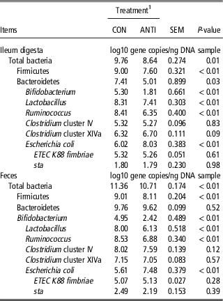
During the entire experiment, no clinical signs of diarrhea or health impairment were observed. Growth performance was not significantly affected by treatment with AGM (Supplementary Table S3). To understand the quantitative changes in the bacterial community following treatment with AGM, qPCR was performed on some of the main bacterial groups. Before AGM treatment, no differences between CON and ANTI groups were evident in populations of the main bacterial groups (Supplementary Table S4). As shown in Table 1, in the ileal digesta, AGM significantly reduced the abundance of total bacteria, Firmicutes, Bacteroidetes, Bifidobacterium, Lactobacillus and Ruminococcus, in comparison with the CON group (P<0.05). However, AGM significantly increased the abundance of E. coli (P<0.05), and tended to increase the abundance of Clostridium cluster XIVa (P<0.10). No differences were observed between the CON and ANTI groups in Clostridium cluster IV, gene copy numbers of Enterotoxigenic E. coli (ETEC) K88 fimbriae, and the heat-stable enterotoxin 1 (sta) (P>0.05). In feces, AGM significantly reduced the abundance of total bacteria, Firmicutes, Bifidobacterium, Ruminococcus and Lactobacillus, in comparison with the CON group (P<0.05). The abundance of E. coli was also increased following the AGM treatment (P<0.05). No differences were observed between the CON and ANTI groups, in Bacteroidetes, Clostridium cluster IV, Clostridium cluster XIVa and gene copy numbers of ETEC K88 fimbriae and sta (P>0.05). These results indicate that AGM could markedly alter specific bacterial groups in the gut.
Responses of major taxonomic groups of bacteria in growing pigs toward antibiotic treatment

ETEC=Enterotoxigenic Escherichia coli; sta=Heat-stable enterotoxin 1.
1 Pigs were fed a basal diet (CON) or basal diet supplemented with antibiotics (ANTI) (n=8).
Microbial metabolites and microbial CP
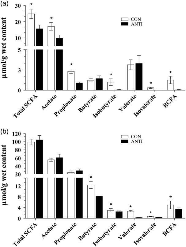
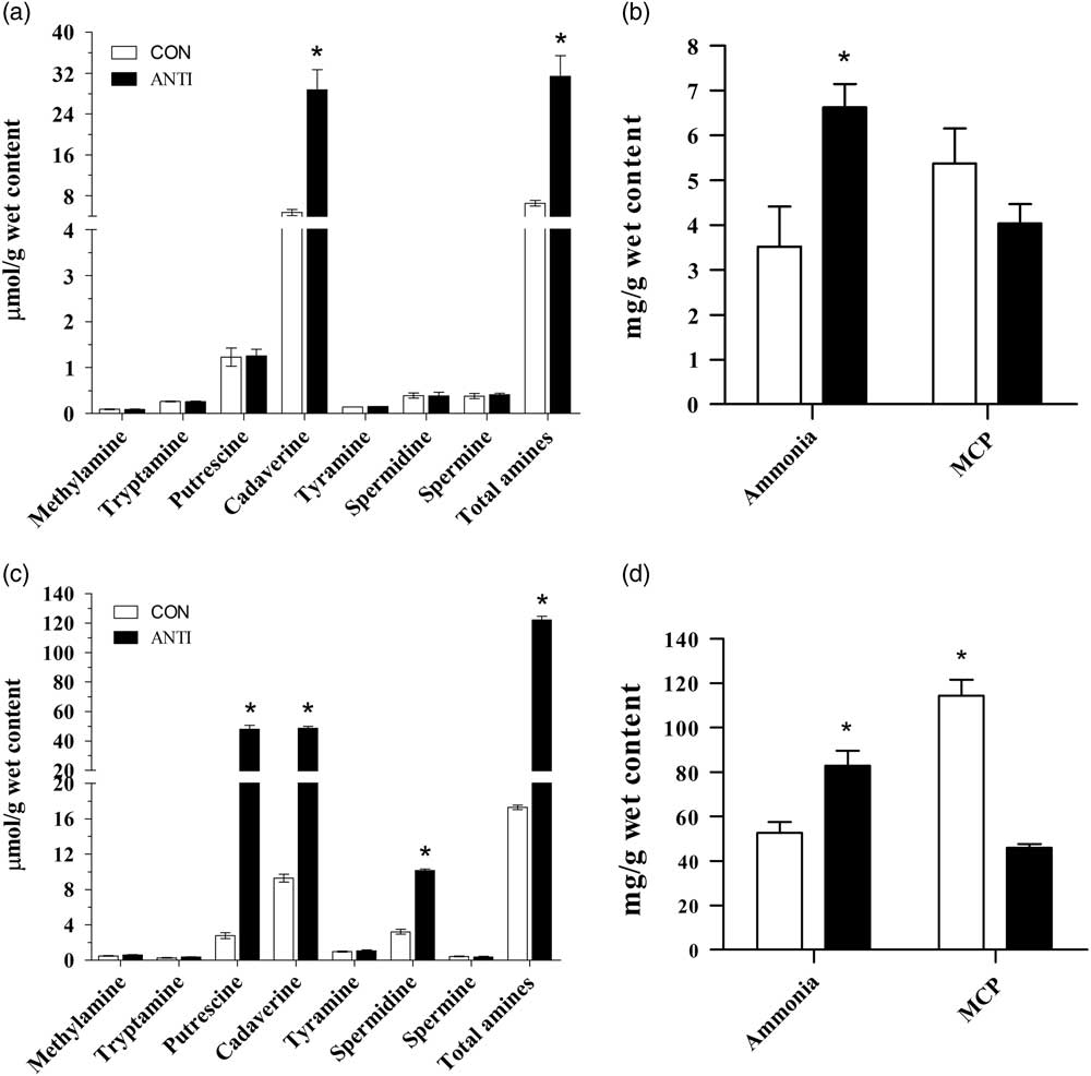
Gut microbes can ferment carbohydrates and nitrogenous compounds, and produce SCFA and biogenic amines, respectively. Before AGM treatment, no differences in the concentrations of microbial metabolites were observed between the CON and ANTI groups (Supplementary Table S5). As shown in Figure 1, in the ileal digesta, AGM significantly reduced the concentrations of acetate, propionate, isobutyrate, isovalerate, branched-chain fatty acids (BCFA), and total SCFA (P<0.05), and had no effect on the concentrations of butyrate and valerate (P>0.05). In feces, the AGM significantly reduced the concentrations of butyrate, isobutyrate, valerate and isovalerate (P<0.05), but had no effect on the concentrations of acetate, propionate and total SCFA (P>0.05). As for the biogenic amines, in the ileal digesta, AGM markedly increased the concentrations of cadaverine and total amines (P<0.05) (Figure 2). In feces, AGM treatment significantly increased the concentrations of putrescine, cadaverine, spermidine and total amines (P<0.05).
Effects of antibiotics on the concentrations of short-chain fatty acids (SCFA). (a) Terminal ileal digesta and (b) feces of growing pigs (n=8). Pigs were fed a basal diet (CON) or basal diet supplemented with antibiotics (ANTI). *Statistically significant differences between CON and ANTI. *P<0.05. BCFA=branched-chain fatty acid.

Effects of antibiotics on the concentrations of amines, ammonia and microbial CP (MCP). (a, b) Terminal ileal digesta and (c, d) feces of growing pigs (n=8). Pigs were fed a basal diet (CON) or basal diet supplemented with antibiotics (ANTI). *Statistically significant differences between CON and ANTI. *P<0.05.

Treatment with AGM significantly increased the concentration of ammonia in the ileal digesta (P<0.05), but had no effect on MCP (P>0.05). In feces, AGM also markedly increased the concentration of ammonia (P<0.05), but markedly reduced the concentration of MCP (P<0.05) (Figure 2). These results indicate that AGM treatment reduces bacterial fermentation of carbohydrates in ileum, and enhances the bacterial fermentation of protein in the large intestine.
Serum parameters, ileal enzyme activity and pH
Regarding serum parameters, the concentrations of glucose, albumin, globulin, TG, TC, HDLC and LDLC remained relatively unchanged (P>0.05) in the ANTI group. The concentration of total protein, a parameter associated with protein metabolism, tended to increase in the ANTI group, in comparison with the CON group (P=0.058) (Supplementary Table S6). Treatment with AGM did not affect blood hormone parameters, such as gastrin and ghrelin (P>0.05) (Supplementary Table S7). In comparison with the CON group, AGM had no effect on enzyme activity in the ileal digesta (P>0.05, Supplementary Table S8). These results indicate that AGM had little effect on the general physiological status of the pigs.
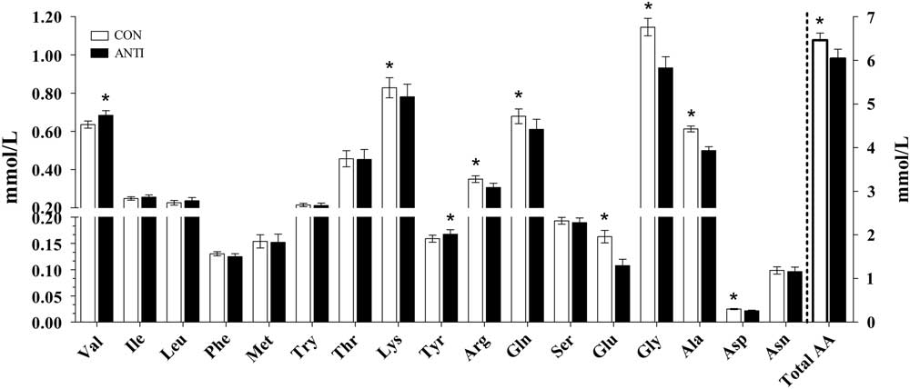
However, AGM treatment had evident effects on the AA profiles in serum. Treatment with AGM significantly increased the concentrations of valine and tyrosine, and reduced the concentrations of lysine, arginine, glutamine, glutamate, alanine, aspartic acid and total AA in serum (P<0.05, Figure 3). In addition, the concentration of urea N was significantly increased in the ANTI group (P<0.05). A time dynamic diagram showed that the pH values of the terminal ileal digesta and feces in the ANTI group were maintained at a higher level than those in the CON group at the time points of 4 and 8 h after the morning feeding. The pH in the ileal digesta and feces was first reduced, and then increased at the time point of 8 h after the morning feeding in the CON group. However, the pH in the ANTI group was first increased, then reduced at the time point of 4 h after the morning feeding (Supplementary Figure S1).
Effects of antibiotics on the concentrations of amino acids (AA) in the serum of growing pigs (n=8). Pigs were fed a basal diet (CON) or basal diet supplemented with antibiotics (ANTI). *Statistically significant difference between CON and ANTI. *P<0.05.

Nitrogen utilization
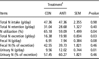
The N utilization efficiency of the growing pigs was determined (Table 2). In Comparison with the CON group, ANTI treatment had no effect on N intake, N retention, or ratios of N excretion in feces and urine (P>0.05). However, in comparison with the CON group, AGM significantly reduced N utilization (P<0.05). Moreover, AGM tended to increase N excretion in feces (P<0.10) and urine (P<0.01). These results indicate that AGM could alter the N balance by reducing N utilization and increasing N excretion in feces and urine.
Effects of antibiotics on nitrogen (N) utilization efficiency in growing pigs

1 Pigs were fed a basal diet (CON) or basal diet supplemented with antibiotics (ANTI) (n=8).
Apparent digestibility
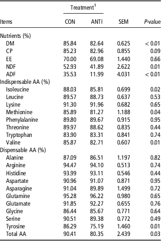
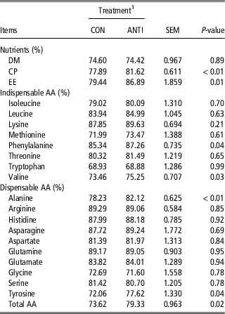
In comparison with the CON group, AGM significantly reduced DM, NDF and ADF TTAD (P<0.05) (Table 3). Moreover, AGM tended to reduce CP TTAD (P<0.10). As for AA TTAD, AGM significantly reduced isoleucine, methionine, valine, tyrosine and total AA TTAD (P<0.05). These results indicate that AGM reduced N utilization and increased N excretion, by reducing CP and AA TTAD. Treatment with AGM also significantly increased CP and EE AID (P<0.05), but had no effect on DM AID, when compared with the CON group. As for AA AID, AGM significantly increased phenylalanine, valine, alanine, tyrosine and total AA AID (P<0.05) (Table 4). These results suggest differences in the apparent digestibility of CP and AA, between small intestine and large intestine segments following treatment with AGM.
Effects of antibiotics on total tract apparent digestibility (TTAD) of nutrients and amino acid (AA) in growing pigs

DM=dry matter; EE=ether extract.
1 Pigs were fed a basal diet (CON) or basal diet supplemented with antibiotics (ANTI) (n=8).
Effects of antibiotics on apparent ileal digestibility (AID) of nutrients and amino acid (AA) in growing pigs

DM=dry matter; EE=ether extract.
1 Pigs were fed a basal diet (CON) or basal diet supplemented with antibiotics (ANTI) (n=8).
Correlation analysis
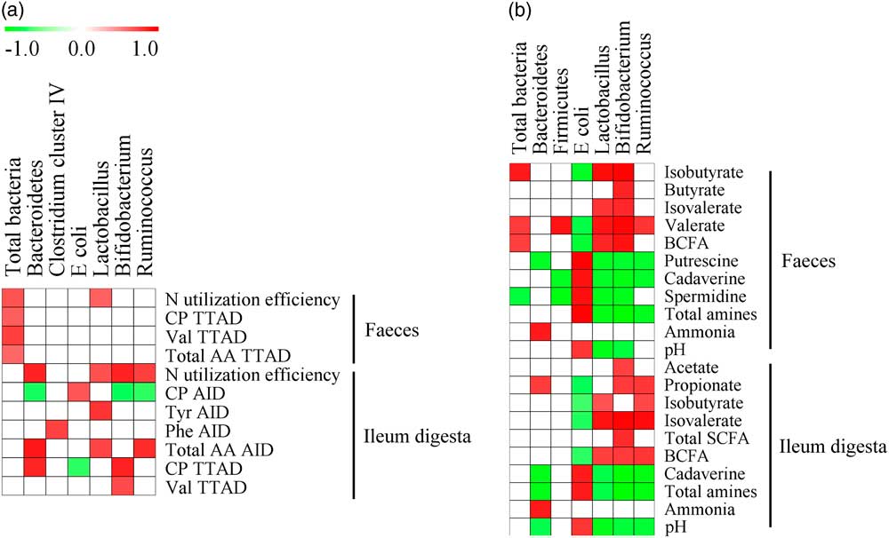
To understand whether intestinal bacterial groups were related to apparent digestibility and microbial metabolites, Pearson’s correlation analysis was performed among the populations of bacteria, and indicators of N utilization and microbial metabolites. As shown in Figure 4a, the N utilization efficiency, CP, valine and total AA TTAD in fecal samples were all positively correlated with total bacteria quantities. In addition, N utilization efficiency was also positively correlated with Lactobacillus quantity. In the ileal digesta, N utilization efficiency was positively correlated with the quantities of Bacteroidetes, Lactobacillus, Bifidobacterium and Ruminococcus. The CP AID was negatively correlated with quantities of Bacteroidetes, Bifidobacterium and Ruminococcus, but positively correlated with E. coli quantity. The total AA AID was positively correlated with the populations of Bacteroidetes, Lactobacillus and Ruminococcus. As shown in Figure 4b, the population of E. coli in feces and ileal contents was negatively correlated with the concentrations of SCFA, and positively correlated with the concentration of amines. The populations of Lactobacillus, Bifidobacterium and Ruminococcus were negatively correlated with the concentration of amines, but positively correlated with SCFA concentration.
(Colour online) Pearson correlation analysis between bacteria and nitrogen utilization, total tract apparent digestibility (TTAD) and terminal ileum apparent digestibility (AID) of amino acids and CP (a) and the concentrations of ammonia, short-chain fatty acids (SCFA), biogenic amines, and pH (b) of growing pigs. Only the bacteria that were significantly associated with nitrogen metabolism and microbial metabolites are presented. The cells are colored based on the Pearson’s correlation coefficient. Red represents significant positive correlation (P<0.05), green represents significant negative correlation (P<0.05), and white represents no significant correlation (P>0.05). AA=amino acids; BCFA=branched-chain fatty acids; n=nitrogen; Tyr=tyrosine; Phe=phenylalanine; Val=valine.

Discussion
The components of the AGM mixture used in the present study have been widely used as broad-spectrum antibiotics in treating human and animal intestinal infections (Willing et al., Reference Willing, Russell and Finlay2011). Previous studies have shown that intravenous or oral administration of AGM could induce disruption of gut microbiota (Cilieborg et al., Reference Cilieborg, Boye, Molbak, Thymann and Sangild2011; Puiman et al., Reference Stein, Shipley and Easter2013; Gao et al., Reference Geypens, Claus, Evenepoel, Hiele, Maes, Peeters, Rutgeerts and Ghoos2018). In the present study, we combined the analysis of the microbiota, microbial metabolites and N metabolism to explore the impact of AGM on host N metabolism and nutrition. AGM altered the N balance by increasing N excretion in the feces and urine, and altered the N apparent digestibility by reducing the TTAD of CP and AAs and increasing the AID of CP and AAs. AGM markedly altered the gut microbiota by reducing the populations of Bacteroidetes, Bifidobacterium, Lactobacillus and Ruminococcus and increasing the population of E. coli. In addition, AGM altered microbial N metabolites, a finding that could partially explain the changes observed in host N metabolism.
Antibiotics affected gut microbiota and increased microbial fermentation of protein
In the present study, the AGM antibiotics markedly altered the populations of certain key bacterial groups in the ileal digesta and feces. These findings are consistent with those of our previous study in which AGM reduced the relative abundance of Lactobacillus, Bifidobacterium and Ruminococcus, and increased the abundance of Escherichia/Shigella, based on the results of 16S rRNA high-throughput sequencing technology (Gao et al., Reference Geypens, Claus, Evenepoel, Hiele, Maes, Peeters, Rutgeerts and Ghoos2018). Similarly, the phyla Firmicutes and Bacteroidetes have also been reportedly reduced by therapeutic antibiotics in humans (Ferrer et al., Reference Gao, Pi, Peng, Mu and Zhu2014). Among bacterial genera, Lactobacillus and Bifidobacterium in the small intestine are generally considered beneficial bacteria, as they exert health-promoting effects on gut physiology. In addition, a rapid reduction in the abundance of these bacteria in the large intestine of humans could facilitate the colonization of opportunistic pathogens (Jonkers et al., Reference Macfarlane, Allison, Gibson and Cummings2012). In contrast, in the present study, the population of E. coli in the ileum and feces was higher in the ANTI group than in the CON group. Although certain strains of E. coli have been associated with intestinal inflammation in the host (Caesar et al., Reference Caesar, Reigstad, Backhed, Reinhardt, Ketonen, Lunden, Cani and Backhed2012), many strains of E. coli may reside as harmless residents in the intestine and participate in the metabolism of nutrients in the gut. In the present study, the abundance of potentially pathogenic E. coli (ETEC K88 fimbriae and sta) showed no change after antibiotic treatment. Thus, the health implications of the changes in the microbial community need further investigation.
Short-chain fatty acids are the main products of carbohydrate fermentation by bacteria in the gut. The phylum of Firmicutes consists of many bacteria that ferment carbohydrates, such as those within the genera Ruminococcus, Lactobacillus and Clostridium. The marked reduction in the population of Ruminococcus and Lactobacillus following antibiotic treatment might explain the lower SCFA concentration observed in the ileal digesta, in comparison with CON group. However, the main site of bacterial carbohydrate fermentation is the large intestine, and in the present study, the concentrations of total SCFA in the ileum were far lower than those in feces (20.22 v. 101.88 μmol/g wet mass). Even though the concentrations of SCFA were markedly altered in the ileum, the impact of the change in these SCFAs in the small intestine might be limited and remains unclear. In the feces, the effects of AGM on the concentrations of SCFA were mainly through the influence of the concentrations of BCFA. The BCFAs are formed from branched-chain amino acids (BCAA), such as valine and leucine that originate exclusively from the breakdown of protein (Geypens et al., Reference Geypens, Claus, Evenepoel, Hiele, Maes, Peeters, Rutgeerts and Ghoos1997), and thus serve as indicators of microbial BCAA deamination. The reduction in the populations of Lactobacillus and Bifidobacterium was positively correlated with BCFA concentration in the feces (Figure 4b). Therefore, the reduction in BCFA concentration in feces might have been due to the effects of AGM on microbial N metabolism in the large intestine.
The luminal ammonia concentration in the large intestine is the net result of microbial production, microbial utilization of ammonia to synthesize MCP, and epithelial ammonia absorption. The increase in ammonia concentration and the parallel reduction in MCP and AA deamination in the feces after antibiotic treatment, suggest an increase in urea hydrolysis in the large intestine. A previous study on 15N perfusion showed that ammonia in the gut lumen did not originate from host urease activity, but from proteolytic and microbial urease activity alone (Wrong et al., Reference Wrong, Vince and Waterlow1985). The main bacteria with proteolytic activity in the large intestine include Bacteroides, Clostridium, Propionibacterium, Fusobacterium and Streptococcus (Davila et al., Reference Davila, Blachier, Gotteland, Andriamihaja, Benetti, Sanz and Tome2013). Bacteroides spp. are known to secrete proteases in the intestine with presumed activity near the brush border of absorptive cells. Thus, the increased relative abundance of Bacteroides in the feces of the ANTI group in our previous study (Gao et al., Reference Geypens, Claus, Evenepoel, Hiele, Maes, Peeters, Rutgeerts and Ghoos2018), might have enhanced microbial proteolytic activity, thereby leading to higher concentrations of ammonia. Moreover, proteases are more active at a neutral or slightly alkaline pH than an acidic pH (Macfarlane et al., Reference Makkar, Sharma, Dawra and Negi1988). In the present study, the pH in the terminal ileal digesta was relatively stable and maintained at a slightly alkaline level after feeding, for 4 h and 8 h in the ANTI group (mean of 6.86 and 7.68 in CON and ANTI groups, respectively) as compared with a sharp decline in pH in the CON group (Supplementary Figure S1). The stable pH in the ANTI group could therefore be more suitable for protease activity that would further support the conclusion that a higher concentration of ammonia in the ANTI group originated from microbial proteolytic activity. Thus, our findings suggest that the AGM might enhance microbial proteolytic activity in the intestine; however, the underlying mechanism needs further investigation.
Biogenic amines are microbial nitrogenous metabolites that are derived from AA decarboxylation by bacteria, such as Escherichia/Shigella, Enterococcus and Klebsiella (Blachier et al., Reference Blachier, Mariotti, Huneau and Tome2007). The results of the present study show that AGM markedly increased the population of E. coli, both in the ileum and feces. This finding is consistent with those of previous studies on AGM in pigs (Puiman et al., Reference Stein, Shipley and Easter2013; Jensen et al., Reference Jonkers, Penders, Masclee and Pierik2014), and could explain the higher levels of amines observed in the gut. Indeed, the population of E. coli, both in the ileum and feces, was positively correlated with the concentration of amines (Figure 4b). In addition, the concentrations of cadaverine, putrescine and total amines were markedly increased in the ileal digesta and feces of the ANTI group (Figure 2). Lysine and arginine are precursor AAs for cadaverine and putrescine, respectively (Zhang et al., Reference Zhang, Yu, Yang, Mu, Su and Zhu2016). The intestinal digestibility of arginine and lysine showed no differences between the CON and ANTI groups, as revealed by their TTAD and AID, whereas their serum levels were lower in the ANTI group than in the CON group. In addition, AGM had no effect on feed intake, indicating that the quantities of these AA entering the gut were equal. Thus, these results suggest that levels of the substrates in the gut for these nitrogenous compounds in bacterial fermentation (decarboxylation) were increased and more amines were produced following AGM administration.
Antibiotics affect host nitrogen utilization efficiency
Nitrogen utilization efficiency can be reflective of digestion, absorption and metabolism of N in the host. In the present study, the AGM-induced increase in AID of total AA and specific AAs, and in N excretion in the feces and urine suggests that AGM reduced host N utilization efficiency. In general, the small intestine is the main site of nutrient digestion and absorption. A previous study showed that antibiotics could increase the intestinal absorption of nutrients, suppress the growth and metabolic activities of harmful gut microbiota, and increase TTAD of nutrients, with simultaneous alterations in intestinal morphology, intestinal epithelium thickness and epithelial cell turnover in pigs (Choi et al., Reference Choi, Kim, Ingale, Kim, Shinde, Kwon and Chae2011). The activity of digestive enzymes plays an important role in the process of nutrient digestion. In the present study, AGM treatment did not affect the activity digestive enzymes. This finding is consistent with those of a previous study, in which dietary supplementation with an antibiotic (tylosin) had no effect on depeptidase activity in pigs (Collington et al., Reference Collington, Parker and Armstrong1990). The antibiotic-induced changes in microbiota composition and microbial metabolism in the gut lumen might have led to the changes observed in host N metabolism, because of the competitive use of N nutrients by gut bacteria. This is further supported by the positive correlation observed between the populations of Lactobacillus, Bifidobacterium and Ruminococcus and N utilization in the ileum (Figure 4). Therefore, the changes in host N utilization efficiency could be partly explained by the action of AGM on gut microbiota.
The AGM also affected urea kinetics. In the present study, oral administration of AGM influenced N metabolism by increasing N excretion in urine and increasing the concentration of blood urea N in comparison with the CON group. However, in a previous study on neonatal piglets, intravenous administration of AGM suppressed urea kinetics, by reducing blood urea N and eventually reducing the N excretory load as urea (Puiman et al., Reference Stein, Shipley and Easter2013). The liver plays an important role in host metabolism and detoxification. Thus, the intravenous administration of AGM may directly influence liver function when the antibiotic enters hepatic tissue, leading to the suppression of urea kinetics. However, in the present study with oral administration of AGM, intestinal AA metabolism, such as AA deamination and decarboxylation, might have contributed significantly to the impact of the antibiotics on host urea kinetics. For example, in the present study, the concentrations of some AAs (lysine, arginine, glutamine and glutamate) in serum were reduced in the AGM group, whereas the AID of these AAs were either increased or remain unchanged. This could indicate increased AA fermentation by bacteria in the gut, as revealed by the increase in amine concentrations in the ANTI group. These findings are similar to those of a previous study that reported that in-feed antibiotics could increase AA decarboxylation in the gut (Mu et al., Reference Nicholson, Holmes, Kinross, Burcelin, Gibson, Jia and Pettersson2017). Collectively, the oral AGM could affect host N utilization, and these effects could be partially due to the AGM-induced changes in gut microbiota AA fermentation. However, the underlying mechanisms need further investigation.
Differences between the small intestine and large intestine in amino acid apparent digestibility, in response to antibiotics administration
An interesting finding of the present study is the pattern of alteration in the AA profile. Particularly, the BCAA and aromatic amino acids (AAA), and apparent digestibility after oral administration of AGM, showed marked difference between the small and large intestines. The AIDs of phenylalanine, valine, tyrosine and total AA were significantly increased following AGM treatment, whereas their TTAD values and values of Ile and Met were reduced in the ANTI group, as compared with the CON group. The small intestine is the main site for nutrient digestion and absorption, whereas the large intestine is the main site for bacteria fermentation. A previous study showed that antibiotics could increase intestinal absorption (Choi et al., Reference Choi, Kim, Ingale, Kim, Shinde, Kwon and Chae2011). Our previous study also reported that in-feed antibiotics upregulated the gene expression of AA transporters in the small intestine (Yu et al., Reference Yu, Zhang, Yang, Mu, Su, Yu and Zhu2017). These findings might explain the increased AIDs of phenylalanine, valine, tyrosine and total AA following AGM treatment. The BCFA is used as an indicator for bacterial AA deamination (Davila et al., Reference Davila, Blachier, Gotteland, Andriamihaja, Benetti, Sanz and Tome2013). However, in the large intestine, the BCFA concentration was reduced, possibly indicating a reduction in the utilization efficiency of BCAA and AAA following antibiotic treatment. Thus, the differences in AA apparent digestibility between the small intestine and large intestine, in response to AGM might have been due to differences in microbial AA fermentation (N metabolism) in the small and large intestines.
Conclusions
Antibiotics (AGM) significantly alters specific bacterial populations in the swine ileal and fecal microbiota. Treatment with AGM can also alter microbial N fermentative metabolites, and these alterations parallel the changes in host N balance and AA metabolism in the intestine and urine of growing pigs. Further correlation analyses showed significant correlations among the altered microbiota, microbial metabolites and host N metabolism. These results suggest that AGM can partially affect host N metabolism by influencing the microbial fermentation of protein in pigs. These findings provide insight into the contribution of gut microbiota to host N metabolism, and provide guidelines for the development of antibiotic alternatives for pigs.
Acknowledgements
This research was supported by National Natural Science Foundation of China (NSFC) (31430082) and research programs from the National Key Basic Research Program of China (2013CB127300). The authors thank Chris R Stokes from the University of Bristol for his assistance with language editing of this paper. W.Y.Z. also thanks Jiangsu Collaborative Innovation Center of Meat Production and Processing, Quality and Safety Control for support.
Declaration of interest
The authors declare no conflicts of interest.
Ethics statement
The experimental design and procedures were approved by the Animal Care and Use Committee of Nanjing Agricultural University following the requirements of the Regulations for the Administration of Affairs Concerning Experimental Animals (The State Science and Technology Commission of P. R. China 1988).
Software and data repository resources
Data are not deposited in an official repository.
Supplementary materials
To view supplementary material for this article, please visit https://doi.org/10.1017/S1751731118001416