Introduction
The rising of bacterial resistance in the last decades urges to pursue novel antimicrobial strategies. Among potential compounds, the set of antimicrobial peptides (AMPs), or host defense peptides, and their derivatives have several advantageous properties (Gordon et al., Reference Gordon, Romanowski and McDermott2005; Hancock and Sahl, Reference Hancock and Sahl2006; Fjell et al., Reference Fjell, Hiss, Hancock and Schneider2012; Haney et al., Reference Haney, Straus and Hancock2019). It is particularly promising that AMPs show collateral sensitivity to antibiotic-resistant bacterial strains (Lázár et al., Reference Lázár, Martins, Spohn, Daruka, Grézal, Fekete, Számel, Jangir, Kintses and Csörgő2018). Majority of AMPs are polycationic, belonging to the innate immune system of all multicellular organisms. Their positive charge, short sequence, and amphipathic nature provide specificity to biomembranes of pathogen microbes. Although some have intracellular targets, the action mechanism of most AMPs is based on the disruption of cell membranes (Hancock and Sahl, Reference Hancock and Sahl2006; Ageitos et al., Reference Ageitos, Sánchez-Pérez, Calo-Mata and Villa2017). This process is often associated with a disorder-to-order conformational transition upon interaction with lipid membranes (Ageitos et al., Reference Ageitos, Sánchez-Pérez, Calo-Mata and Villa2017), but also observed for interactions with biomacromolecules (Otvos et al., Reference Otvos, I, Rogers, Consolvo, Condie, Lovas, Bulet and Blaszczyk-Thurin2000; Kragol et al., Reference Kragol, Lovas, Varadi, Condie, Hoffmann and Otvos2001; Barańska-Rybak et al., Reference Barańska-Rybak, Sonesson, Nowicki and Schmidtchen2005; Bandyopadhyay et al., Reference Bandyopadhyay, Lee, Sivaraman and Chatterjee2013), or even smaller compounds such as endogenous metabolites or drug molecules (Zsila et al., Reference Zsila, Bősze, Horváti, Szigyártó and Beke-Somfai2017a, Reference Zsila, Juhász, Bősze, Horváti and Beke-Somfai2017b, Juhász et al., Reference Juhász, Mihály, Kohut, Németh, Liliom and Beke-Somfai2018). AMPs often show a broad-spectrum activity toward bacteria, viruses, fungi, protozoa, and even cancer cells (Hancock and Sahl, Reference Hancock and Sahl2006; Hancock et al., Reference Hancock, Haney and Gill2016). Due to the above properties, it is difficult for the pathogenic targets to develop resistance to AMPs, though some cases have recently been reported (Cheung et al., Reference Cheung, Fisher, McCausland, Choi, Collins, Dickey and Otto2018; Lai et al. Reference Lai, Villaruz, Li, Cha, Sturdevant and Otto2007).
All the above also suggest that strategies targeting microbial membrane integrity have an increasing relevance (Hurdle et al., Reference Hurdle, O'neill, Chopra and Lee2011), where natural AMP sequences may be optimal candidates. However, many of them have significantly lower in vitro activity and also high biodegradability compared to synthetic antibiotics. There are several promising directions to amend lower activity of certain antibiotics, among others, light-induced cis-trans transition in azo compounds (Velema et al., Reference Velema, Van Der Berg, Hansen, Szymanski, Driessen and Feringa2013), or the use of synergistic mixtures where AMPs are employed together with antibiotics or adjuvants, such as e.g. silver ions (Morones-Ramirez et al., Reference Morones-Ramirez, Winkler, Spina and Collins2013). In the latter case, the different compounds act simultaneously on separate vital processes of the target organism (Fan et al., Reference Fan, Reichling and Wink2013; Pollini et al., Reference Pollini, Brunetti, Sennati, Rossolini, Bracci, Pini and Falciani2017). Alternatively, enhancing peptide activity by aiding the formation of their active conformation via inserted constrains, e.g. stapled peptides, may also represent a viable solution (Morrison, Reference Morrison2018).
In this respect, a better understanding of AMP action mechanisms, especially exploring their molecular level interactions with other small molecules present in the biological environment, and understand their effects on AMP activity may be very important to offer improved strategies for future developments. However, very little is explored related to the nature of such AMP interactions. It has recently been demonstrated that some biologically active small molecules, such as therapeutic drugs, azo dyes with sulphonyl groups, endogenous molecules, and lipid mediators, are able to affect the secondary structure of AMPs (Zsila et al., Reference Zsila, Bősze, Horváti, Szigyártó and Beke-Somfai2017a, Reference Zsila, Juhász, Bősze, Horváti and Beke-Somfai2017b, Reference Zsila, Kohut and Beke-Somfai2019; Juhász et al., Reference Juhász, Mihály, Kohut, Németh, Liliom and Beke-Somfai2018). These compounds induce a disorder-to-order conformational transition similarly to what is regularly observed for AMP–membrane interactions (Ageitos et al., Reference Ageitos, Sánchez-Pérez, Calo-Mata and Villa2017). All these AMPs were highly cationic, with their small molecule counterparts commonly having negative charges, preferably in terminal positions, and a hydrophobic or aromatic region in the middle segment. Although complex formation between cationic and anionic species driven by electrostatic forces seems straightforward, several previously studied combinations showed neither structure- nor oligomerization-inducing effects, suggesting a sequence-dependent interaction (Zsila et al., Reference Zsila, Bősze, Horváti, Szigyártó and Beke-Somfai2017a). To address this direction, here we demonstrate that DHVAR4 (see also Table S1), a salivary peptide-derived AMP (Helmerhorst et al., Reference Helmerhorst, van't Hof, Breeuwer, Veerman, Abee, Troxler, Amerongen and Oppenheim2001) that can kill both regular and multidrug-resistant species with a similar activity as histatins (Melino et al., Reference Melino, Santone, Di Nardo and Bibudhendra2013), can form non-covalent molecular complexes with several food colors, such as tartrazine (TZ) or amaranth (AmTH) (see also Fig. S1a and Table S2). DHVAR4 is derived from histatins, its sequence, KRLFKKLLFSLRKY-NH2, contains a net charge of +7 and has high amphipathicity. It has a disordered conformation in water and interacts with lipid membranes adopting an amphipathic α-helical structure (Den Hertog et al., Reference Den Hertog, Sang, Kraayenhof, Bolscher, Van't Hof, Veerman and Amerongen2004). The antimicrobial mechanism might be related to the impairment of the cell membrane integrity and the decrease of the metabolic activity. Indeed, it has been observed that DHVAR4 causes severe membrane damage and morphological changes in MonoMac6 cells (Horváti et al., Reference Horváti, Bacsa, Mlinkó, Szabó, Hudecz, Zsila and Bősze2017), leakage of ATP and NADH in C. albicans (Veerman et al., Reference Veerman, Nazmi, van ’t Hof, Bolscher, den Hertog and Nieuw Amerongen2004), and collapse of the mitochondrial organization (Ruissen et al., Reference Ruissen, Groenink, Helmerhorst, Walgreen-Weterings, Van't Hof, Veerman and Nieuw Amerongen2001). TZ is a synthetic water-soluble orange-yellow coloring, broadly used in drinks, foods, cosmetics, and drug capsules, whereas AmTH has a dark red color with applications also ranging from food to cosmetics (see also Fig. S1a and Table S2). Both contain one azo (N = N) group, where TZ has two sulphonate groups and a carboxylic group, AmTH has three sulphonate groups. At the beginning of the alimentary canal, these compounds could interact with numerous biomolecules, including AMPs such as histatins, which are salivary histidine-rich cationic peptides, mainly secreted by the human parotid salivary gland (Tenovuo, Reference Tenovuo1989). The formation of peptide–dye molecular complexes can result in enhanced antimicrobial activity against both Gram-positive and Gram-negative bacteria. The complexes do not show increased cytotoxicity against mammalian cells, thus reaching increased specificity toward bacteria. Our data indicate an altered membrane–AMP interaction in the presence of the negatively charged food dyes, with a higher helical character for the AMP. This results in more controlled hemifusion of lipid bilayers and bleb formation of mammalian membranes. At the same time, the cationic AMP–food color assemblies attract bacteria, resulting in larger cell cluster formation and severe impairment of the cell integrity. These together may provide the mechanistic reasons underlying the increased antimicrobial activity.
Methods
Antibacterial and cytotoxicity tests
The antibacterial effect of the compounds was measured on Escherichia coli (ATCC 35033) and on Streptococcus pneumoniae (ATCC 49619) strains. Bacterial lyophilizates were resuspended in Bouillion broth and cultured in blood agar plate for 24 h. To test peptide efficacy, 0.5McFarland was diluted 100-times and then 100 µl of bacterial suspension was plated on a 96-well U-bottom plate. For E. coli, Luria-Bertrani (LB) broth was used, and for S. pneumoniae, Mueller Hinton Broth (MH) was used. DHVAR4 peptide solution was added to the wells in a final concentration of 40, 20, 10, and 5 µM. To test the effect of TZ on the antibacterial effect of DHVAR4, 40 µM dye was added to the peptide solutions. Plates were incubated at 37 °C for 24 h, then the optical density OD600 was measured on a UV-VIS spectrophotometer (iEMS, Labsystems, Finland ). All samples were measured in triplicates. A representative set of raw OD data is shown in Table S3.
Cytotoxic effect of the compounds was measured on MonoMac6 human monocytic cell line (Ziegler-Heitbrock et al., Reference Ziegler-Heitbroc, Thiel, Futterer, Herzog, Wirtz and Riethmüller1988). Prior to treatment, cells were cultured in serum-free RPMI-medium and plated (15 000 cells, 100 µl well−1) in a flat-bottom 96-well plate. DHVAR4 peptide in the presence or absence of TZ was dissolved in serum-free medium and added to the cells to achieve 1–100 µM final concentration. The ratio of DHVAR4 to TZ was 1:1. Cells were incubated with the compounds for 4 h, then the cell viability was tested using MTT assay (Liu et al., Reference Liu, Peterson, Kimura and Schubert1997). Briefly, 45 µl MTT solutions were added to each well (2 mg ml−1, solved in serum-free medium). Following 4 h of incubation, plates were centrifuged at 2000 rpm for 5 min, and the supernatant was carefully aspirated with a G30 needle. The precipitated purple crystals were dissolved in 100 µl DMSO, and after 10 min agitation, the absorbance was determined at λ = 540 and 620 nm using ELISA plate reader (iEMS Reader, Labsystems, Finland). Cytotoxicity, expressed in percentage as the function of peptide concentration, was represented graphically and IC50 values were determined. All measurements were performed in triplicates.
Cellular uptake evaluation by flow cytometry
Internalization of Cf-labeled DHVAR4 was measured on HeLa human cervix carcinoma cell line by using a BD LSR II flow cytometer (BD Biosciences, USA). Cells were harvested in the logarithmic phase of growth and plated on a 24-well tissue culture plate (5 × 10⁴ cells per 1 ml medium per well) 24 h prior to the experiment. DHVAR4 in the presence or absence of TZ was dissolved in serum-free DMEM medium and added to the cells at 5, 10, and 25 µM final concentrations. Cells were incubated with compounds for 15 and 60 min (37 °C, 5% CO2 atmosphere). After centrifugation (1000 rpm, 5 min) and washing with DMEM medium, the supernatant was removed and 100 µl 1 mM trypsin was added to the cells. After 2 min incubation at 37 °C, 0.8 ml 10% FCS/HPMI medium was added, then cells were washed and re-suspended in 0.15 ml HPMI. The intracellular fluorescence intensity of the cells was measured on channel PE LP550 (emission at λ = 550 nm) and data were analyzed with FACSDiva 5.0 software. All measurements were performed in triplicates.
Circular dichroism (CD) spectroscopy
For CD investigation, samples were dissolved in deionized water and in 10 mM, pH 7.3 potassium phosphate buffer (0.05 M Na2SO4). CD spectra were collected using a JASCO J-715 spectropolarimeter at 25 ± 0.2 °C in a 0.1 cm path length rectangular quartz cuvette (Hellma, USA). Temperature was controlled by a Peltier thermostat. CD data of the peptide were collected in continuous scanning mode between 190 and 270 nm at a rate of 50 nm min−1, with a step size of 0.1 nm, response time of 4 s, three or four accumulations, and 1 nm bandwidth. The CD curves were corrected by the spectral contribution of the blank buffer solution. JASCO CD spectropolarimeters record CD data as ellipticity (Θ) in units of millidegrees (mdeg). The Θ values were converted into molar circular dichroic absorption per residues (Δɛ/n, for DHVAR4 n is 14). Δɛ=Θ/(32982cl), where ‘c’ is the molar concentration of the peptide (mol L−1), and ‘l’ is the optical path length expressed in cm.
Attenuated total reflection Fourier-transform infrared spectroscopy (ATR-FTIR)
ATR-FTIR measurements were performed using a Varian 2000 FTIR Scimitar Series spectrometer (Varian Inc., USA) equipped with a liquid nitrogen cooled mercury-cadmium-telluride (MCT) detector and with a ‘Golden Gate’ single reflection diamond ATR accessory (Specac Ltd, UK). Three microliters of sample was pipetted onto the diamond ATR crystal and spectra were acquired from the thin dry film obtained by slow evaporation of the buffer solvent (within approximately 15 min). The peptide concentration used for IR investigation was 74 µM. The measurement was performed at room temperature. Sixty-four scans were co-added at a nominal resolution of 2 cm−1. After each data acquisition, ATR correction was performed. Samples were measured in triplicates. Spectra were analyzed using Origin software package (OriginLab, Northampton, MA, USA).
Freeze-fracture transmission electron microscopy (FF-TEM)
In order to prevent freezing artefacts, samples for TEM investigation were prepared mixing the vesicles with glycerol (sample:glycerol at 3:1 volume ratio). A small amount of sample (~1 µl) was placed onto a golden sample holder, subsequently frozen in liquid Freon at −194 °C and stored in liquid nitrogen. The peptide concentration used was 74 µM. A Balzers freeze fracture device (Balzers BAF 400D, Balzers AG, Liechtenstein) was used to fracture the samples at −100 °C. From the fractured surfaces, replicas were obtained by carbon-platinum shadowing followed by washing with surfactant solution and distilled water. FF-TEM images were obtained with a MORGAGNI 268D (FEI, The Netherlands) transmission electron microscope, placing the replicas on 200 mesh copper grid.
Fluorescence microscopy
HeLa cells obtained from ATCC were maintained in DMEM (Gibco) medium supplemented with 10% FBS (Sigma), 200 mM L-glutamine (Gibco), and 1% penicillin/streptomycin (Gibco) at 37 °C and 5% CO2. Before the experiments, the cells were plated onto eight-well imaging chambers (Ibidi) and cultured for 24 h. Then the cells were washed with pre-warmed PBS (Sigma) and the culturing media was replaced with Ca2+ and Mg2+ free HBSS (Sigma) containing 5, 10, and 25 µM Cf-labeled peptide or peptide-TZ (molar ratio 1:1). The imaging was carried out immediately after this step at room temperature on a Leica TCS SP8 system using a 40 × PLAN APO objective (oil, 1.3NA). For assessing apoptosis, cells were treated as above, then Alexa647 conjugated annexin V (ThermoFisher Scientific) was used according to the manufacturer's instructions. For visualization, the LAS X 3.3.1 software was used. The excitation wavelength used for Cf-DHVAR4 and Alexa647-annexin V was 488 and 633 nm, and the detection window was 500–550 and 650–800 nm, respectively.
E. coli cells were cultivated in LB broth at 37 °C shaken at 180 rpm overnight. The optical density at 600 nm (OD600) of the overnight cultured bacteria was measured with a Shimadzu UV-2600 spectrophotometer. Bacterial suspension was diluted with PBS to OD600 = 0.5, and mixed with DHVAR4 or DHVAR4–TZ solutions prepared in PBS, yielding final peptide and dye concentrations of 20 and 40 µM, respectively. The mixtures were imaged immediately or after incubation for 40 min at 37 °C. The bacterial cell imaging was performed using a microscope specified above with a 100× oil immersion objective. The fluorescence was detected between 510 and 590 nm at 488 nm excitation.
An extended method section is reported in Supplementary Information.
Results and discussion
Selection strategy for the investigated compounds and their complexes
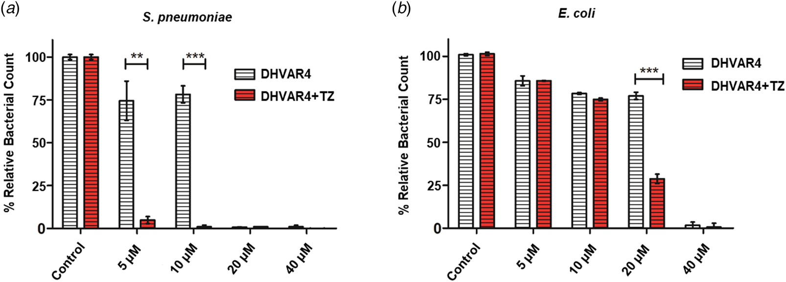
Among food additives approved for human use, azo compounds are negatively charged at physiological pH and are widely used as food colorants in a large variety of foodstuff and pharmaceuticals (Saleh et al., Reference Saleh, Hashem and Salahi2016). They are characterized by aromatic rings in conjunction with N = N linkages and several are known to interact with biomacromolecules (Basu and Kumar, Reference Basu and Kumar2015; Basu and Suresh Kumar, Reference Basu and Suresh Kumar2017; Al-Shabib et al., Reference Al-Shabib, Khan, Alsenaidy, Alsenaidy, Khan, Husain, Khan, Naseem, Sen, Alam and Khan2018). Primarily, six dyes were selected for the combined evaluation of the antibacterial activity. These tests have demonstrated that in principle all food colors have a positive effect on the peptide activity at a 1:1 molar ratio (see also Fig. S1b). Based on the first bacterial assays, TZ has shown the highest impact. To select an additional test compound for comparison, we checked the general physicochemical properties of all dyes, such as charge, the structural distribution of hydrophobic and polar regions, and the nature of the functional groups present. According to these, AmTH showed the closest resemblance to TZ in terms of the above chemical parameters, both having three negative charges in physiological conditions (Saleh et al., Reference Saleh, Hashem and Salahi2016), and thus AmTH was also selected for further studies. Notably, the highest toxicity was detected when DHVAR4 was mixed with TZ. Indeed, the addition of 5 µM TZ already improved the antibacterial effect of the peptide on S. pneumoniae (Fig. 1a). It is worth noting that TZ itself has no antibacterial effect even at the highest concentration. The same tests were repeated on a Gram-negative strain, E.coli, where a similar effect was observed (Fig. 1b).
Antibacterial efficacy of the DHVAR4–TZ complex. Antibacterial activity of DHVAR4 in the presence and absence of 40 µM TZ was measured on S. pneumoniae (a) and on E. coli (b) bacteria. (a) The addition of TZ to the peptide solution significantly improved the antibacterial effect of the peptide at 5 µM concentration. (b) Similar effect was detected on E.coli, namely the addition of TZ significantly improved the antibacterial effect of DHVAR4 at 20 µM. Note that TZ itself has no antibacterial effect even at the highest concentration. (For analysis of statistical significance unpaired t-test was used. ***p < 0.001; **p < 0.005.)

No alteration on cytotoxicity to mammalian cells
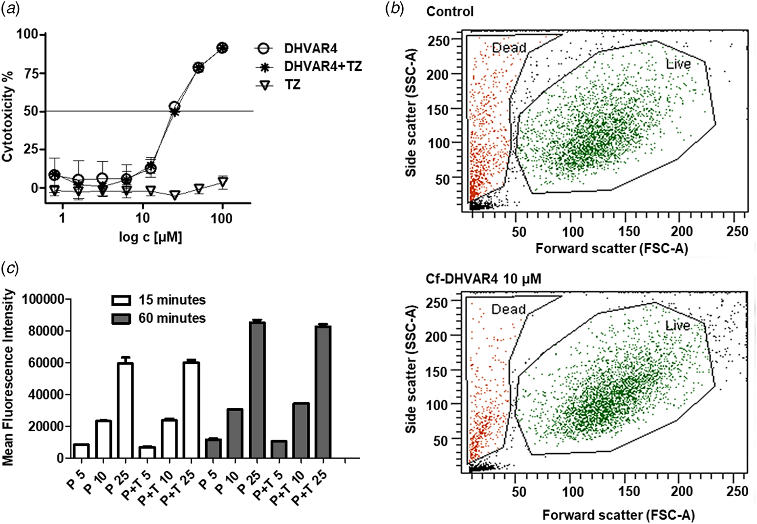
The activity of DHVAR4 alone and its 1:1 mixture with TZ was measured on the human model cell line (MonoMac6) where cytotoxicity was the same for both cases (Fig. 2a). Similarly, cellular uptake was measured on HeLa cells. Even though the peptide internalization is very efficient (see also Fig. S2a), it does not cause a significant increase in the dead cell population in the investigated time frame (Fig. 2b). Results show that peptide uptake depends on the concentration and, to a lesser extent, on the incubation time (Fig. 2c). Notably, TZ did not affect the peptide cellular uptake in HeLa cells.
Cytotoxicity and cellular uptake of DHVAR4 and TZ. (a) Cytotoxicity of DHVAR4 alone and in the presence of TZ (molar ratio 1:1) measured on MonoMac6. Cytotoxicity of the compounds, presented by the IC50 values showed no significant difference (IC50 (DHVAR4) = 25.9 ± 2.1 µM; IC50 (DHVAR4 + TZ) = 26.4 ± 1.7 µM). (b) Flow cytometry investigation of HeLa cells treated with the labeled peptide (Cf-DHVAR4). (c) Cellular uptake of Cf-DHVAR4 in HeLa cells after 15 and 60 min. P5, P10, and P25 refer to 5, 10, and 25 µM Cf-DHVAR4, while P+T5, P+T10, P+T25 to 5, 10, and 25 µM Cf-DHVAR4–TZ.

The lack of increased toxic effect on mammalian cells is important as this way the food color increases the relative selectivity of the AMP toward bacterial cells. This is surprising, since the formation of the peptide–dye complex should result in a partial charge neutralization, and AMPs with less cationic, but more hydrophobic character are typically more toxic to mammalian cells.
Molecular mechanism: food color–DHVAR4 interactions
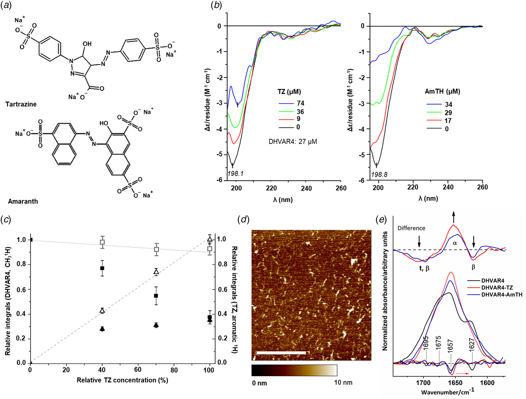
To better understand the nature of the interaction between DHVAR4 and food colors, TZ and AmTH were considered as best candidates, because, despite identical charge and similar distribution of apolar and polar groups, when compared with the other tested molecules (Fig. 3a and see also Table S2), they showed markedly different effect on AMP bioactivity (see also Fig. S1b). Initially, CD and ATR-IR spectroscopic measurements were performed on DHVAR4 alone. The far-UV CD curve recorded in buffer solution shows that DHVAR4 adopts a disordered state (Fig. 3b). The spectrum is constituted by a negative band located in the range 195–210 nm and a weaker shoulder above 215 nm (Woody, Reference Woody2010). Upon consecutive addition of TZ, the main CD peak loses intensity associated with the progressive deterioration of the signal-to-noise ratio (Fig. 3b, left panel). In the current case, the reduced CD signals may arise from the intermolecular aggregation of the peptide chains triggered by the dye molecules. The interaction between DHVAR4 and AmTH shows similar behavior as increasing AmTH concentrations lead to signal reduction with the concomitant deterioration of the signal-to-noise ratio (Fig. 3b, right panel). The anionic groups of TZ and AmTH can neutralize the positive charges of the side chains enhancing the overall hydrophobicity of the peptide–food color complexes. Dynamic light scattering (DLS) measurements also suggest the formation of large DHVAR4–TZ aggregates as TZ concentration increases (see also Fig. S6). To further corroborate this process, atomic resolution studies were conducted using 1H, 13C as well as 2D homo (1H,1H)- and heteronuclear (1H,13C; 1H,15N) NMR methods which resulted in the full assignment of DHVAR4 (see SI text and Fig. S4). NMR investigation confirmed the highly mobile, disordered nature of DHVAR4, a property also supported by its translational diffusion coefficient (Dudás and Bodor, Reference Dudás and Bodor2019). Interaction with TZ was followed by 1D 1H measurements at increasing TZ concentrations, until DHVAR4:TZ = 1:1 molar ratio was reached. No variation in chemical shifts (1H, 13C, 15N) or translational diffusion coefficients (Table S4), and no intermolecular NOEs were detected. At first glance, this observation may suggest that the two molecules coexist in solution without any complex formation. However, a significant intensity decrease of the signals is detected for both partners (Fig. 3c), and above 40% TZ content, a visible yellow precipitate is formed in the NMR tube. A quantitative evaluation showed that the amount of DHVAR4 decreases and the TZ amount is less than expected (Fig. 3c). In contrast, control experiments with samples containing only TZ in the same medium in the investigated concentration range showed no precipitation. When separating and redissolving the precipitate, the existence of both the peptide and TZ was detected. Based on these results, we propose that electrostatic interactions between the positively charged peptide and the negatively charged TZ are the cause of precipitation. Morphology of the aggregates was further investigated using AFM, analyzing dried samples of 1:1 DHVAR4–TZ solution (Fig. 3d). The picture obtained suggests the formation of globular or irregularly-shaped aggregates, with a maximum height of 10 nm and connected by thin fibril-like structures. The presence of different morphologies can be related to the co-existence of distinct conformations (Caruso et al., Reference Caruso, Placidi, Gatto, Mazzuca, Stella, Bocchinfuso, Palleschi, Formaggio, Toniolo and Venanzi2013). In order to address the peptide structure in DHVAR4–TZ aggregates, ATR-IR spectroscopy was performed on dried film samples (Fig. 3e). Analyzing the peptide amide I region, it is possible to observe contributions from various secondary structures (see SI for further details). In the presence of TZ, the concurrence of different conformations is preserved; however, it is possible to note a sensible loss of intermolecular β-sheets and a slight increase of the α-helix fraction (Fig. 3e).
DHVAR4-food color interaction: formation of aggregates. (a) Chemical structure of the dyes. (b) Far-UV CD spectra of DHVAR4 in the buffer, and after addition of TZ (left panel) or AmTH (right panel). (c) NMR analysis. The (0.7 −1.0)ppm –CH3 region of DHVAR4 was used for quantitative analysis: for the pure DHVAR4 sample, this value was considered 1.0, and integrals of the same region for samples with increasing TZ content were normalized accordingly (filled squares); DSS (open squares) was used as internal standard and its value is unchanged at increasing TZ additions; theoretically calculated integrals (solid line) for DSS and DHVAR4 in case all peptide remains in solution. Samples containing only TZ in the same medium showed no precipitation and calculated relative integrals in the aromatic (7.6–8.1)ppm range show the expected values (open triangles, dashed line). (d) Representative AFM micrograph of the dried DHVAR4–TZ (1:1 ratio). The micrograph size is 5.0 µm × 5.0 µm x-y and the total z range is 10 nm. Scale bar: 2 µm. Color scale refers to the sample height. Sample was prepared in water and measured after drying on a silicon wafer. (e) ATR-IR spectra in the amide region of DHVAR4 in the presence of TZ and AmTH (molar ratio DHVAR4:dye is 1:1). Samples were prepared in a buffer solution and measured after drying on the ATR crystal. Spectra are normalized by the area. Second derivative is shown below the spectra and was used for peak identification. Difference spectra show the variation of the peptide conformation when in complex with TZ or AmTH. α, β, and t refer to α-helix, β-sheet, and turn, respectively.

Molecular mechanism: interactions with lipid membranes
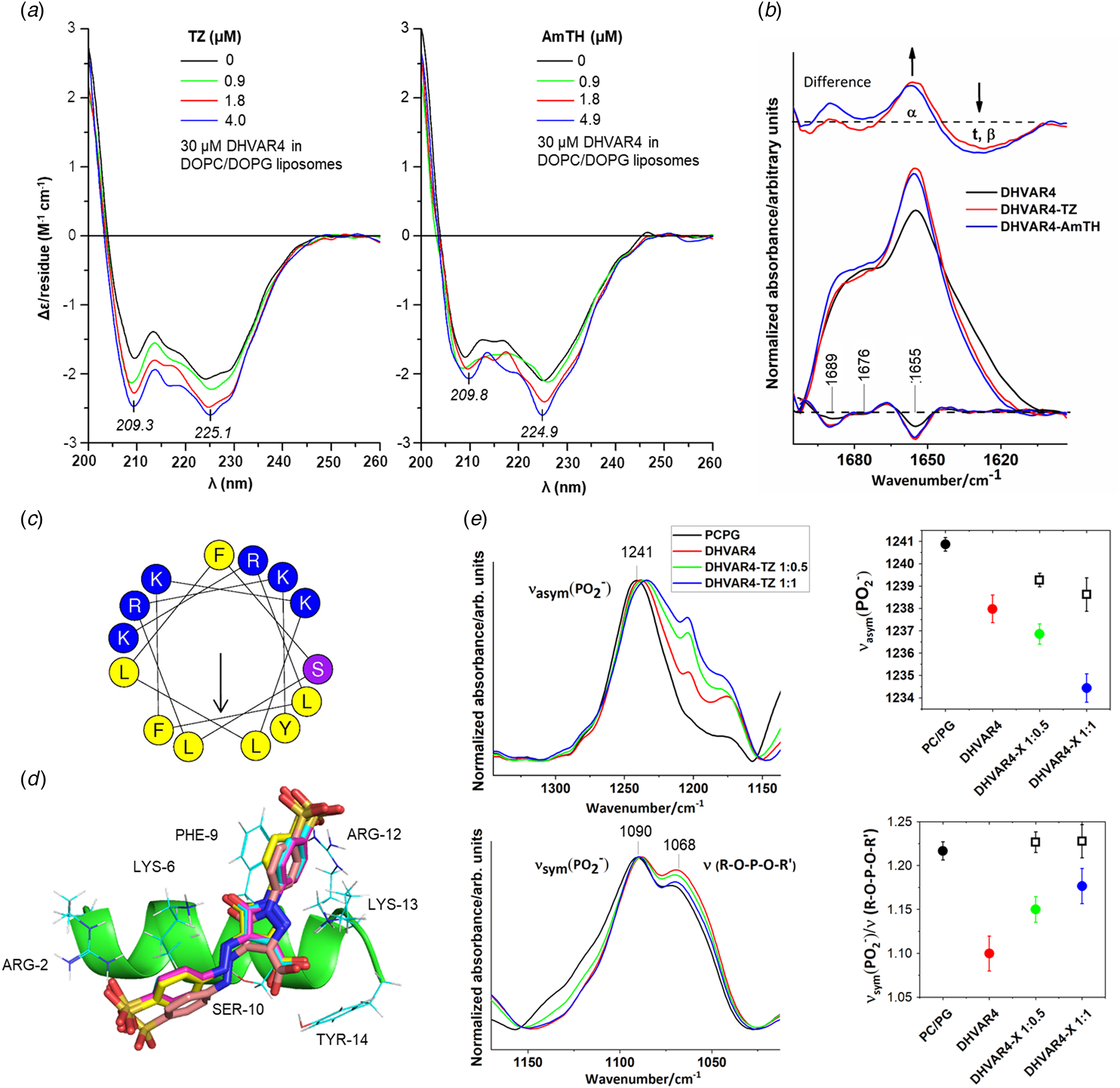
To further address the structure-inducing effect of food colors on the AMP, CD measurements were conducted also in the presence of lipid membranes. In particular, to study conformational variations, negatively charged, dipalmitoylphosphatidylcholine (PC)/dioleoylphosphatidylglycerol (PG), and also neutral, PC, unilamellar liposomes were investigated. In the presence of PC/PG liposomes, the CD spectrum of DHVAR4 exhibits two negative peaks at 209 and 225 with a zero-crossing around 204 nm (Fig. 4a). This double-minima pattern is typical for the α-helical conformation and suggests its prevalence in line with the general conformational change of cationic AMPs upon interaction with negatively charged membranes (Vogt et al., Reference Vogt, Ducarme, Schinzel, Brasseur and Bechinger2000; Sato and Feix, Reference Sato and Feix2006; Chou et al., Reference Chou, Wen, Kuo, Lin and Chen2010; Wiradharma et al., Reference Wiradharma, Khoe, Hauser, Seow, Zhang and Yang2011). Note, however, that a significant fraction of the peptide is still in the disordered state as indicated by the remarkably higher molar CD values of DHVAR4 measured in the helix promoting solvent trifluoroethanol (Fig. S8). Considering the ratio of the Δϵ values of n→π* minima obtained with liposomes and in TFE (−2.1 at 224 nm and −6.7 at 222 nm, respectively) suggests that apparently ~30% of the total peptide adopt a helical conformation in the presence of lipid membranes. Adding only 1 µM of TZ to this system significantly increased the magnitude of both the n→π* and the π→π* peak at 225 and 209 nm, respectively. Further signal intensification was observed upon the stepwise raising of the dye concentration to 4 µM (Fig. 4a, left panel). By using the rough approximation presented above, TZ increased the α-helical contribution from ~30 to ~40%. At higher TZ concentrations, however, the n→π* CD band displayed no additional changes and the amplitude of the π→π* peak started to decrease (data not shown) which is related to the growing impact of the non-specific, aggregation provoking ionic binding of TZ to the free peptide chains (Fig. 3b). Very similar CD spectral alterations were induced by low concentrations of AmTH in the same system (Fig. 4a, right panel). These results refer to a moderate, dye-mediated enhancement of the helicity of membrane-associated DHVAR4 population that is in stark contrast with the aggregation-inducing effect observed under membrane-free conditions. It seems that the partially folded secondary structure ensured by the lipid bilayer allows a new, helix-promoting binding mode of the dyes.
Interaction with lipid membranes. (a) Far-UV CD spectra of DHVAR4 in the presence of PC/PG liposomes (peptide to lipid ratio 1:45) and at 0.9, 1.8, and 4.0/4.9 µM TZ/AmTH. (b) ATR-IR spectra in the amide region of DHVAR4 in PC/PG liposomes (peptide to lipid ratio 1:45), in the presence of TZ and AmTH. DHVAR4:dye molar ratio is 1:1. Spectra are normalized by the area. Second derivatives are shown below and were used for peak identification. Difference spectra show the variation of the peptide conformation. α, β, and t refer to α-helix, β-sheet, and turn, respectively. (c) Helical wheel projection of DHVAR4 obtained with HeliQuest (http://heliquest.ipmc.cnrs.fr/). Polar (positively charged) residues are shown in blue, polar uncharged residues in violet, non-polar (hydrophobic) residues in yellow. (d) Energetically most preferred cluster of binding orientations of TZ on DHVAR4 (see SI for more details). TZ molecule is shown as licorice. Side chains interacting with TZ are shown as lines. For TZ only heavy atoms are displayed. (e) Upper-left insets: IR spectra in the ν asym(PO2−) region of PC/PG liposomes and in the presence of DHVAR4 at different peptide:TZ molar ratio (1:0.5 and 1:1). Spectra are normalized by the intensity at 1241 cm−1. Bottom-left inset: IR spectra in the ν sym(PO2−) region. Spectra are normalized by the intensity at 1090 cm−1. Upper-right inset: shift of ν asym(PO2−) peak position of PC/PG liposomes (black dot) in the presence of DHVAR4 (red dot) and DHVAR4–TZ (green and blue dots) or DHVAR4–AmTH (empty squares). Bottom-right inset: variation of the relative intensity at 1090 cm−1 and ~1068 cm−1. Peptide to lipid ratio is 1:45. Peak picking was performed using a mathematical fitting analysis (Peak Analyzer, Origin-Lab software). Error bars indicate the standard deviation.

Analyzing IR spectra, a major variation occurs in the shape of the amide I band upon interaction of the peptide with PC/PG phospholipid molecules (Fig. 4b) and the addition of both TZ and AmTH causes a slight increase of the α-helix fraction (Fig. 4b, difference spectra). Therefore, despite different measurement setup, both CD and ATR-IR experiments suggest very similar tendencies. In the buffer solution, the disordered AMP and the dyes form ionic complexes, leading to intermolecular aggregation in a concentration-dependent manner. Negatively charged lipid model membranes induce the α-helical folding of the peptide that is fostered by TZ and AmTH, both in the hydrated and in dry film state.
To estimate the potential binding orientations of TZ molecules to the helical DHVAR4 conformation (Fig. 4c), molecular docking simulations were performed. In the most populated cluster, ARG-2, LYS-5, LYS-6, and PHE-9 form hydrogen bonds and a π-π stacking hydrophobic interaction with TZ, indicating that both polar and apolar regions of the two molecules contribute to the formation of the complex (Fig. 4d) (see also Supporting Information for more details).
Molecular details on the interaction of DHVAR4 and DHVAR4–TZ with lipid membranes were also obtained with ATR-IR. For both PC/PG and the PC liposomes, vibrational modes characteristic of the acyl chains were affected neither with DHVAR4 nor with the DHVAR4–TZ complex (see also SI and Fig. S9a). Conversely, significant spectral modifications occur in the hydrophilic region (Fig. 4e and see SI for further details). When the peptide is added to PC/PG liposomes, the phosphate asymmetric stretching ν asym(PO2−) band shifts to lower wavenumbers (Fig. 4e, upper-left and upper-right insets). A further red shift can be observed upon TZ addition, indicating that the formation of DHVAR4–TZ complexes enhances the capacity of the peptide to form more established H-bonds with the components of the lipid bilayers. Furthermore, the addition of DHVAR4 to PC/PG liposomes causes the increase of the shoulder of the phosphate symmetric stretching ν sym(PO2−) (Fig. 4e, lower-left and lower-right insets), which is suggestive of modification of the internal packing in the lipid head-group region (Schibli et al., Reference Schibli, Epand, Vogel and Epand2002). Adding TZ to DHVAR4, the packing of the bilayer is forced back close to a similar state of a normal lipid bilayer, where analysis shows that for PC/PG systems this effect is mainly due to TZ (Fig. 4e and see also Fig. S10). In contrast to the effect observed with TZ, AmTH does not promote the interaction of DHVAR4 with the phosphate groups irrespective of its concentration (Fig. 4e, upper-right and bottom-right insets). Note that the observed spectral variations are preserved at a higher peptide to lipid ratio (see also Fig. S11).
In overall, it is clear that the AMP alone is localized on the surface of negatively charged membranes whereas TZ induces more established H-bonds between the lipid phosphate groups and the AMP and also renders a more ordered head-group region of the bilayer.
Change in the morphology of lipid vesicles
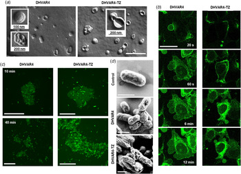
Besides the above molecular level insight, the effect of the DHVAR4–TZ system on lipid vesicle morphology was also investigated (Fig. 5a and see also Fig. S12). The electron micrographs obtained by FF-TEM show that already in the presence of DHVAR4 alone, the regular liposome morphology is altered (Fig. 5a, left panel). Besides single unilamellar liposomes with a diameter of 80–140 nm, oligo- and multilamellar vesicles are frequently present. The interaction of DHVAR4 with the anionic membranes may promote the contact between lipid vesicles by neutralizing the surface charge; as a result, DHVAR4 facilitates the fusion of proximal membranes, leading to hemifused intermediates with diverse morphology. We observed groups of closely packed small vesicles embedded in larger ones (Fig. 5a, lower inset of left panel) and vesicles with shell structures (Fig. 5a, upper inset of left panel). Interestingly, when TZ is also present, it is possible to note clusters of two or three vesicles with an expanding hemifusion diaphragm (Fig. 5a, right panel); however, the shell structures observed in the presence of the peptide alone can barely be seen. In contrast to previous fusion models (Warner and O'Shaughnessy, Reference Warner and O'Shaughnessy2012), the interaction of anionic membranes with DHVAR4–TZ promotes a more controlled growth process of the hemifused intermediates with only two or three vesicles involved. The development of the fusion stalk, the contact area between proximal monolayers involved in the fusion, is affected by the spontaneous membrane curvature induced by lipid molecules (Warner and O'Shaughnessy, Reference Warner and O'Shaughnessy2012), which is in turn influenced by the interaction of amphipathic helices with the head-group region (Schmidt and Wong, Reference Schmidt and Wong2013). It is thus reasonable to hypothesize that the presence of TZ introduces a further curvature variation, leading to different fusion stalk evolution. In the light of the above, the altered interaction of DHVAR4 with the lipid bilayer when in complex with TZ, as observed by IR spectroscopy, is likely to cause a more controlled fusogenic activity. DLS investigation supports the results obtained with FF-TEM as the presence of DHVAR4 alone and in combination with food colors induces a shift of the correlation function toward higher decay times indicating larger particles than seen for the liposomes alone (see also Fig. S12e). The modified fusogenic activity can be also observed when DHVAR4 is in complex with AmTH. However, in this case, beside the hemifused intermediates detected in the presence of the peptide alone, significantly larger and more complex multilamellar objects also appear (see also Fig. S12d and S12e).
Change of membrane morphology in the model system, mammalian and bacterial cells. (a) FF-TEM picture of 100 nm PC/PG unilamellar liposomes in the presence of DHVAR4 and DHVAR4–TZ. Scale bar: 400 nm. (b) Representative time-lapse confocal microscopy images of HeLa cells acquired at 20 s, 60 s, 6 min, and 12 min after addition of 5 µM Cf-DHVAR4 or Cf-DHVAR4–TZ (molar ratio 1:1). The focus was set to the middle of the cell. Scale bar: 10 µm. (c) Confocal microscopy images of aggregated E. coli cells treated for 10 and 40 min with Cf-DHVAR4 and Cf-DHVAR4–TZ. Scale bars: 25 µm. (d) FESEM micrographs of untreated (control) and treated E. coli cells for 40 min with Cf-DHVAR4 and Cf-DHVAR4–TZ. Scale bars: 1 µm.

Cellular membrane remodeling and agglutination
Finally, the effects of the changes observed at molecular as well as lipid morphology level were further investigated on cellular systems. First, HeLa cells, used frequently for morphological studies (Zhong et al., Reference Zhong, Busetto, Fededa, Buhmann and Gerlich2012), were employed as these are much larger and more deformable compared to bacteria, thereby offering a more suitable setup for visual inspection of rapid mechanistic events. The real-time detection of HeLa cells treated with DHVAR4 and DHVAR4–TZ (molar ratio 1:1) shows similar uptake mechanism at all studied concentrations, 5, 10, and 25 µM, as previously observed for other AMPs (Duchardt et al., Reference Duchardt, Fotin-Mleczek, Schwarz, Fischer and Brock2007). In more detail, the peptide first accumulates on specific areas of the cell membrane that immediately starts blebbing. The peptide rapidly spreads inside the cytoplasm and, eventually, reaches the nucleus with a preferential localization in the nucleoli (Fig. 5b). Flow cytometry investigation confirms that the peptide is able to enter HeLa cells with high efficiency (Fig. 2b and c and see also Fig. S2) and at first no significant difference can be detected in the presence of TZ. However, the dye influences the bleb development and fate over time. In the case of cells treated with the AMP alone (Fig. 5b left panel), blebbing starts with the formation of 1–2 µm vesicles that evolve until the final stage characterized by bleb fusion. Annexin V affinity assays show that bleb formation is related to an early apoptotic event (see also Fig. S13). Interestingly, when cells are treated with the DHVAR4–TZ complex (Fig. 5b right panel), the size of these blebs is markedly smaller and the terminal blebs at the end of the studied time range are less prone to fusion. The latter observation is closely related to the results obtained on the effect of DHVAR4–TZ on PC/PG model membranes (Fig. 5a). In general, the amphipathic AMP helices inserted between the lipid head-groups force an increased membrane curvature (McMahon and Gallop, Reference McMahon and Gallop2005). In contrast, for the DHVAR4–TZ complexes, this interaction is further modified. Although, due to the different molecular composition and level of complexity, the features observed for the model liposomes and for HeLa cell membranes are different; however, in both cases, the DHVAR4–TZ systems result in a more controlled fusogenic activity when compared to the AMP alone.
When considering bacterial cells, E. coli have the propensity to agglutinate after addition of the peptide or peptide–dye similar to PC/PG liposomes described above. Upon interaction with bacterial cells, both DHVAR4 and DHVAR4–TZ preferentially accumulate on the bacterial membrane (see also Fig. S14a), likely causing the neutralization of the surface charge, which results in clustering of numerous cells closely packed. For DHVAR4, peptide fluorescence can be detected mainly in cell clusters and in the proximity (see also Fig. S14b), while DHVAR4–TZ complexes accumulate also in solitary cells (see also Fig. S14c), indicating a higher peptide uptake. Moreover, a different evolution of the cluster morphology can be detected during the followed time course of the experiment (Fig. 5c). After 10 min of exposure to DHVAR4 or DHVAR4–TZ, bacterial cells form clusters of similar size and morphology. More heterogeneous morphologies can be detected after 40 min. Indeed, a larger number of cells interact with the peptide, forming clusters that spread out irregularly. On the other hand, bigger and more compact clusters can be observed upon addition of TZ. DHVAR4–TZ increases the cell–cell contact and attraction between E. coli cells. This effect might be related to the molecular level action seen on negatively charged liposomes (Fig. 5a), that results in higher uptake and more controlled fusion compared to DHVAR4 alone. Further on, it is likely that while the cationic aggregates of DHVAR4–TZ enhance closer cellular packing, individual AMP–food color complexes can directly ‘dissolve’ into the bacterial cell wall. As a consequence, the morphology of the bacterial cells is drastically altered (Fig. 5d). E. coli cells treated with DHVAR4 show roughening of the cell surface, pore formation, blebbing, and wrinkling. Exposure to DHVAR4–TZ compromises even more the integrity of the bacterial cells, provoking severe damage, with rupture of the membrane, consequent cellular leakage, and cell aggregation. Note, however, that the latter could not be observed during the antibacterial and cytotoxicity tests, suggesting that altered timeframe and the environmental conditions have an effect on this equilibrium process. Thereby, TZ enhances not only the uptake of DHVAR4 in E. coli cells, but also the ability of the peptide to induce cell damage and potential agglutination, which can be connected to bacterial cell death (Pulido et al., Reference Pulido, Moussaoui, Andreu, Nogués, Torrent and Boix2012; Gopal et al., Reference Gopal, Seo, Song and Park2013).
Conclusions
We have shown that AMP activity can be modulated by food color molecules and also provided insight into this mechanism at molecular and cellular levels. In particular, when TZ forms complexes with the synthetic salivary peptide DHVAR4, toxicity against both Gram-positive and Gram-negative bacteria is enhanced, but not against mammalian cells. Biophysical results indicate that the underlying molecular mechanism is related to the membrane interaction of DHVAR4–TZ complexes, where the peptide has increased helical character, presumably due to the TZ acting as a non-covalent stapling agent by coordinating the basic and hydrophobic residues. The resulting more established AMP–phospholipid hydrogen bonds perturb the bilayer integrity leading to a more regulated fusion of bacterial-mimicking model membranes. The model membrane studies and the microscopy results together indicate that in the presence of lipid bilayers, DHVAR4–TZ complexes more likely associate with the membrane surface, causing severe cell damage. At this point, however, several mechanistic questions remain open along with other aspects, such as bactericidal activity at significantly different molar ratios.
Manipulation of AMP activity in saliva or intestines by small molecules which have already undergone stringent approval processes for human use is an exciting new direction with a potential to offer improved host defense against e.g. food-based bacterial infections, in an optimal scenario by simple diet considerations. Moreover, the sequence similarity of cationic AMPs and the structure-inducing effect observed recently for additional endogenous and synthetic compounds on other AMPs (Zsila et al., Reference Zsila, Bősze, Horváti, Szigyártó and Beke-Somfai2017a, Reference Zsila, Juhász, Bősze, Horváti and Beke-Somfai2017b; Juhász et al., Reference Juhász, Mihály, Kohut, Németh, Liliom and Beke-Somfai2018) postulate that there may be a general mechanism, where in vivo bioactivity of AMPs and host defense peptides are regularly altered by small molecule agents. Exploring these connections is hoped to provide efficient future strategies to combat microbial infections.
Speculation
Controlled increase of immune system performance receives worldwide interest due to its potential as a natural solution for many formidable diseases. The presented example outlines that enhanced host defense peptide activity may also be called forth by simple natural compounds or by regularly employed food additives approved for human use. The future understanding of the mechanistic aspects of further examples on small molecule–peptide interactions may ultimately lead us to manipulate in vivo bioactivity of host defense peptides on a rational basis by relatively simple diet considerations, an aspect with direct practical benefits in immunosuppressive therapies and in medical cases challenged by a compromised immune system.
Supplementary material
The supplementary material for this article can be found at https://doi.org/10.1017/S0033583520000013
Acknowledgements
This study has been financially supported by the Momentum Program (LP2016-2), the National Competitiveness and Excellence Program (NVKP_16-1-2016-0007), the GINOP grant (BIONANO_GINOP-2.3.2-15-2016-00017). We are also grateful to the National Research Development and Innovation Office, Hungary (grants OTKA 104275, 115431, 124077) and the grants VEKOP-2.3.3-15-2017-00020, VEKOP-2.3.2-16-2017-00014. K.H. was supported by the János Bolyai Research Scholarship of the Hungarian Academy of Sciences. S.B. thanks the ELTE Institutional Excellence Program (783-3/2018/FEKUTSRAT) of the Hungarian Ministry of Human Capacities. The contribution of A.Bodor was completed in the ELTE Institutional Excellence Program supported by the National Research, Development and Innovation Office (NKFIH-1157-8/2019-DT). Support by the NKFI Grant K124900 is also acknowledged. Finally, L.H. was supported by the National Research, Development and Innovation Office (grant number: OTKA_K 128123). The authors thank Teréz Kiss for performing FF-TEM and Mayra Quemé-Peña for helping with the fluorescence imaging of E.coli cells.
Conflict of interest
The authors declare no competing interests.