Introduction
Human gnathostomiasis is a food-borne zoonosis caused by a tissue nematode of the genus Gnathostoma. The disease is highly endemic in Asia, including Thailand (Daengsvang, Reference Daengsvang1980, Reference Daengsvang and Sucharit1986). Clinical features of gnathostomiasis are mostly cutaneous and/or visceral larva migrans. A serious clinical manifestation is eosinophilic meningitis with myelo-encephalitis (Daengsvang, Reference Daengsvang and Sucharit1986; Nawa et al., Reference Nawa, Maleewong, Intapan, Diaz-Chamacho, Xiao, Ryan and Feng2015). Dogs, cats, tigers and leopards are natural definitive hosts of Gnathostoma species, while humans are an accidental host. The nematode requires two intermediate hosts: Cyclops as first intermediate host; and freshwater fish, frogs, snakes, freshwater swamp eels, chicken or birds as second intermediate host (Daengsvang, Reference Daengsvang1980, Reference Daengsvang and Sucharit1986). In Thailand, at least 48 species of vertebrates in natural habitats are reported to serve as second intermediate and/or paratenic hosts for Gnathostoma spinigerum. These include various species of fish (20), reptiles (11), amphibians (2), birds (11) and mammals (4) (Daengsvang, Reference Daengsvang and Sucharit1986; Saksirisampant & Thanomsub, Reference Saksirisampant and Thanomsub2012). Among these, freshwater swamp eels (Monopterus alba, previously known as Fluta alba) had the highest prevalence of G. spinigerum larval infection (Daengsvang, Reference Daengsvang and Sucharit1986; Saksirisampant & Thanomsub, Reference Saksirisampant and Thanomsub2012).
Humans become infected by eating raw or improperly cooked meat or fish containing living infective third-stage larvae. In Thailand, the freshwater swamp eel (Monopterus albus) has an important role in transmitting the infection either to humans or among reservoir animals (Setasuban et al., Reference Setasuban, Nuamtanong, Rojanakittikoon, Yaemput, Dekumyoy, Akahane and Kojima1991). In freshwater swamp eels, gnathostome larvae can be found in the muscle tissue but most are in the liver (Rojekittikhun et al., Reference Rojekittikhun, Pubampen and Waikagul1988, Reference Rojekittikhun, Pubampen, Hiranyachattada, Chiamratana and Setasuban1989; Nuamtanong et al., Reference Nuamtanong, Waikagul and Anantaphruti1998; Xuan & Rojekittikhun, Reference Xuan and Rojekittikhun2000; Saksirisampant et al., Reference Saksirisampant, Kulkaew, Nuchprayoon, Yentakham and Wiwanitkit2002; Saksirisampant & Thanomsub, Reference Saksirisampant and Thanomsub2012). Many other studies on the prevalence and intensity of G. spinigerum third-stage larvae in eels sold in local markets in Thailand were reported by using the liver as the target organ to quantify the prevalence and intensity of the G. spinigerum third-stage larva population (Rojekittikhun et al., Reference Rojekittikhun, Pubampen and Waikagul1988, Reference Rojekittikhun, Pubampen, Hiranyachattada, Chiamratana and Setasuban1989; Setasuban et al., Reference Setasuban, Nuamtanong, Rojanakittikoon, Yaemput, Dekumyoy, Akahane and Kojima1991; Nuamtanong et al., Reference Nuamtanong, Waikagul and Anantaphruti1998; Saksirisampant et al., Reference Saksirisampant, Kulkaew, Nuchprayoon, Yentakham and Wiwanitkit2002; Saksirisampant & Thanomsub, Reference Saksirisampant and Thanomsub2012).
Although surveys on the infective larvae of G. spinigerum based on morphological features in freshwater swamp eels have been performed continuously and reported, there is still limited molecular data/information on intra-species variations of this parasite from Thailand. Gnathostoma infective larvae were found to be concentrated in the liver of freshwater swamp eels and the eel livers were offered for sale in Tavej Market, a large metropolitan market in Bangkok. In our study, the third-stage larvae recovered from the eel livers were used for molecular investigation.
In the present study, the morphologically identified third-stage larvae were confirmed by the mitochondrial partial cytochrome c oxidase subunit I (COI) gene sequence. This molecular approach has been used to determine the species status of G. spinigerum (Ngarmamonpirat et al., Reference Ngarmamonpirat, Waikagul, Petmitr, Dekumyoy, Rojekittikhun and Anantapruti2005; Aozasa et al., Reference Aozasa, Tsunemi, Oto, Kai, Morimura, Kakinuma, Tamaki, Sato, Maeda and Yamasaki2011; Jongthawin et al., Reference Jongthawin, Intapan, Sanpool, Sadaow, Janwan, Thanchomnang, Sangchan, Visaetsilpanonta, Keawkong and Maleewong2015, Reference Jongthawin, Intapan, Sanpool, Janwan, Sadaow, Thanchomnang, Laymanivong and Maleewong2016).
Materials and methods
During the period May to July 2014, a total of 437 viscera from freshwater swamp eels, M. albus, were purchased from a large wholesale shop at Tavej Market in Bangkok, Thailand. According to the shop owner, eels were harvested and sent to the shop from many different provinces in central Thailand. The shop had over 1000 customers per day. The eel livers were separated from the viscera and digested with artificial gastric juice at 37°C for 4 h, then washed several times by sedimentation in normal saline. A total of 19 advanced third-stage larvae of gnathostome nematodes were collected from the digested livers. All the gnathostome larvae were morphologically identified as third-stage larvae of G. spinigerum, based on keys and illustrations developed by Daengsvang (Reference Daengsvang1980, Reference Daengsvang and Sucharit1986). The worms were preserved in absolute ethanol and kept at −40°C for DNA extraction.
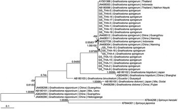
Genomic DNA was extracted directly from tissues of individual third-stage larvae of Gnathostoma using a QIAamp Tissue Kit (QIAGEN, Hilden, Germany) according to the manufacturer's instructions. The molecular method was carried out according to Ando et al. (Reference Ando, Tsunemori, Akahane, Tesana, Hasegawa and Chinzei2006) using a portion of the COI gene. The partial COI gene was amplified by the oligonucleotide primer pairs (F) 5′-TTTTTTGGGCATCCTGAGGTTTA-3′ and (R) 5′-TAAAGAAAGAACATAATGAAAATGAGC-3′. Amplification products were purified using QIAquick PCR Purification Kit (QIAGEN). The purified polymerase chain reaction (PCR) products were sent to a commercial company for sequencing. Samples were sequenced using an ABI PrismDyeTerminator Cycle Sequencing Core Kit (Thermo Fisher Scientific, Waltham, Massachusetts, USA) and analysed on an ABI PRISMH 377 Genetic Analyzer. Of the 19 infective larvae collected in this study, 16 specimens were sequenced successfully and compared with different worldwide G. spinigerum isolates (with COI sequences over 380 bp) and other Gnathostoma species available in GenBank (see fig. 1) to elucidate their phylogenetic relationship.
Maximum likelihood (ML) tree based on the cytochrome c oxidase subunit I (COI) gene of Gnathostoma spinigerum and congeners, with Spiroxys spp. as outgroup taxa. Numeric values at the nodes are Bayesian posterior probabilities/ML bootstrap.

The total length of the aligned sequences of the COI gene was 382 bp and the selected models used for maximum likelihood (ML) and Bayesian Inference (BI) analyses were TN + Gamma and HKY85 + Gamma, respectively. Alignment of the COI sequences, determination of the best-fit nucleotide substitution models and reconstruction of the phylograms were as described in Yong et al. (Reference Yong, Eamsobhana, Song, Prasartvit and Lim2015). The median joining (MJ) network (Bandelt et al., Reference Bandelt, Forster and Röhl1999) was used to estimate the genealogical relationships of the haplotypes. The MJ network was calculated using NETWORK v.5 (http://www.fluxus-engineering.com).
Representative sequences obtained in this study were deposited in the GenBank database under the accession numbers KX648447, KX648448 and KX648449.
Results and discussion
Among 437 investigated eel livers, 19 infective third-stage larvae of the Gnathostoma nematode were observed, with average intensity of infection of 0.04 larvae/liver examined (larvae were counted in samples prepared from pools of livers, not in individual livers).
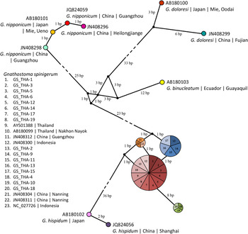
Phylogenetic analysis indicated that the present samples of Gnathostoma parasites from Thailand belonged to G. spinigerum (fig. 1). Both the ML and BI trees had similar topology and revealed the presence of three distinct haplotypes for the Thai samples of G. spinigerum (figs 1 and 2). Eight of the 16 samples were genetically similar to the two reported samples of G. spinigerum from Thailand, and one sample each from China (Guangzhou) and Indonesia (figs 1 and 2). The remaining eight samples formed two other haplotypes, while two samples from China (Nanning) and one from Indonesia formed another haplotype (figs 1 and 2). Four COI haplotypes were present in the G. spinigerum samples from dogs in Thailand, and two haplotypes in the samples from snakes in Laos (Jongthawin et al., Reference Jongthawin, Intapan, Sanpool, Janwan, Sadaow, Thanchomnang, Laymanivong and Maleewong2016).
Haplotype networks of Gnathostoma spinigerum and congeners based on cytochrome c oxidase subunit I (COI) sequences generated by NETWORK software. Circles represent haplotypes and numbers represent individuals sharing the specific haplotype.

The present phylogenetic analysis of Gnathostoma parasites revealed monophyly of the genus, with each species forming a distinct lineage (fig. 1). Gnathostoma nipponicum formed the basal group while G. hispidum formed a sister group with G. spinigerum. This differs from the study conducted using MEGA (Molecular Evolutionary Genetics Analysis) based on a much shorter COI sequence (205 bp) and without outgroup taxa, which indicated G. doloresi as the basal group (Jongthawin et al., Reference Jongthawin, Intapan, Sanpool, Janwan, Sadaow, Thanchomnang, Laymanivong and Maleewong2016). The analysis, based on the ITS-2 gene, indicated G. spinigerum and G. binucleatum forming a sister lineage and [G. doloresi – G. hispidum] forming the basal group (Jongthawin et al., Reference Jongthawin, Intapan, Sanpool, Janwan, Sadaow, Thanchomnang, Laymanivong and Maleewong2016). Studies based on multigene analysis of an extensive taxon sampling are needed to elucidate the phylogenetic relationship of the component species of Gnathostoma.
Acknowledgements
We thank Adisak Yoolex and Sudarat Boonyong for their excellent technical assistance. The constructive comments and valuable suggestions from the editor and one anonymous reviewer have helped to improve the manuscript.
Financial support
This work was supported in part by the Department of Disease Control, Ministry of Public Health, Thailand, and University of Malaya research grant H-5620009 to H.S.Y.
Conflict of interest
None.