Introduction
Neospora caninum is the causative agent of neosporosis which is one of the most important diseases of bovine abortion (Dubey et al., Reference Dubey, Buxton and Wouda2006). The present study characterized a peptide encoded by a cytoplasmic dynein gene which was identified to be downregulated in a non-pathogenic clone of N. caninum in a previous study (Li and Tuo, Reference Li and Tuo2011). The cytoplasmic dyneins in mammals belong to the microtubule minus-end-directed motor proteins and have been shown to be involved in many essential cellular processes (Pfister et al., Reference Pfister, Fisher, Gibbons, Hays, Holzbaur, McIntosh, Porter, Schroer, Vaughan, Witman, King and Vallee2005, Reference Pfister, Shah, Hummerich, Russ, Cotton, Annuar, King and Fisher2006). This multi-functional family of proteins also include the cytoplasmic dynein light chain LC8 (DYNLL), which has recently been recognized to have two distinctive members in mammals, the DYNLL1 and 2 (Naisbitt et al., Reference Naisbitt, Valtschanoff, Allison, Sala, Kim, Craig, Weinberg and Sheng2000; Wilson et al., Reference Wilson, Salata, Susalka and Pfister2001; Pfister et al., Reference Pfister, Shah, Hummerich, Russ, Cotton, Annuar, King and Fisher2006). It was shown that DYNLL1 is a protein inhibitor (PIN) of the neuronal nitric oxide synthase, suggesting a role of this protein in immunoregulation (Jaffrey and Snyder, Reference Jaffrey and Snyder1996). DYNLL1 is abundant in brain and much of it does not appear to be associated with the dynein complex. DYNLL1 may also function to control axonemal dynein motor function as a subunit of the flagellar radial spokes (Yang et al., Reference Yang, Diener, Rosenbaum and Sale2001). DYNLL1 serves as a substrate for a p21-activating kinase, contributing to maintaining cell survival (Vadlamudi et al., Reference Vadlamudi, Bagheri-Yarmand, Yang, Balasenthil, Nguyen, Sahin, den and Kumar2004). DYNLL1 was identified to interact with a number of cytoplasmic factors including proapoptotic factor Bim (Puthalakath et al., Reference Puthalakath, Huang, O'Reilly, King and Strasser1999), Drosophila swallow (Schnorrer et al., Reference Schnorrer, Bohmann and Nusslein-Volhard2000), and rabies virus P protein (Raux et al., Reference Raux, Flamand and Blondel2000), NK-κB inhibitor IκBa (Jung et al., Reference Jung, Kim, Min, Rhee and Jeong2008), oestrogen receptor (Rayala et al., Reference Rayala, den, Balasenthil, Yang, Broaddus and Kumar2005) and zinc-finger protein ASCIZ (Jurado et al., Reference Jurado, Gleeson, O'Donnell, Izon, Walkley, Strasser, Tarlinton and Heierhorst2012) and has been proposed as a dimerization hub important in different protein networks (Barbar, Reference Barbar2008). Mammalian DYNLL1 and DYNLL2 have a protein sequence identity of 93% (Naisbitt et al., Reference Naisbitt, Valtschanoff, Allison, Sala, Kim, Craig, Weinberg and Sheng2000; Wilson et al., Reference Wilson, Salata, Susalka and Pfister2001), suggesting that both molecules may share similar functions. Indeed, limited studies have shown that mammalian DYNLL2 also interacts with the GKAP (Naisbitt et al., Reference Naisbitt, Valtschanoff, Allison, Sala, Kim, Craig, Weinberg and Sheng2000) and Bmf (Puthalakath et al., Reference Puthalakath, Villunger, O'Reilly, Beaumont, Coultas, Cheney, Huang and Strasser2001; Day et al., Reference Day, Puthalakath, Skea, Strasser, Barsukov, Lian, Huang and Hinds2004).
Protozoan DYNLL has not been studied extensively. DYNLL was reported in Plasmodium falciparum (Githui et al., Reference Githui, De Villiers and McArthur2009) and Tetrahymena thermophila in which six orthologues were described (Wilkes et al., Reference Wilkes, Rajagopalan, Chan, Kniazeva, Wiedeman and Asai2007), but no functional analysis was performed. Interestingly, Leishmania DYNLL2 was identified as one of the protective vaccine candidates in a vaccine trial, suggesting a role of Leishmania DYNLL2 in parasitic virulence and pathogenesis (Stober et al., Reference Stober, Lange, Roberts, Gilmartin, Francis, Almeida, Peacock, McCann and Blackwell2006). Most recently, it was demonstrated that DYNLL2 in Toxoplasma gondii is required for parasite growth (Qureshi et al., Reference Qureshi, Hofmann, Arroyo-Olarte, Nickl, Hoehne, Jungblut, Lucius, Scheerer and Gupta2013). Our previous microarray studies revealed that NcDYNLL was downregulated in the non-pathogenic clone Ncts-8 when compared with the wild-type NC1 isolate (Dreier et al., Reference Dreier, Stewarter, Kerlin, Ritter and Brake1999; Lindsay et al., Reference Lindsay, Lenz, Blagburn and Brake1999; Ritter et al., Reference Ritter, Kerlin, Sibert and Brake2002; Li and Tuo, Reference Li and Tuo2011). The present study cloned and expressed the NcDYNLL2 and characterized its differential expression by pathogenic and non-pathogenic N. caninum isolates and its function in regulating host immune responses.
Materials and methods
Animals
Eight to ten week old female BALB/c mice and a 2-year-old ewe were used. Mice were housed in our rodent facility with free access to water and feed and sheep were maintained on pastures with free access to water.
NcDYNLL2 gene cloning and sequencing
Gene alignment and phylogenetic tree construction was done using the ClustalW2 software using neighbour-joining tree method without distance corrections (http://www.ebi.ac.uk/Tools/msa/clustalw2/). Specific primers were designed based on NcDYNLL2 gene sequence XM_003880113 for amplification by polymerase chain reaction (PCR). The forward primer sequence is 5′-GGATAGAATTCATGGCTGACAGGAAGG-3′ with an EcoR I restriction site at the 5′ end and the reverse primer sequence is 5′-CGACGAAGCTTTCAACCGCTCTTAAAG-3′ with a Hind III restriction site at the 5′ end. A forward primer was designed to produce recombinant NcDYNLL2 with a polyhistidine (HIS) tag at the N-terminus for purification purposes. Total N. caninum tachyzoite (NC1 isolate) RNA was isolated using the RNeasy Mini Kit (Qiagen, MD, USA). cDNA was synthesized from total RNA (2 µg per reaction) using the M-MuLV reverse transcription enzyme (NEB, Ipswich, MA, USA) with the reverse primer. The PCR reaction was performed as follows: 95 °C for 10 min followed by 35 cycles of 95 °C for 40 s, 55 °C for 30 s, 72 °C for 40 s with a final 10 min extension at 72 °C. The PCR product was purified by the PCR purification kit (Promega, Madsion, WI, USA) and ligated into the pGEM-T vector (Promega), followed by transformation into Escherichia coli DH5α. The recombinant plasmid was extracted by the Wizard® Plus SV Minipreps kits (Promega) from the overnight liquid culture of LB medium containing ampicillin (50 µg mL−1) and sequenced (Functional Biosciences, Inc., Madison, WI, USA).
Expression, endotoxin removal and purification of recombinant NcDYNLL2 and antiserum preparation
Recombinant NcDYNLL2 (rNcDYNLL2) was produced by subcloning the NcDYNLL2 gene from the pGEM-T (Promega) to pET28a vector (EMD Millipore, San Diego, CA, USA). The rNcDYNLL2 was expressed in E. coli BL21(DE3) (Novagen, Madison, WI, USA) and induced at 37 °C for 3 h with 1 mm isopropyl thiogalactopyranoside. The cultures were harvested by centrifugation at 4000 g for 30 min at 4 °C and the pellet was resuspended in lysis buffer consisting of 50 mm NaH2PO4, 300 mm NaCl, 10 mm imidazole, pH 8.0. The cells were lysed by five freeze–thaw cycles, followed by digestion with 10 µg mL−1 DNase/RNase for 1 h at 37 °C. The soluble fraction of the bacterial lysate was obtained by centrifugation at 20 000 g for 30 min at 4 °C.
Non-recombinant control protein (NR) used as a negative control in assays was prepared from E. coli BL21(DE3) using the procedures similar to those for the preparation of rNcDYNLL2, except that the E. coli for NR was transformed with pET28a vector without the NcDYNLL2 coding sequence (Tuo et al., Reference Tuo, Zhao, Zhu and Jenkins2011).
Prior to purification, Triton X-114 (Sigma, St. Louis, MO, USA) was used to remove endotoxin from the bacterial lysate as previously described (Aida and Pabst, Reference Aida and Pabst1990; Qu et al., Reference Qu, Fetterer, Jenkins, Leng, Shen, Murphy, Han, Bucala and Tuo2013). In brief, Triton X-114 was added to the soluble lysate to a final concentration of 1%. The mixture was vortexed for 10 s and incubated on ice for 5 min before being vortexed again and then incubated at 37 °C for additional 10 min. The mixture was centrifuged at 20 000 g for 2 min at 38 °C and the upper aqueous phase containing rNcDYNLL2 was collected. This procedure was repeated seven times for complete removal of endotoxin. rNcDYNLL2 then was purified by a Ni-NTA column under native conditions according to manufacturer's instructions (Qiagen Inc., Valencia, CA, USA).
Purified rNcDYNLL2 was used to immunize a sheep to produce anti-NcDYNLL2 antiserum. Briefly, purified recombinant NcDYNLL2 was mixed with adjuvant (ImmuMax-SR Adjuvant System, Zonagen, Inc., Woodlands, TX, USA) and injected s.c. to the sheep twice at 30-day intervals. Thirty days following the second injection, blood was collected and serum prepared and stored at −20 °C until used.
Parasite culture and preparation of excretory and secretory antigen (ESAg) and whole parasite soluble antigen (NcAg) from the N. caninum (NC1 and Ncts-8 isolates) and T. gondii tachyzoites
Neospora caninum (NC1 and Ncts-8 isolates) and T. gondii (RH strain) tachyzoites were cultured, harvested and Percoll-purified as described previously (Tuo et al., Reference Tuo, Fetterer, Jenkins and Dubey2005; Li and Tuo, Reference Li and Tuo2011; Yin et al., Reference Yin, Qu, Cao, Li, Fetterer, Feng, Liu, Wang, Qi, Zhang, Miramontes, Jenkins, Zhang and Tuo2012). Briefly, purified tachyzoites were resuspended to 108 parasites mL−1 in RPMI 1640 (25 mm glutamine and 50 µg mL−1 gentamicin) and cultured at 37 °C for NC1 and T. gondii tachyzoites and 32 °C for Ncts-8 clone and 4 °C for all isolates with or without the treatment by 5 µ m calcium ionophore (Sigma) or 1% ethanol for 1 h. The supernatant was collected by centrifugation at 20 000 g for 20 min at 4 °C and stored at −70 °C until use. The soluble (NcAg) and insoluble fractions of the Neospora lysate were prepared on ice by sonicating the tachyzoites with three 15 s pulses at maximal power using the micro-tip (Sonifier 250, Branson Sonic Power, Danbury, CT, USA), followed by centrifugation at 20 000 g for 30 min at 4 °C and the supernatant and the pellet were collected separately and stored at −70 °C until use.
Analysis of NcDYNLL2 by SDS-PAGE and Western blotting
ESAg of N. caninum or T. gondii was concentrated 20-fold by trichloroacetic acid precipitation prior to analysis (Yin et al., Reference Yin, Qu, Cao, Li, Fetterer, Feng, Liu, Wang, Qi, Zhang, Miramontes, Jenkins, Zhang and Tuo2012). CV1 and RAW267.6 cells (ATCC, Manassas, VA, USA) were cultured as previously described (Qu et al., Reference Qu, Fetterer, Jenkins, Leng, Shen, Murphy, Han, Bucala and Tuo2013) and cell lysate prepared using 1 × SDS-PAGE sample loading buffer. SDS-PAGE and Western blotting were performed as described previously (Tuo et al., Reference Tuo, Fetterer, Jenkins and Dubey2005). Briefly, samples were separated on a 4–12% NuPAGE (Invitrogen, Carlsbad, CA, USA) or 15% SDS-PAGE gels under reducing conditions. After electrophoresis, protein was either stained with Coomassie blue or transferred to a PVDF membrane for Western blotting (Millipore, Bedford, MA, USA). The PVDF blot was incubated in blocking solution (3% skim milk in PBS) for 1 h. The sheep anti-NcDYNLL2 antibody (1:500 in blocking buffer) and rabbit anti-sheep IgG-HRP (KPL, Gaithersburg, MD, USA) (1:5000 in blocking buffer) were used as first and secondary reagents, respectively. A chemiluminescence substrate (SuperSignal® West Dura Exrended Duration Substrate, Thermo-Fisher, Rockford, IL, USA) was used to develop the blot according to the manufacturer's instructions. The image was captured using a ChemiImager™ 4400 Low Light Imaging system (Alpha Innotech Corporation, San Leandro, CA, USA).
Immunolocalization of NcDYNLL2 in N. caninum tachyzoites and recombinant NrDYLL2 in RAW264.7 cells pre-incubated with rNrDYLL2
Localization of NcDYNLL2 in Percoll-purified Neospora tachyzoites was performed using indirect immunoflourescence assay (IFA) (Qu et al., Reference Qu, Fetterer, Jenkins, Leng, Shen, Murphy, Han, Bucala and Tuo2013). In Brief, purified N. caninum tachyzoites were pipetted onto individual wells (104 tachyzoites well−1) of multi-well glass slides (Erie Scientific Co., Portsmouth, NH, USA) and allowed to air-dry. After drying, the slides were immersed for 5 min in cold methanol and washed briefly with PBS, then blocked by 2% skim milk in PBS containing 0.05% Tween-20 (PBS-T) at room temperature for 30 min. After washing, slides were incubated for 2 h at room temperature with a sheep anti-NcDYNLL2 or pre-immune sera (1:500 in blocking buffer). Following five washes with PBS-T, rabbit anti-sheep IgG (H + L) labelled with DyLight 488 (KPL; 1:1000 in blocking buffer) was added and incubated for 1 h at room temperature. Following washes, slides were overlaid with Vectashield mounting medium (Vector Laboratories, Burlingame, CA, USA) and cover-slipped. The images were taken using a fluorescence microscope (Carl Zeiss MicroImaging, Inc., Thornwood, NY, USA).
To localize native NcDYNLL2 in tachyzoites of NC1-infected host cells, CV1 cells were allowed to reach 80% confluency prior to infection by NC1 N. caninum tachyzoites overnight using 24-well plates. Infected cells were then fixed with methanol/acetone (1:1) at −20 °C for at least 20 min. The fixative was discarded and cells were allowed to air-dry prior to rehydration with PBS. Cells were blocked and permeabilized with blocking/permeability buffer (1% BSA, 0.3% Triton X-100, 3% non-fat milk and 5% FBS in PBS, pH 7.2) for 1 h at room temperature. Sheep anti-rNcDYNLL2 serum diluted (1:200) with blocking/permeability buffer was then added to the wells and incubated overnight at 4 °C. Following washing with PBS, rabbit anti-sheep IgG (H + L) labelled with DyLight 488 (1:1000; KPL) diluted in blocking/permeability buffer was added to wells and incubated for 2 h at room temperature. Plates were washed three times with PBS prior to addition of 1 µg mL−1 of DAPI (Life Technologies, Grand Island, NY, USA) in PBS and incubated at room temperature for 10 min. Then plates were washed for three times with PBS. Following complete removal of PBS, Vectashield mounting medium was gently overlaid onto the stained cells prior to imaging or storage at 4 °C. Images were captured and analysed using an Olympus IX71 fluorescence microscope equipped with QCapture Pro 6.0 software.
To determine uptake/internalization of rNcDYNLL2 by immune cells, RAW264.7, a murine macrophage cell line, were seeded in 24-well plates and grown to 50–70% confluency prior to addition of 10 µg mL−1 of the purified rNcDYNLL2. Following adding rNcDYNLL2, the culture was allowed to continue overnight at 37 °C. At the completion of incubation, the plates were washed five times with PBS-T and cells were then fixed with methanol/acetone (1:1) at −20 °C for at least 20 min. After fixation, the fixative was discarded and cells were air-dried and stored at 4 °C. The cells were then rehydrated and stained for rNcDYNLL2 using the same procedure for localization of intracellular NcDYNLL2 in tachyzoite described in previous paragraph of this section (Qu et al., Reference Qu, Fetterer, Leng, Du, Zarlenga, Shen, Han, Bucala and Tuo2014).
NcDYNLL2-regulated tumour necrosis factor-α and interleukin-12 production by murine dendritic cells and nitric oxide production by macrophages
Murine dendritic cells (DCs) were prepared for the tumour necrosis factor-α (TNF-α) and interleukin-12 (IL-12) production assay, as described elsewhere (Feng et al., Reference Feng, Zhang and Tuo2010). In brief, bone marrow cells were collected by flushing mouse femurs with RPMI-1640-EDTA, followed by red blood cell lysis for 3 min at room temperature in a lysis buffer (0.15 M ammonium chloride, 10 mm potassium bicarbonate, 0.1 mm EDTA, pH 7.4). The remaining cells were washed three times and resuspended in complete medium supplemented with 10% FBS, 25 mm glutamine, 5 µg mL−1 gentamicin, and 5 ng mL−1 murine GM-CSF (Prospec, East Brunswick, NJ, USA). Cells (2 × 10⁶) in 1 mL complete medium were plated in 24-well plates and cultured at 37 °C for 2 days. Non-adherent cells were gently removed, the adherent cells were gently washed with RMPI-1640 and fresh complete medium was added. The cells were allowed to culture for additional 2 days; all non-adherent cells were collected, transferred to a new plate and continue to culture for additional 2–3 days. Enriched DCs were seeded in 48-well plates at 0.5 × 10⁶ cells per well in 0.5 mL complete medium and treated with rNcDYNLL2 for 24 h. At the end of the culture, supernatant was collected by centrifugation and stored at −70 °C until assayed. Murine IL-12 and TNF-α in cell culture supernatants were determined using IL-12 ELISA kit following manufacturer's instructions (eBiosciences).
Murine RAW264.7 macrophages were used for nitric oxide assay. The cells at ~80% confluency were treated with rNcDYNLL2 (0.1, 1 or 10 µg mL−1) alone or in the presence of 10 ng mL−1 of lipopolysaccharide (LPS) overnight using 24-well plates. Nitric oxide content in cell culture supernatants was determined by the Griess reagent as described previously (Qu et al., Reference Qu, Fetterer, Jenkins, Leng, Shen, Murphy, Han, Bucala and Tuo2013). In brief, 150 µL of the cell culture supernatants was added into the wells of 96-well plates, followed by addition of 20 µL of 1 µ m nicotinamide adenine dinucleotide phosphate (Sigma) and 30 µL of the master mixture consisting of glucose-6-phophate (Sigma), glucose-6-phosphate dehydrogenase (Sigma) and nitrate reductase (Roche Diagnostics, Indianapolis, IN, USA). The plate was incubated for 30 min at room temperature and then 20 µL 1% of sulfanilamide and 20 µL of 0.1% N-(1-Naphthyl) ethylenediamine dihydrochloride (Sigma) were added. After incubation for 5 min at room temperature, the plate was read at 550 and 650 nm using a microplate reader (Molecular Devices SpectraMax Plus384, Ramsey, MN, USA). Total nitric oxide in samples was calculated using a nitrate (Sigma) standard curve diluted ranging from 1.6 to 50 µ m.
Statistics
Cytokine levels and amounts of NcDYNLL2 in ESAg were analysed by one-way ANOVA with a Bonferroni Multiple Comparisons Test and levels of NcDYNLL2 in NC1 and Ncts-8 isolate whole cell lysate were analysis by two-tailed t-test (GraphPad InStat). Probability values of P < 0.05 were considered statistically significant.
Results
NcDYNLL2 sequence analysis
The EST sequence CF273809 (265 bp) was identified as one of the downregulated dynein light chain motor proteins in our previous study (Li and Tuo, Reference Li and Tuo2011) and this sequence was used to BLAST-search the Genbank and the GeneDB (http://genedb.org) for identification of similar gene sequences. The BLASTN-search in the Genbank EST database produced two additional EST sequences encoding putative dynein light chains (CF598536 and CF423006) with 93–96% identity to CF273809. Search of the GenBank nucleotide collection (nr/nt) identified one putative dynein light chain gene XM_003880113 or NCLIV_006030 with 95% identity, or protein ID CBZ50127 (N. caninum Liverpool complete genome, chromosome II, FR823382.1). The BLASTN-search in the GeneDB revealed one putative dynein light chain protein (NCLIV_006030) with an amino acid sequence identity of 95% to CF273809. Then, BLASTN-search of GeneDB with NCLIV_006030 did not result in identification of additional sequences that are highly similar to NCLIV_006030. Three genes (NCLIV_016950, NCLIV_024890, NCLIV_068030) coding for putative dynein light chains of similar molecular mass were also identified, but the identity between these three genes and their identity to NCLIV_006030 is relatively low (<40% at the amino acid sequence level). These results suggest that the EST sequence CF273809 may contain some errors; thus, XM_003880113 (GenBank) which is the same as NCLIV_006030 (GeneDB) was used to design primers to PCR-amplify NcDYNLL2.
To demonstrate similarity across different species, DYNLL2 sequences representing those of protozoa including N. caninum DYNLL2 (encoded by XM_003880113) and T. gondii DYNLL2 (encoded by XM_002370046); algae such as Micromonas pusilla flagellar outer dynein arm light chain 8 (encoded by XM_003056050); the purple sea urchins such as Strongylocentrotus purpuratus dynein light chain LC6 (encoded by XM_790626); mammals including Bos taurus (a natural host for N. caninum) dynein light chain LC8-type 2 (encoded by NM_001113303), Homo sapiens dynein light chain LC8-type 2 (encoded by NM_080677) and Mus musculus dynein light chain LC8-type 2 (encoded by NM_001168472); insects such as Apis florea dynein light chain 2 (encoded by XM_003693250) and Drosophila melanogaster cytoplasmic dynein light chain 2 (encoded by NM_001258907), and Megachile rotundata dynein light chain 2 (XM_003705036); and birds including Gallus gallus dynein light chain LC8-type 2 (encoded by XM_415908).
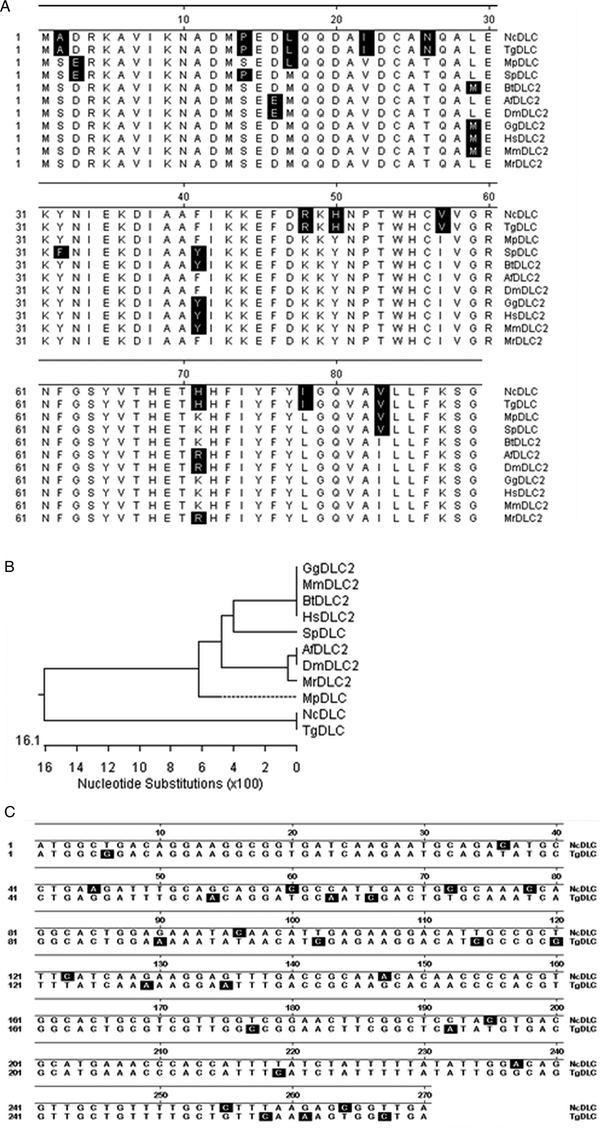
NcDYNLL2 mature protein has 89 amino acids and does not have a predicted conventional signal peptide. It has a calculated molecular mass of 10.3 kDa and pI of 7.1. Overall, DYNLL2s across different species from mammals to protozoa are highly conserved with an identity of at least 85% (Fig. 1A and B). At the amino acid sequence level, DYNLL2s among mammals and chicken are identical (Fig. 1A and 1B). Based on the sequence alignment, NcDYNLL is most similar to mammalian, chicken, drosophila and dwarf honey bee DYNLL2. Thus, NcDYNLL identified and characterized in the present study is named NcDYNLL2, and similarly, TgDYNLL2 for T. gondii. In addition, sequence alignment revealed that although NcDYNLL2 and TgDYNLL2 are divergent at the genetic level (89% identity) (Fig. 1C), the amino acid sequences of NcDYNLL2 and TgDYNLL2 are identical (Fig. 1A).
Dynein LC8 light chain 2 (DYNLL2) amino acid sequence alignment across 11 different species (A), a phylogenetic tree showing the evolutionary relationship among these genes (B), and a comparison between NcDYNLL2 and TgDYNLL2 cDNA coding sequences (C). Nc DYNLL2, Neospora caninum DYNLL2 (XM_003880113); TgDYNLL2, Toxoplasma gondii DYNLL2 (XM_002370046); MpDLC, Micromonas pusilla flagellar outer dynein arm light chain 8 (XM_003056050); SpDLC, Strongylocentrotus purpuratus dynein light chain LC6 (XM_790626); BtDYNLL2, Bos taurus dynein, light chain, LC8-type 2 (NM_001113303); AfDLC2, Apis florea dynein light chain 2 (XM_003693250); DmDLC2, Drosophila melanogaster cytoplasmic dynein light chain 2 (NM_001258907); GgDYNLL2, Gallus gallus dynein, light chain, LC8-type 2 (XM_415908); HpDYNLL2, Homo sapiens dynein, light chain, LC8-type 2 (NM_080677); MmDYNLL2, Mus musculus dynein light chain LC8-type 2 (NM_001168472); MrDLC2, Megachile rotundata dynein light chain 2 (XM_003705036).

Recombinant NcDYNLL2 production in E. coli, native NcDYNLL2 production by N. caninum and T. gondii tachyzoites and Western blotting
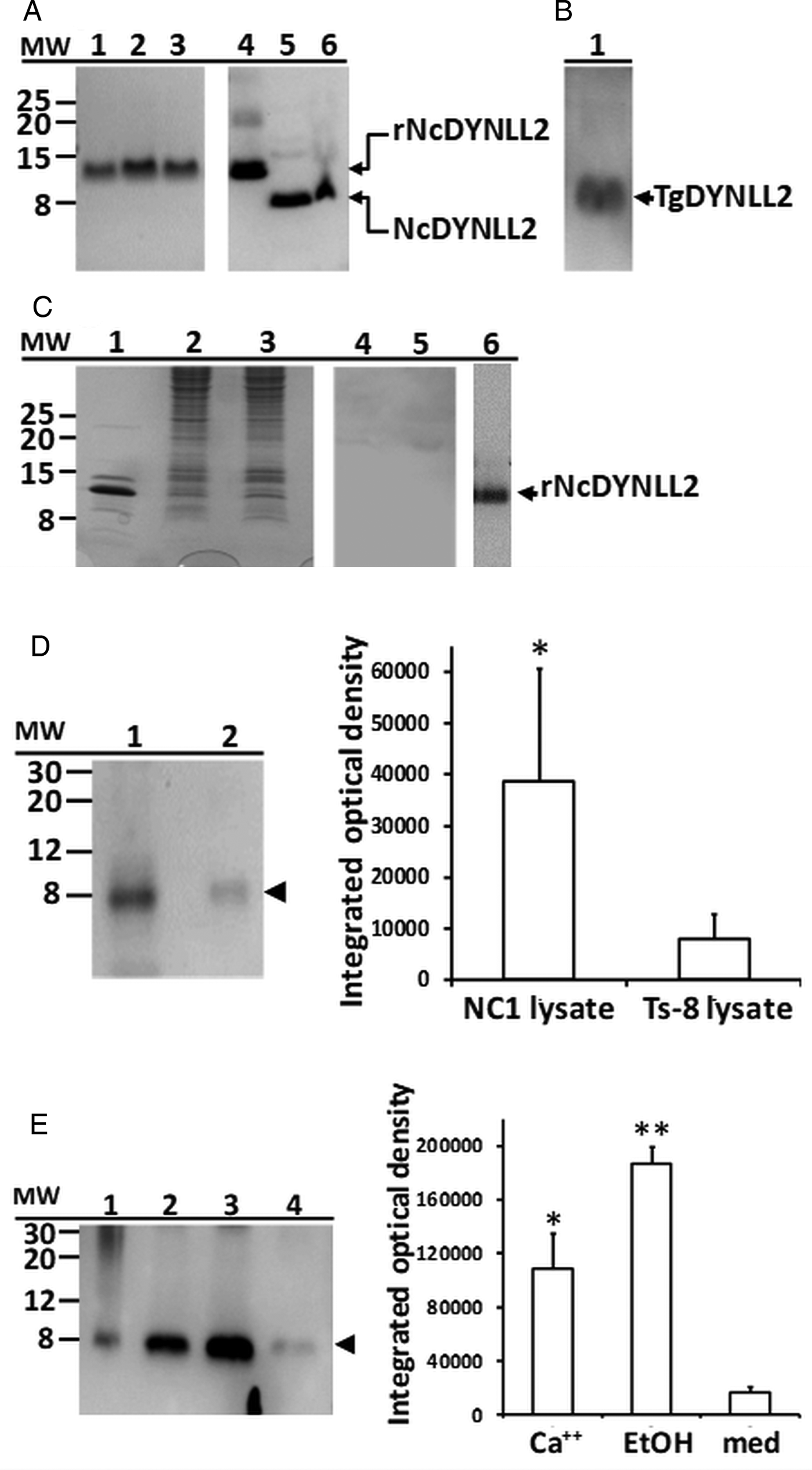
rNcDYNLL2 was expressed as a HIS-tagged, soluble protein in E. coli and purified by the Ni-NTA (Fig. 2A, lanes 1, 2 and 3 representing three different clones) as well as size-exclusion chromatography (data not shown) with comparable purity of >98%. rNcDYNLL2 had an apparent molecular mass of 14 kDa (Fig. 2A). rNcDYNLL2 (Fig. 2A, lane 4) and native NcDYNLL2 (Fig. 1A, lanes 5 and 6) were specifically recognized by the sheep anti-rNcDYNLL2 antibody. NcDYNLL2 was present in both soluble (Fig. 2A, lane 5) and insoluble (Fig. 2A, lane 6) fractions of the NC1 tachyzoite lysate prepared by sonication and centrifugation. As expected, TgDNYLL2 in T. gondii ESAg prepared by treating T. gondii tachyzoites with 5 µ m calcium ionophore at 37 °C for 1 h was also detected by the sheep anti-rNcDYNLL2 serum (Fig. 2B, lane 1). As a control, the DYLLs of mammalian host cells were not detectable by the sheep anti-rNcDYNLL2 antibody (Fig. 2C, lanes 4 and 5). Semi-qualitative analysis using Western blotting showed that NcDYNLL2 was 5-fold higher (P < 0.05) (Fig. 2D, right panel) in NC1 lysate than in Ncts-8 lysate (Fig. 2D, Western blot lanes 1 and 2, left panel). Low levels of NcDYNLL2 were released by the NC1 tachyzoites (Fig. 2E, Western blot lane 4, left panel); however, NcDYNLL2 release was enhanced (P < 0.05) (Fig. 2E, right panel) when the NC1 tachyzoites were treated with either 5 µ m calcium ionophore or 1% ethanol at 37 °C for 1 h (Fig. 2E, Western blot lanes 2 or 3, left panel). Ethanol treatment (1%) appeared to be more effective in enhancing NcDYNLL2 release, but the amounts of NcDYNLL2 released in response to calcium ionophore and ethanol treatments were not significantly different (P > 0.05; Fig. 2E right panel). NcDYNLL2 or TgDYNLL2 was not detected by either pre-immune sera or secondary antibody alone in controls (data not shown).
SDS-PAGE and Western blot analysis of rNcDYNLL2, NcDYNLL2 and TgNYNLL2. (A) rNcDYNLL2 purified by Ni-NTA column from three different clones of recombinant Escherichia coli (lanes 1 through 3); Western blot analysis of rNcDYNLL2 (lane 4) and native NcDYNLL2 in soluble (lane 5) and insoluble (lane 6) fractions of the NC1 tachyzoite lysate. (B) Western blot analysis of TgDYNLL2 in ESAg preparation from Toxoplasma gondii tachyzoites treated with 5 µ m calcium ionophore (lane 1). (C) Coomassie blue staining of rNcDYNLL2 (lane 1) and CV1 (lane 2) and RAW264.7 (lane 3) cell lysates separated by SDS-PAGE; Western blot analysis of cross-reactivity of sheep anti-rNcDYNLL2 serum with host DYLLs in CV1 (lane 4) and RAW264.7 (lane 5) cell lysates, rNcDYNLL2 was used as a positive control (lane 6). (D) Representative Western blotting (left panel) of native NcDYNLL2 in NC1 (lane 1) and Ncts-8 (lane 2) soluble tachyzoite lysates, and the mean integrated optical density ± standard deviation of four independent experiments (right panel). (E) Representative Western blotting (left panel) of native NcDYNLL2 in soluble NC1 tachyzoite lysate (lane 1) and in ESAg preparations from NC1 tachyzoites treated for 1 h at 37 °C with 5 µ m calcium ionophore (lane 2), 1% ethanol (lane 3) and RPMI 1640 alone (lane 4), and the mean integrated optical density ± standard deviation of two independent experiments (right panel). Arrow head indicates NcDYNLL2. Negative controls also included secondary antibody alone or pre-immune serum (data not shown). rNcDYNLL2, recombinant NcDYNLL2; Ca++, 5 µ m calcium ionophore; EtOH, 1% ethanol; med, RPMI 1640 alone. *P < 0.05; **P < 0.01.

Western blotting in Fig. 2D and 2E was performed with four and two biological replicates, respectively, resulting from N. caninum soluble antigen or ES antigen produced in independent experiments. The replicates for Fig. 2E were limited to 2 because of difficulties in obtaining sufficient highly purified tachyzoites for ES antigen production.
Immunolocalization of native NcDYNLL2 in tachyzoites and rNcDYNLL2 in immune cells
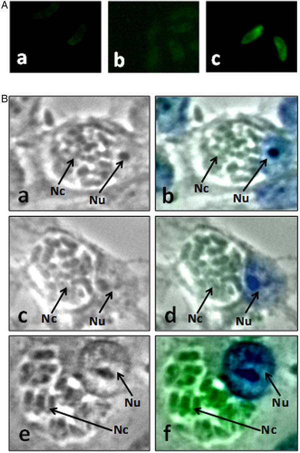
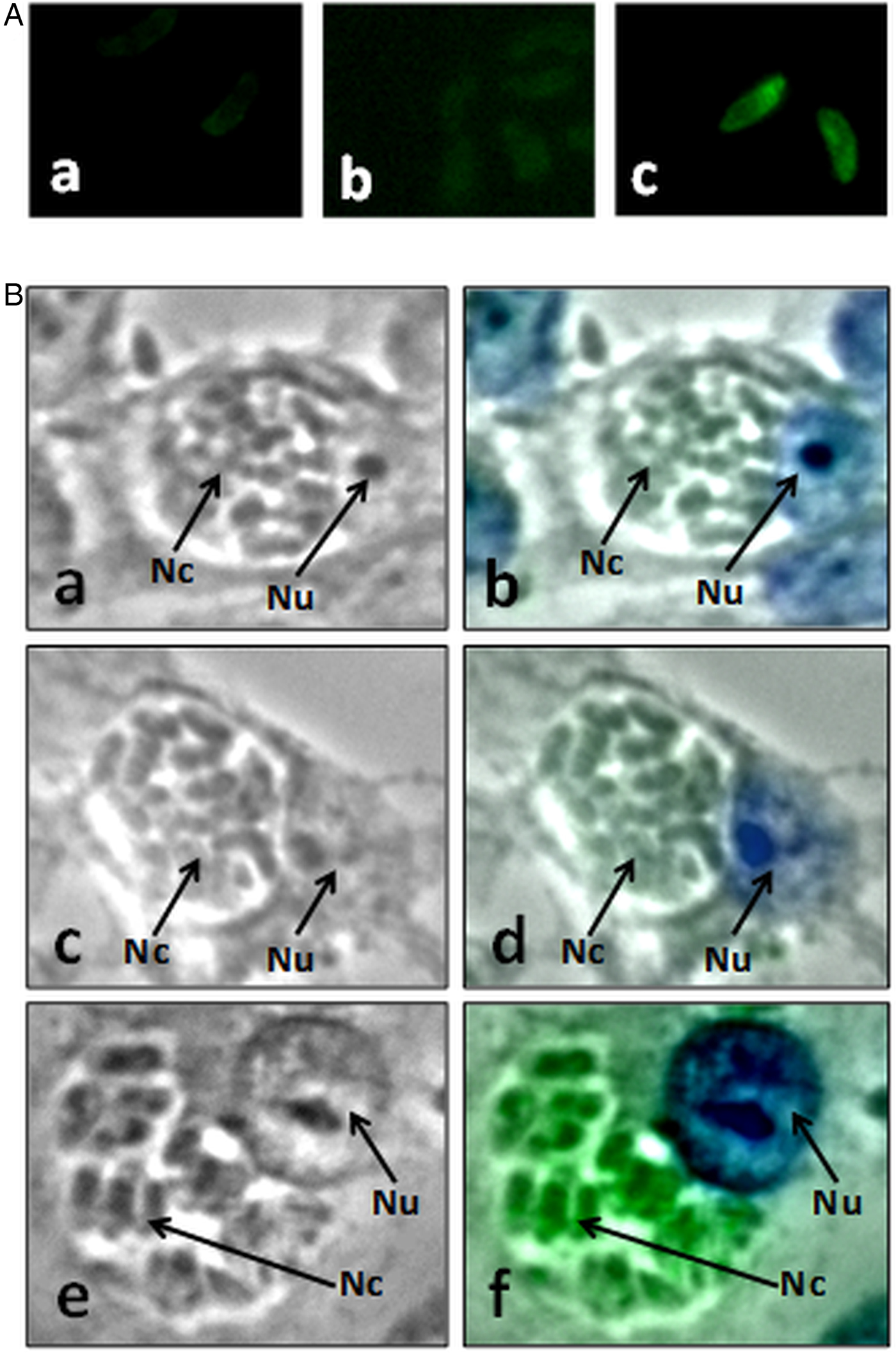
NcDYNLL2 was specifically, but diffusely, localized in air-dried, methanol-fixed, purified tachyzoites using IFA (Fig. 3A, c). Secondary antibody alone (Fig. 3A, a) and pre-immune serum plus secondary antibody (Fig. 3A, b) showed negative staining. Similar specific staining was clearly evident in intracellular tachyzoites of NC1 tachyzoite-infected CV1 cells which were methanol/acetone-fixed (Fig. 3B, f), but in tachyzoite-infected cells stained with second antibody alone (Fig. 3B, b) or pre-immune serum plus secondary antibody (Fig. 3B, d).
NcDYNLL2 localization in Percoll-purified NC1 tachyzoites (A) and in intracellular NC1 tachyzoites in infected host cells (CV1) (B). (A) a: Tachyzoites were stained with rabbit anti-sheep IgG-Alexa 488 alone control; b: sheep pre-immune serum at 1:100 dilution; c: sheep anti-NcDYNLL2 serum at 1:100 dilution. (B) a, c and e: phase-contrast images; b, d and f: phase-contrast and fluorescence image overlay. a and b: Neospora caninum-infected cells stained with rabbit anti-sheep IgG-Alexa 488 alone; c and d: N. caninum-infected cells stained with pre-immune serum at 1:200 dilution; e and f: N. caninum-infected cells were stained with sheep anti-rNcDYNLL2 serum at 1:200 dilution. Blue colour represents host cell nuclear staining by DAPI, green colour represents NcDYNLL2 staining by Alexa 488. Nc, N. caninum tachyzoites; Nu, host cell nucleus. Magnification, 400×.
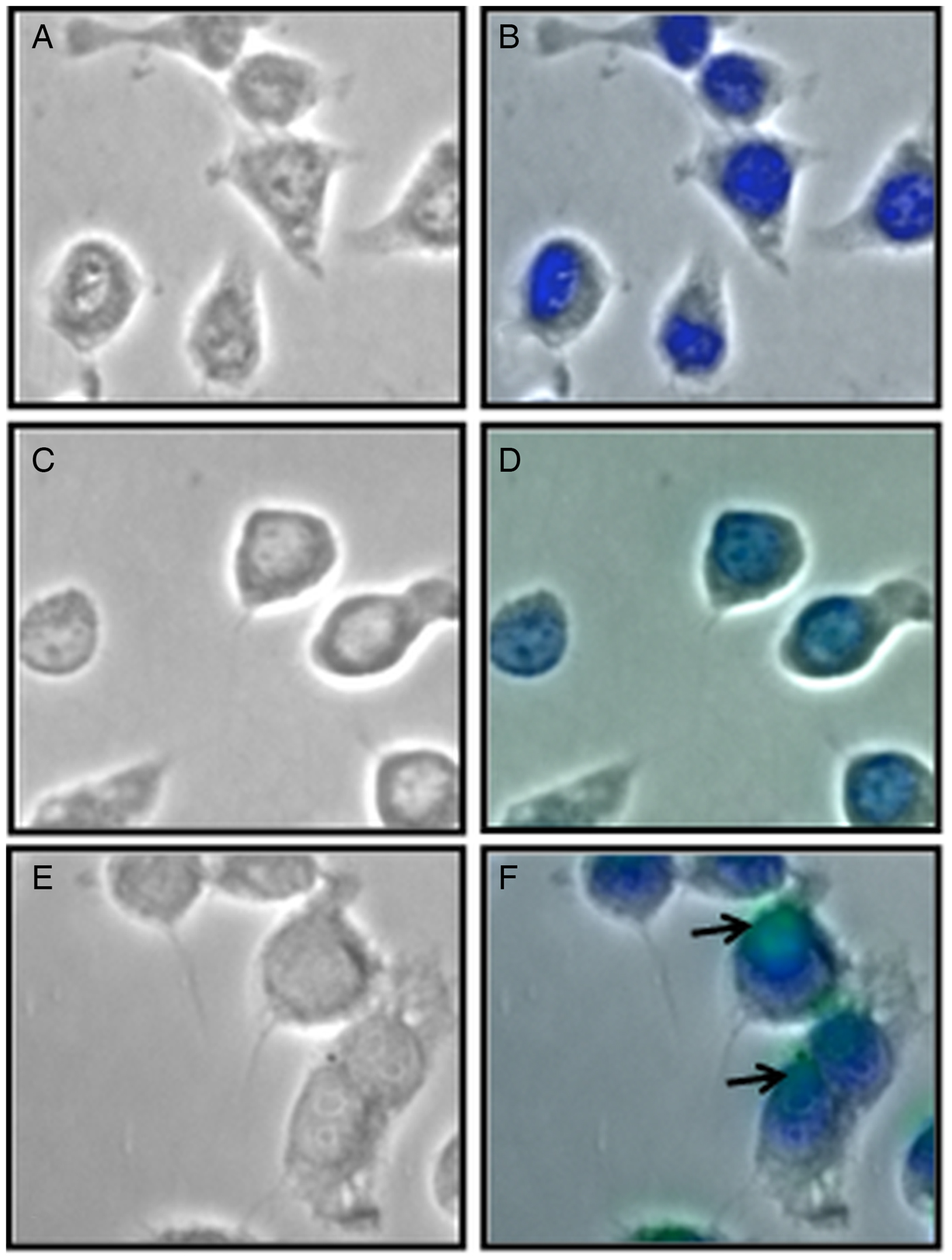
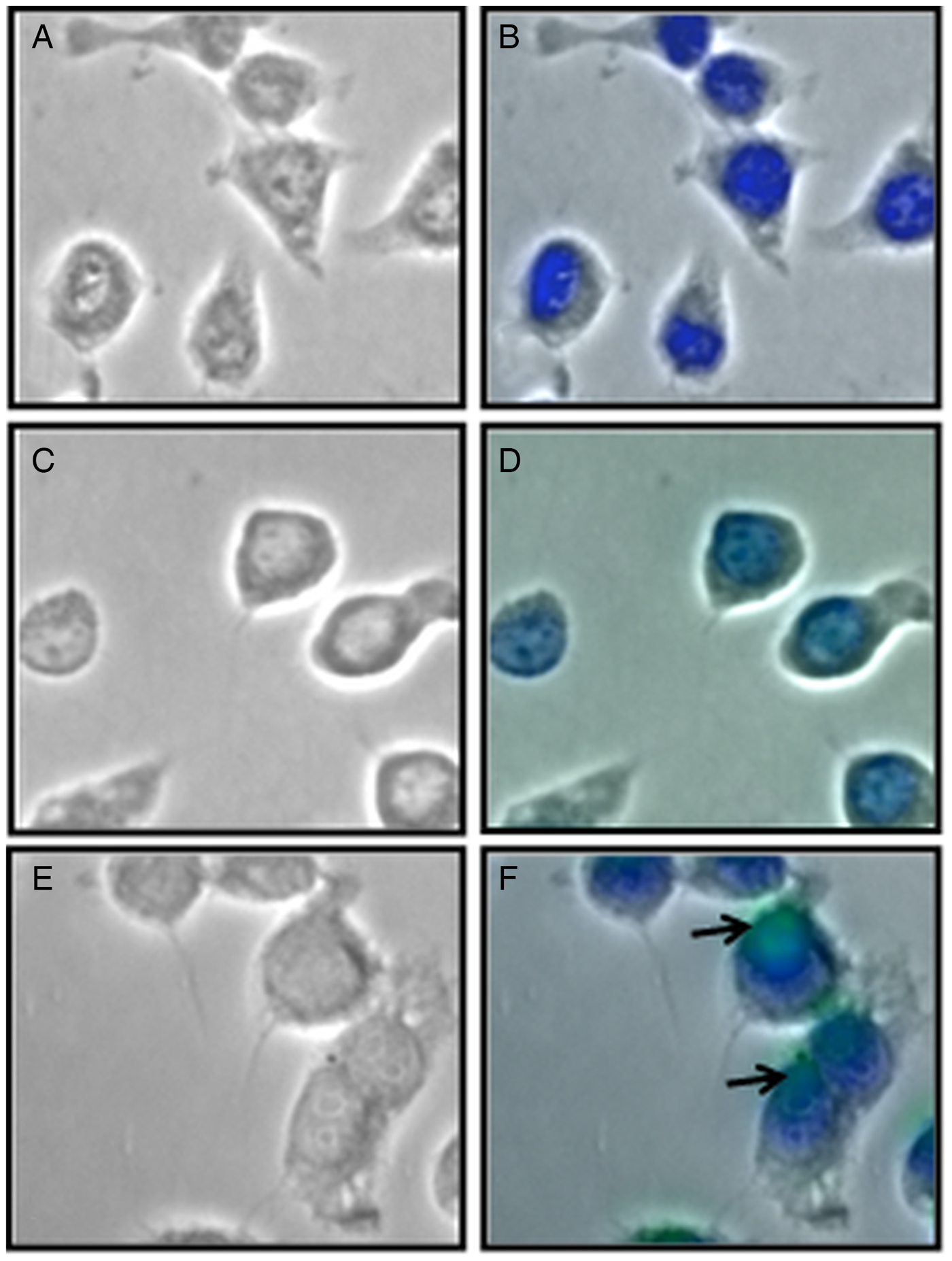
Immunolocalization of rNcDYNLL2 RAW264.7 macrophages co-cultured with rNcDYNLL2 was demonstrated (Fig. 4). Immunoreactive rNcDYNLL2 was not detected by pre-immune serum in cells co-cultured with rNcDYNLL2 (Fig. 4b), or in cells without being co-cultured with rNcDYNLL2 by specific anti-rNcDYNLL2 serum (Fig. 4d). Staining by specific anti-rNcDYNLL2 serum was evident in macrophages co-cultured with rNcDYNLL2 (Fig. 4f).
Immunolocalization of rNcDYNLL2 in RAW264.7 macrophages co-cultured with 10 µg mL−1 of rNcDYNLL2. (a, c and e) Phase-contrast images; (b, d, and f) phase-contrast and fluorescence staining image overlay. (a and b) RAW264.7 cells were cultured in the presence of rNcDYNLL2 and stained with pre-immune sera; (c and d) RAW264.7 cells were cultured in the absence of rNcDYNLL2 and stained with anti-rNcDYNLL2 serum; (e and f) cells were cultured in the presence of rNcDYNLL2 and stained with anti-rNcDYNLL2 serum. Blue colour represents nuclear staining with DAPI and green colour represents specific staining of rNcDYNLL2 with Alexa 488. Magnification, 400×.
The immunoregulatory function of rNcDYNLL2
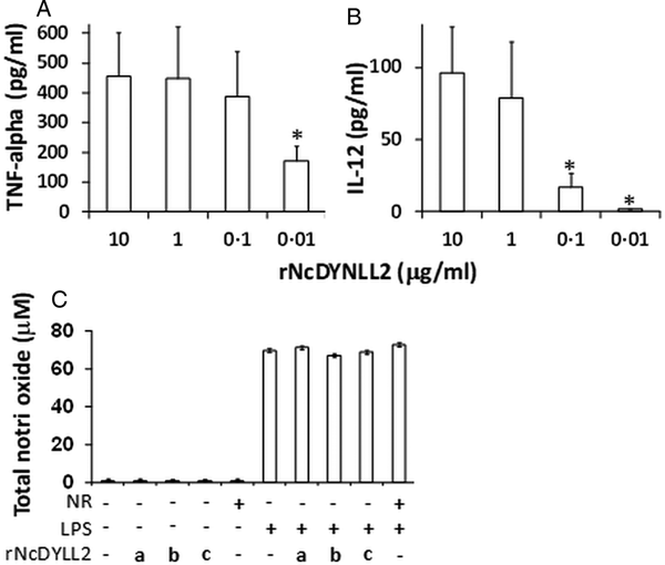
rNcDYNLL2 stimulated both TNF-α (Fig. 5A) and IL-12 (Fig. 5B) production by mouse DCs in a dose-dependent manner. TNF-α production was maximized when rNcDYNLL2 concentrations higher than 0.1 µg mL−1 were used (Fig. 5A); while at least 1 µg mL−1 of rNcDYNLL2 was required to stimulate maximal IL-12 production by DCs (Fig. 5B). Nitric oxide production by murine macrophages (RAW264.7) was not affected by rNcDYNLL2 either in the absence or presence of LPS, although LPS alone at 10 ng mL−1 induced high levels of nitric oxide (Fig. 5C). rNcDYNLL2 preparations had undetectable levels of endotoxin as confirmed using the LAL Assay kit (Lonza, Walkersville, MD, USA) (data not shown). Low levels of cytokine-inducing activity were detected in non-recombinant protein preparation (NR) and were subtracted from those induced by rNcDYNLL2. In average, DCs produced 99.1 ± 74.1 pg mL−1 of TNF-α and 32.8 ± 14.1 pg mL−1 of IL-12 in the presence of NR alone. NR had no effect on nitric oxide production (Fig. 5C).
Effect of rNcDYNLL2 on TNF-α (A) and IL-12 (B) production by murine bone marrow-derived dendritic cells (DCs) and nitric oxide (C) production by murine RAW264.7 macrophages. For the nitric oxide assay (C), cells were treated with three concentrations of rNcDYNLL2, 10 µg mL−1 (a), 1 µg mL−1 (b) and 0.1 µg mL−1 (c), with or without LPS (10 ng mL−1). Non-recombinant protein (NR) was used as a negative control in all assays. Data represent mean ± standard deviation of two independent experiments. *P < 0.05.

Discussion
In a previous microarray study determining differential gene expression by pathogenically different N. caninum isolates, an EST sequence (CF273809) coding for N. caninum DYNLL was identified to be downregulated in a chemically mutated, temperature-sensitive, low pathologic clone, Ncts-8 (Dreier et al., Reference Dreier, Stewarter, Kerlin, Ritter and Brake1999; Lindsay et al., Reference Lindsay, Lenz, Blagburn and Brake1999; Ritter et al., Reference Ritter, Kerlin, Sibert and Brake2002; Li and Tuo, Reference Li and Tuo2011). As N. caninum genomics and transcriptomics data become available recently, the original EST sequence (CF273809) used in the previous microarray experiment was used to BLASTN the GeneBank to confirm the sequence identity. The closest match to this gene, XM_003880113, which has been confirmed by several sources, was used in this study. The sequence alignment revealed that this NcDYNLL, encoded by XM_003880113 (Fig. 1A and C), shares the highest identity with the vertebrate DYNLL2, so it is named NcDYNLL2 according to the rules of cytoplasmic dynein nomenclature (Pfister et al., Reference Pfister, Fisher, Gibbons, Hays, Holzbaur, McIntosh, Porter, Schroer, Vaughan, Witman, King and Vallee2005). Three additional putative dynein light chains identified using the XM_003880113 sequence have low identity with NcDYNLL2. Based on the high identity (93%) shared between the mammalian DYNLL1 and 2, these three putative N. caninum dynein light chains are unlikely to be the candidates for NcDYNLL1. Thus, the relationship between NcDYNLL2 and these three putative proteins remains to be determined.
Naturally occurring non-pathogenic N. caninum isolates have not been reported. A NC1 clone, the Ncts-8, is a temperature-sensitive mutant exhibiting a non-pathogenic phenotype (Dreier et al., Reference Dreier, Stewarter, Kerlin, Ritter and Brake1999; Lindsay et al., Reference Lindsay, Lenz, Blagburn and Brake1999; Ritter et al., Reference Ritter, Kerlin, Sibert and Brake2002). Studies indicate that vaccination with live, but not killed, Ncts-8 provided significant protection against NC1 challenge infection, suggesting that loss of virulence did not compromise its immunogenicity (Dreier et al., Reference Dreier, Stewarter, Kerlin, Ritter and Brake1999; Lindsay et al., Reference Lindsay, Lenz, Blagburn and Brake1999). Our recent investigation on comparative gene expression between the wild-type NC1 and the avirulent mutant Ncts-8 indicate that NcDYNLL2 is among the 111 repressed transcripts in Ncts-8. It may be speculated that some of these downregulated genes are associated with virulence (Li and Tuo, Reference Li and Tuo2011). The present study further confirmed that NcDYNLL2 protein is also significantly downregulated in Ncts-8. Furthermore, NcDYNLL2 was shown to be secreted, although it does not have a conventional signal peptide, and the secretion of this peptide was regulated by intracellular calcium levels. The similar mechanism of secretory regulation shared by microneme proteins and NcDYNLL2 may suggest that NcDYNLL2 is associated with the microneme and may be discharged upon calcium mobilization and play a role during invasion (Carruthers and Tomley, Reference Carruthers and Tomley2008). However, the mechanisms by which this peptide, without a predicted signal sequence, is secreted remains to be determined in future studies. NcDYNLL2 present in high levels in the insoluble fraction of the parasite lysate may suggest that a proportion of this protein is in a bound form on the large dynein motor complex (Pfister et al., Reference Pfister, Shah, Hummerich, Russ, Cotton, Annuar, King and Fisher2006). The soluble portion of the NcDYNLL2 may participate in functions other than as a motor protein in the parasite. NcDYNLL2 was localized diffusely to the entire tachyzoites and no association with any particular parasitic cellular organelles such as micronemes was evident. Nonetheless, additional studies should be conducted to define NcDYNLL2 localization and potential mechanisms by which its release is regulated.
Previous reports indicate that DYNLL1 is a PIN for nNOS (PIN, or DYNLL1) (Jaffrey and Snyder, Reference Jaffrey and Snyder1996) and showed that DYNLL1 affects nNOS dimerization and inhibits nNOS activity. Additional research showed that DYNLL was a likely substrate for TRP14 in the process of modulating TNF-α signalling (Jeong et al., Reference Jeong, Chang, Boja, Fales and Rhee2004, Reference Jeong, Jung, Kim, Park and Rhee2009;Jung et al., Reference Jung, Kim, Min, Rhee and Jeong2008). However, there is no evidence that PIN or DYNLL1 may be associated with the modulation of IL-12 production. The use of exogenous DYNLL for the regulation of immune cell function has not been reported. The present study treated murine bone marrow-derived DCs with endotoxin-free rNcDYNLL2 and determined the TNF-α and IL-12 released. The secretion of both IL-12 and TNF-α by DCs was enhanced by rNcDYNLL2 in a dose-dependent fashion. We speculate that rNcDYNLL2 may be taken up by the DCs and function to interact with components of various pathways in cytokine production regulation. This hypothesis was in part confirmed in the present study that rNcDYNLL2 was detected in murine macrophages pre-cultured with rNcDYNLL2. It may be speculated that, when DCs are infected by N. caninum, NcDYNLL2 released by the tachyzoites within the cell will cross-regulate host functions, in favour of its survival of the parasite. However, the exact means by which NcDYNLL2 regulates host cell functions remains to be elucidated. It is noteworthy that the Leishmania DYNLL2 was recently confirmed in a vaccine trial as a protective vaccine candidate, suggesting that this molecule may play a role in pathogenesis of the parasite (Stober et al., Reference Stober, Lange, Roberts, Gilmartin, Francis, Almeida, Peacock, McCann and Blackwell2006). It is intriguing that rNcDYNLL2 had no effect on nitric oxide release by macrophages stimulated with or without LPS (Fig. 5C). However, these results may suggest that, unlike the mammalian PIN for nNOS or DYNLL1, NcDYNLL2 may not play a role in regulating iNOS.
The present study cloned and expressed NcDYNLL2 and characterized this molecule in cellular localization, differential production by different isolates of N. caninum and its ability to regulate cytokine production in immune cells. Further studies are warranted to understand the mechanisms by which NcDYNLL2 regulates host immune responses and demonstrate its potential as a protective vaccine candidate.
Acknowledgements
The authors wish to thank Mr Eli Miramontes and Ms Ruth Barfield for technical assistance.
Financial support
This research received no specific grant from any funding agency, commercial or not-for-profit sectors.
Conflict of interest
None.
Ethical standards
All animals were cared for by trained animal care takers and an attending veterinarian. Animal care and use was approved by the Beltsville Agricultural Research Center (BARC) Animal Care and Use Committee.
Author ORCIDs
Wenbin Tuo 0000-0002-3764-8981