I. INTRODUCTION
In the recent years, there was a growing interest for electronic devices operating in very harsh environments, at elevated temperatures, high power, high frequency, and high radiation fields.Reference Park, Willardson and Weber1, Reference Matsunami2 In that context, silicon carbide (SiC) is one of the most promising wide band gap materials for these devices due to its high breakdown electric field, high electron saturation velocity, high operating temperature, and high radiation hardness properties. Over the last few years, considerable effort has been concentrated on better understanding the performance of SiC-based radiation detectors to be used in high temperatures and high radiation conditions under which conventional semiconductors detectors cannot be adequately used.Reference Nava, Bertuccio, Cavallini and Vittone3
SiC detectors were used with good results for neutrons,Reference Ruddy, Dulloo, Seidel, Das, Ryu and Agarwal4 for x-rays,Reference Bertuccio, Casiraghi, Gatti, Maiocchi, Nava, Canali, Cetronio and Lanzieri5 and for charged particles emitted by alpha sources up to 5.48 MeV.Reference De Napoli, Raciti, Rapisarda and Sfienti6 In particular, the possibility to use x-ray SiC detectors operating at room or high temperature is of large interest as an alternative to conventional detectors that must operate at cryogenic temperatures.Reference Bertuccio, Casiraghi, Gatti, Maiocchi, Nava, Canali, Cetronio and Lanzieri5
Recently, SiC detectors have been used to monitor the ionizing radiation emitted from laser-generated plasma,Reference Torrisi, Foti, Giuffrida, Puglisi, Wolowski, Badziak, Parys, Rosinski, Margarone, Krasa, Velyhan and Ullschmied7, Reference Margarone, Krása, Giuffrida, Picciotto, Torrisi, Nowak, Musumeci, Velyhan, Prokůpek, Láska, Mocek, Ullschmied and Rus8 permitting prompt measurements of x-rays and ions emitted during the short life of pulsed plasma (typically picosecond or nanosecond scale).Reference Borghesi, Fuchs, Bulanov, Macckinnon, Patel and Roth9, Reference Margarone, Torrisi, Cavallaro, Milani, Verona-Rinati, Marinelli, Tuvè, Láska, Krása, Pfeifer, Krousky, Ullshmied, Ryć, Mangione and Mezzasalma10 The use of SiC detectors in this case is very attractive as they are not sensitive to the visible, soft ultraviolet (UV), and infrared light. Optical photons are not able to produce electron–hole pairs because their energies are below the 3.2 eV of the 4H–SiC gap energy. Therefore, SiC detectors can operate under high intensity visible light to which SiC is transparent.
The main advantages of SiC-based detectors are the radiation hardness,Reference Nava, Bertuccio, Cavallini and Vittone3 the possibility to operate at high voltage, the fast collection time, room temperature operation with low dark current,Reference Moscatelli11 and high detection efficiency. The photons, electrons, and ions absorbed in the sensitive volume of the detector generate e–h pairs, losing 7.8 eV for a pair production,Reference Lo Giudice, Fizzoti, Manfredotti, Vittone and Nava12 which results in voltage signal at the device electrodes that is proportional to the deposited radiation energy. The lack of sensitivity to the visible light allows the use of the SiC detector without any shielding filters to detect x-ray, electron, and ion beams simultaneously. Moreover, by using a time-of-flight (TOF) configuration, these detectors permit the measurements of the velocity of the particles emitted from the plasma source.
In the present work, single-crystal 4H–SiC Schottky diode detectors are used to monitor proton/ion beams generated and accelerated by high power repetitive laser obtained at Prague Asterix Laser System (PALS) laboratory. The energies of the emitted ions are measured through TOF approach. The response of SiC detectors will be compared with those coming from an Ion Collector and a Thomson Parabola Spectrometer (TPS).
II. EXPERIMENTAL SETUP AND DETECTORS
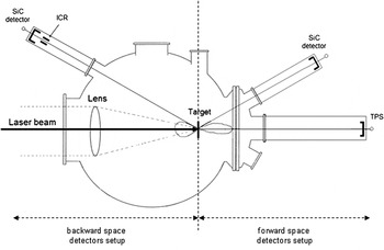
The experimental measurements are carried out with the Asterix Iodine laser of PALS Laboratory operating in single pulse at 1315 nm, 300 ps pulse duration, 70 μm laser spot diameter, 1016 W/cm2 pulse intensity, and using laser energies ranging from 480 to 600 J. Measurements were performed irradiating in normal incidence targets of Au, Al, and Si and monitoring the plasma radiation emission both in forward direction (rear face of the irradiated target) and in backward direction (laser-irradiated front face). SiC detectors were located at 30° angle in forward and backward direction. Ion collector ring (ICR) was placed at 30° in backward direction and a TPS was placed in forward direction along the normal to the target surface. The laser focal position (FP) on the target surface was controlled though a x-ray streak camera permitting to measure its distance from the target surface. Micrometric step motors permitted change of the FP to check the self-focusing conditions for distances of the order of 100 μm in front of the target surface.Reference Laska, Badziak, Gammino, Jungwirth, Kasperczuk, Kasa, Krousky, Kubes, Parys, Pfeifer, Pisarczyk, Rohlena, Rosinski, Ryc, Skala, Torrisi, Ullschmied, Velyhan and Wolowski13
Three main experiments were performed: (i) the first was conducted by irradiating a bulk hydrogen-enriched silicon (SiH) target at 0° incidence angle and at FP = 0; (ii) the second by irradiating a thin Au foil, whose thickness is 0.6 μm, at FP = −100 μm; and (iii) the third by irradiating, in the same conditions, a thin Al foil, 0.8-μm thick, at FP = −100 μm FP. A schematic view of the experimental setup is shown in Fig. 1.
Schematic of the experimental setup for the target irradiation in the vacuum chamber at PALS laboratory.

The SiC detectors were built on a 4H–SiC epitaxial layer grown with chemical vapor deposition technique in HCl atmosphere, which allows to obtain very thick layer (about 100 μm) with low dopant concentration (about 1014 cm-3) and with very low defect concentration.Reference Calcagno, Izzo, Litrico, Foti, La Via, Galvagno, Mauceri and Leone14, Reference La Via, Izzo, Mauceri, Pistone, Condorelli, Perdicaro, Abbondanza, Calcagno, Foti and Crippa15 The point defect concentration, as measured by Deep Level Transient Spectroscopy, was below 1011 cm−3.
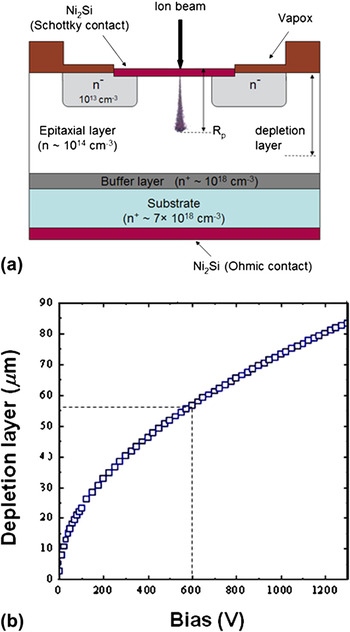
Schottky diodes were fabricated on 4H–SiC n-type epitaxial layers with a carrier concentration of 1.5 × 1014 cm−3, a thickness of 80 μm, and grown on a highly doped n-type (∼6 × 1018 cm−3) substrate. The backside ohmic contact was formed by a rapid thermal annealing of a 100-nm-thick Ni film at 950 °C. A Ni layer was then deposited in an ultra high vacuum (UHV) chamber on the front of the wafer and various diodes have been realized using standard photolithography. These diodes were then further annealed at 600 °C to optimize the Schottky barrier properties by the formation of a nickel silicide layer (Ni2Si), with a resulting thickness of about 200 nm. Fig. 2(a) shows a schematic section of the used Schottky diodes, in which the track and the range (R p) of a generic incident ion beam is also reported.
(a) Cross-sectional schematic of the ion SiC detector and of the ion trajectories (R p is the ion range) and (b) depletion layer thickness versus reverse bias polarization.

The as-prepared diodes were characterized by I–V to test the junction characteristic. The measured values of 1.65 eV for the barrier height and n = 1.05 for the ideality factor evidence the good rectifying proprieties of the junction. The reverse I–V characteristics are of particular interest because the detectors must operate in reverse bias with minimum current. The reverse I–V characteristic measured at 21 °C shows that the current density is low and it goes from 10−10 A/cm2 at V = 200 V up to 10−8 A/cm2 at 700 V.
The application of a reverse bias creates a depletion layer that lies entirely in the lightly doped SiC epitaxial layer and Fig. 2(b) shows the depletion thickness versus the inverse bias, which follows roughly the inverse bias square root. In our experiment, we use a reverse bias of 600 V, which produces a depletion layer of about 55 μm, thus allowing the measurement of the entire energy loss of ions whose range (R p) is less than 55 μm.
In such configuration, at 600 V inverse bias, the detector has high linearity with the radiation energy, high resolution [full width half maximum (FWHM) = 34 eV], and 100% charge collection efficiency, as demonstrated detecting alpha particles emitted from radioactive sources.Reference Moscatelli11 The active area of the SiC detector was 2 × 2 mm2 and it was glued onto high temperature resistance holder. TOF technique was used in our measurements, and the spectra are recorded with a fast oscilloscope operating to 20 GS/s recording the laser shot as a start signal and the ion peaks as stop signals.
The SiC detector distance from the target was 150 cm for the backward condition and 102 cm for forward ones, and a full TOF spectrum was recorded to correlate temporally the x-ray emission and the ion emission, as it will be shown in the following section.
To analyze the radiation emitted from laser-generated plasma, we have also used ion collectors and a TPS. The ion collector is a simple Faraday cup of ring geometry (ICR), and in one case, it was shielded with a thin Al foil [ion collector ring shielded (ICRS)].
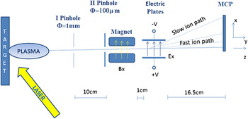
The TPS located in forward direction along the normal to the target surface is preceded by two pinholes, the first 1 mm in diameter and the second 100 μm in diameter, to finely collimate the detected ions from the plasma and to exactly determine the incidence direction. The spectrometer uses a 0.05 T horizontal magnetic deflection and +2 kV (from the top) voltage for the vertical electric deflection. The distance between the electric deflectors and the microchannel plate (MCP) plane detector of the parabola ions is D = 16.5 cm, as in similar apparatuses reported in literature.Reference Harres, Schollmeier and Brambrik16 A schematic of the TPS used is reported in Fig. 3.
Schematic of the used TPS.

Inside the TPS, the ions are deflected by the Lorentz force,  $\vec F = \;q\;{\rm{(}}\vec E + \vec v \times \vec B{\rm{)}}$, with a magnetic (
 $\vec B$) and electric (
 $\vec E$) fields, which are parallel to each other and perpendicular to the ion incidence direction.
 $\vec v$ is the velocity of the ions, and q = Z × e is the ion charge. Assuming nonrelativistic velocities and a field length l << D, the deflections orthogonal to the ion incidence direction, z, are given by the following equations:
 $$x = {{qEID} \over {2{\rm{\varepsilon }}_{{\rm{kin}}} }};\,y = {{qBID} \over {\sqrt {2m{\rm{\varepsilon }}_{{\rm{kin}}} } }};\,y^2 = {{qB^2 IDx} \over {mE}},$$
where x and y are the deflections due to the electric and magnetic fields, respectively, B and E are the field amplitudes, and q, m, and εkin are the charges, the masses, and the kinetic energies of the ions, respectively. The parabolic equation is a combination of the two deflection equations and shows that TPS provides a separation of all ion species and charge states according to q/m ratio. Every single parabola on the detector belongs to a different ion charge-to-mass state ratio. The deflection along the parabola contains the information about the ion energy.
III. RESULTS AND DISCUSSION
The TOF spectra acquired with ion collectors are usually composed by a relatively fast photopeak due to photons coming from the plasma generating photoelectron on the collector metallic surface, sometimes a negative signal due to the detection of plasma electrons, and a relatively long and structured peak due to the detection of plasma ions, from light to heavy ones depending on their velocity and charge state distributions.
Due to self-generated electric fields in the laser plasma, sometimes, the velocity (i.e., TOF) of protons is close to the one of highly charged heavier ions, thus, if the detector is not fast enough, it becomes difficult to distinguish both contributions in the TOF spectra.
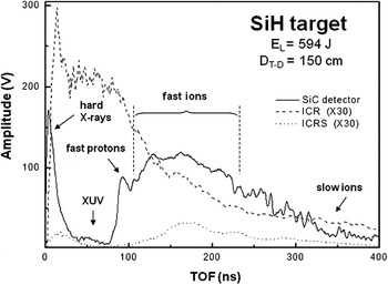
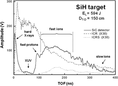
The last statement is demonstrated by the TOF spectrum shown in Fig. 4, which was obtained by an ICR detector when a hydrogen-enriched silicon target was irradiated at a laser energy of 594 J. The ICR spectrum (dashed line) shows a broad peak between 0 and 250 ns related to x-rays, UV radiation, and fast ions, followed by a broad structure with some peaks between 250 and 400 ns related to slow ions. The ICR resolution is very poor and it does not permit a good separation of the different ion contributions. To filter out the XUV component, a simple method based on the application of thin absorbers has been developed: a thin (2 μm) Al foil has been placed in front of the ion collector to absorb the heavy ions and to reduce the photon background. The crucial parameters of the foils, i.e., material choice and thickness, have been selected through analysis aimed at optimization of the radiation transmission and the ion stopping range by a filter transmission database and the SRIM Monte Carlo code, respectively.Reference Ziegler, Biersack and Ziegler17
TOF spectra detected by an unshielded ICR, the shielded one (ICRS), and the SiC detector. The spectrum is obtained using a SiH target and a laser energy of 594 J. D T–D is the target–detector distance.

The TOF spectrum of the shielded detector (ICRS), reported in the same Fig. 4, shows a reduction of the XUV peak.
An alternative method to evidence the plasma fast protons/ions with respect to the XUV photopeak in the TOF spectrum is the use of SiC semiconductor detector. The current response of such devices is proportional to the incident radiation energy released into the detector active layer,Reference Margarone, Krása, Giuffrida, Picciotto, Torrisi, Nowak, Musumeci, Velyhan, Prokůpek, Láska, Mocek, Ullschmied and Rus8 thus the photopeak (usually consisting of the response to UV/soft x-rays) is strongly lowered in comparison to the fast (high energy) ion peak. Moreover, the wide band gap (3.2 eV) allows a high attenuation of the plasma-emitted UV and soft x-ray radiation (XUV). Then, the SiC main advantage consists in the possibility to detect simultaneously the hard x-rays and fast ions emitted from the laser-generated plasma. This statement is evidenced by the spectrum obtained by SiC detector reported in the same Fig. 4. The spectrum points out the presence of three main structures:
(i) A peak in the time region 0–20 ns related to hard x-ray radiation (few keV up to few tens of keV) emitted during and after the laser–target interaction until the plasma is too “cold” due to its expansion in vacuum. Such a “harder” radiation component is also detectable by the filtered ion collector even if its sensitivity is low in such a photon energy range.
(ii) A broad peak around 50 ns related to XUV due to electron recombination mechanisms occurring during the plasma expansion into vacuum or to plasma electrons.
(iii) A broad structure with different peaks from 80 to 400 ns, due to the energetic ions contained in the plasma. The first peak of this structure is ascribable to protons. The SiC detector shows a high sensitivity to the fast protons since they have the highest velocity and the detector signal is not proportional to the ion charge state. The proton energy has been calculated to be 1.2 MeV. The proton peak is followed by the peaks related to Sin+ ions with different charge states. The calculated maximum energy of the Sin+ ions is 13 MeV, and considering that the Si atoms can be full ionized (n = 14), the corresponding energy for charge state should be about 1 MeV.
SiC detectors appear very useful to the real-time monitoring of the laser-generated plasma due to their high response velocity, high sensitivity, and well proportionality to the ion energy. The main advantage with respect to ICR is the proportionality to the ion energy instead of the ion charge state.
In particular, the expressions for the current density signals, J, of the ICR and SiC detectors, respectively, are:
 $$J_{{\rm{ICR}}} = dq/dt = ez_{\rm{i}} n_{\rm{i}} v_{\rm{i}} ,$$
 $$J_{{\rm{SiC}}} = e(n_{\rm{i}} E_{\rm{i}} /{\rm{\varepsilon }}){\rm{\mu }}_{{\rm{eff}}} (U_{\rm{d}} /d),$$
where e is the electron charge, z i is the ion charge state, n i is the ion density, v i is the ion velocity, E i is the ion energy, ε is the energy necessary for electron–hole pair creation, μeff is the quantum efficiency that takes into account the energy loss in the detector, U d is the bias applied to the semiconductor detector, and d is the thickness of the semiconductor active layer, as discussed in our previous work.Reference Margarone, Krása, Giuffrida, Picciotto, Torrisi, Nowak, Musumeci, Velyhan, Prokůpek, Láska, Mocek, Ullschmied and Rus8
To have a more complete characterization of the laser-generated plasma, the SiC detector was tested in a different experimental setup by placing it in the forward space.
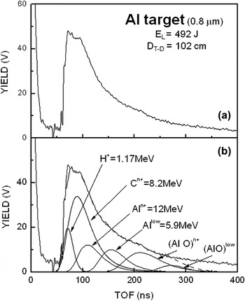
Fig. 5(a) reported a typical SiC–TOF spectrum of ions emitted at 30° forward direction, relative to 492 J pulse energy irradiating a thin Al foil (0.8 μm) at −100 μm FP, with a target–SiC detector distance of 102 cm. The spectrum shows an intense photopeak at low TOF (≈20 ns), followed by a large structured peak extending from 60 to about 300 ns. This structure is due to the fast protons and to the different charge states of the Al ions. The proton kinetic energy is evaluated to be 1.17 MeV, and the maximum energy of the Al ions, due to the full ionized species, is about 12 MeV. Carbon or oxygen ion contaminations might also be present at a maximum energy of about 0.7 MeV/nucleon. The protons, carbon/oxygen, and aluminum ion contributions are reported in the deconvoluted spectra of Fig. 5(b). The deconvolution was performed by using the Coulomb–Boltzmann shifted function distribution reported in literature.Reference Torrisi, Gammino, Andò and Laska18
TOF spectrum achieved by a SiC detector (a) by irradiating a thin Al target with a laser energy of 492 J. D T–D is the target–detector distance. The same spectrum after deconvolution with the use of Coulomb–Boltzmann Shifted function (b).

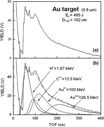
Figure 6(a) shows a typical SiC–TOF spectrum of ions emitted at 30° forward direction, relative to 485 J pulse energy irradiating a thin Au foil (0.6 μm) irradiated at −100 μm FP, with a target–SiC detector distance of 102 cm. The spectrum shows an intense photopeak at low TOF (≈20 ns), followed by a large structured peak from 40 to 300 ns. This last structure is due to the fast protons and to the different charge states of the Au ions. The proton kinetic energy is about 1.87 MeV and the maximum energy of the Au ions, due to the higher ionized specie, is about 100 MeV. Carbon ion contaminations are also present at a maximum energy of about 13.5 MeV. The protons, carbon, and gold ion contributions are reported in the deconvoluted spectra of Fig. 6(b).
TOF spectrum achieved by a SiC detector (a) by irradiating a thin Au target with a laser energy of 485 J. D T–D is the target–detector distance. The same spectrum after deconvolution with the use of Coulomb–Boltzmann Shifted function (b).

These results evidence, as expected, that the Au plasma is hotter than the Al plasma, due to the higher electron density.
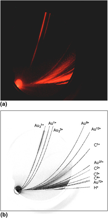
The plasma was also characterized by the TPS, which allows to determine the charge state distributions of the Al and Au ions generated on the forward emitted plasma. Figure 7(a) shows the TPS spectrum obtained by the laser irradiation of the Au thin target during the same SiC detection experiment. The spectrum shows a circular zone due to plasma photons and undeflected neutral particles arriving onto the MCP and different parabolas outgoing from this circular zone.
Original image provided from TPS by irradiating a thin Au target (a) and simulated spectra for the same shot obtained by executing OPERA-3D/TOSCA code (b).

The experimental TPS parabolas were compared with the simulation data obtained by OPERA-3D/TOSCA code.19 The fixed parameters are the geometrical TPS dimensions, the magnetic field value, and the electric voltage applied to the electric deflector plates. Fig. 7(b) shows the simulated parabola and the experimental data. The lower parabola is related to proton deflections and the others to Au ions from high charge state to the single charge state. The more vertically deflected parabola is due to the lower kinetic energy (on the top) and the less vertically deflected is due to the higher kinetic energy (on the bottom). The parabola lying nearest to the circular zone is due to the detection of highest energy ions. The distance between the protons parabola and the center circle is compatible with a proton maximum energy of 1.9 MeV, in agreement with SiC detector TOF measurements.
The comparison between SiC detector and TPS evidences the high resolution of TPS to low energy ions, while at high energy, the parabolas almost overlap. Then, different detectors should be used to have a complete energetic characterization of the laser-accelerated ions and of the plasma proprieties in terms of maximum charge state, temperature, ion current, and kinetic energy.
IV. CONCLUSIONS
In the present work, we have investigated the response of SiC detectors to ions emitted from a laser-generated plasma. The large SiC band gap permits the cutting of the visible and UV radiation allowing the monitoring of high-energy ions in a low background level. The comparison of TOF spectra detected by SiC and Ion Collectors demonstrates the high resolution and sensitivity of SiC detectors for high-energy ions and the high fast response to energetic particle detection. We have revealed protons up to 1.9 MeV and Au ions with energy up to 100 MeV. Moreover, complementary techniques such as TPS permit a high resolution to low energy ions, evidencing that the use of different detectors gives a more complete characterization of the laser-generated plasma.
ACKNOWLEDGMENTS
This work was performed at the PALS Research Infrastructure, benefiting from the Laserlab Europe project No. pals001653, coordinated by Prof. L. Torrisi. The authors thank E. Krousky, M. Pfeifer, and J. Skala for the technical and scientific support provided during the experiments. Thanks also to G. Saccà and M. D’Andrea of the Istituto Nazionale di Fisica Nucleare (Sezione di Catania) for the technical assistance for the SiC detector electronics equipment.