Introduction
Laryngeal cancer is one of the most frequent malignant tumours in the head and neck region and is the second most common tumour of the respiratory tract, claiming more than 1500 lives in 2006 in Germany.1 The incidence of laryngeal cancer in women has progressively risen since 1980,Reference Robin, Reid, Powell and McConkey2, Reference Pantel and Guntinas-Lichius3 while the incidence in men has decreased by a third over the same period.Reference Pantel and Guntinas-Lichius3
Laryngeal cancer originates from squamous cells, which form the majority of the epithelium of the larynx. The standard method of detection of laryngeal cancer and its precursors is laryngoscopy with removal of tissue for histopathological analysis. The biopsy needs to be sufficiently deep to include the basement membrane so as to make a proper diagnosis. Laryngeal cancer and pre-stages of the disease have a great influence on quality of life. The disease itself, the sampling size for diagnosis, and the treatment modality can cause persistent hoarseness.Reference Smith, Johnson, Cognetti, Landsittel, Gooding and Cano4 Therefore, early diagnosis is needed with a detection method that allows a precise biopsy.
Several imaging methods have been proposed to increase the precision of the biopsy. Notable modalities include rigid endoscopy, contact endoscopy, autofluorescence endoscopy, aminolevulinic acid induced fluorescence, Raman spectroscopy, endosonography and optical coherence tomography.Reference Arens, Vorwerk, Just, Betz and Kraft5 Contact endoscopy, introduced in 1995 by Andrea et al.,Reference Andrea, Dias and Santos6 was the first in vivo technique that provided cellular information on the laryngeal epithelium. Using a rigid endoscope in contact mode, methylene blue stained laryngeal mucosa can be visualised under 60- or 150-fold magnification. However, this method only detects cellular abnormalities in the most superficial cell layers. Contact endoscopy cannot monitor the basal cell layer or the underlying basement membrane.
Confocal microscopy is a rapidly evolving imaging modality that provides high-resolution, en-face images of the tissue microanatomy. The availability of confocal microscopy motivated the development of a rigid endoscope that could provide cellular and subcellular information on laryngeal lesions. This technique enabled automatic, in-depth scans (about 80 µm per scan). It has been more than six years since we reported on ex vivo confocal microscopy of the human larynx.Reference Just, Stave, Boltze, Wree, Kramp and Guthoff7, Reference Just, Stave, Kreutzer, Guthoff and Pau8 During that time, the development of a suitable rigid endoscope has been successfully completed. The long-term aim of this project is to apply confocal endomicroscopy as a complementary imaging technique to optical coherence tomography, in order to improve the accuracy of sampling during microlaryngoscopy. The latter method provides optical cross-sections from the tissue. Optical coherence tomography enables the integrity of the basal membrane to be assessed. The severity of dysplasia is defined by measuring the thickness of the laryngeal epithelium. Thicker epithelium is more likely to be dysplastic than epithelium of normal thickness. However, there is an overlap between the average thickness of normal and dysplastic squamous epithelium,Reference Kraft, Lüerssen, Lubatschowski, Woenckhaus, Schoberlein and Glanz9 and so a proper diagnosis cannot be made with certainty. Therefore, there is a need to combine optical coherence tomography with a complementary imaging modality.
Our approach has been to combine optical coherence tomography with confocal endomicroscopy, which provides en-face images of the tissue.10 Recently, we published results from an animal modelReference Farahati, Stachs, Prall, Stave, Guthoff and Pau11 and a clinicopathological pilot study in humans,12 using a rigid confocal endoscope. With regard to our human study, confocal endomicroscopy test outcomes for differentiation between benign laryngeal lesions and dysplasia or carcinoma in situ indicated a sensitivity of 86 per cent, specificity of 87 per cent, positive predictive value of 92 per cent and negative predictive value of 78 per cent, and a predicted accuracy of 83 per cent. However, because of the limited penetration of the laser light primarily in hyperkeratotic lesions, only small suspicious lesions were included.
In this paper, we report our method of applying confocal endoscopy during microlaryngoscopy for the diagnosis of suspicious laryngeal lesions. In contrast to a previously published ex vivo study on laser scanning microscopy of the larynx,Reference Just, Stave, Boltze, Wree, Kramp and Guthoff7 a rigid endoscope was connected to the scanner head of a confocal microscope. Three-dimensional (3D) imaging of small parts of the laryngeal epithelium is possible using the volume mode. The depth range was 80 µm per volume scan and the acquisition time for a single volume scan was 6 seconds.
Materials and methods
This was a conceptual study and did not include pilot study data. The images of patients presented here indicate representative cases which were discussed with a pathologist.
System, imaging and image analysis
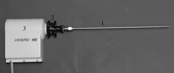
The custom-made endoscope consisted of an endoscope shaft with an integrated rod lens system (Karl Storz, Tuttlingen, Germany). The length and diameter were 23 cm and 5 mm, respectively (Figure 1). A detailed description of the endoscope has been presented elsewhere.12 At its end, the endoscope had a perforation plate with a central hole. This avoided tissue compression in the field of vision. A connector was developed to join the confocal microscope to the endoscope (Karl Storz) (Figure 1). Using the adapter, the starting plane could be set manually. To achieve optical sectioning of the tissue, the scanning mechanism of the scanner of the Heidelberg Retina Tomograph II (Heidelberg Engineering, Heidelberg, Germany) was connected to a rigid endoscope. Illumination was delivered by a 670-nm diode laser. For endoscopic use, the λ/4 plate of the Heidelberg Retina Tomograph II was removed to avoid strong reflections, and the laser power of the tomograph system was increased. The resultant devices (i.e. the Heidelberg Retina Tomograph II without λ/4 plate, the adapter and the endoscope) supplied images of 400 × 400 µm. An average penetration depth of 100–300 µm could be achieved. The lateral and axial resolutions were approximately 1–2 and 2 µm, respectively. The endoscope was autoclavable.
Photograph of the rigid endoscope and the connector for confocal imaging of the larynx. The endoscope (1) is coupled by the connector (2) to the scanner (3) of the confocal microscope (Heidelberg Retina Tomograph II; Heidelberg Engineering, Heidelberg, Germany).

Intra-operative application
During microlaryngoscopy, the larynx was examined with a Kleinsasser operating laryngoscope held by suspension (laryngoscope holder or chest support) (Karl Storz). Using the operating microscope and rigid endoscopes (0°, 30° and 70°), laryngeal lesions were identified and photo-documented. For confocal endoscopy, the rigid endoscope was inserted through the laryngoscope. The endoscope was used in contact mode. The end of the endoscope was gently placed on the lesion under microscopic or endoscopic view. A contact gel was used (Vidisic, Dr Mann Pharma, Berlin, Germany) to avoid strong reflections. In order to allow comparison of confocal endomicroscopic images and volume scans with histopathological assessment at a later stage, a biopsy was taken, as precisely as possible, from the same laryngeal lesion that had been observed by confocal endomicroscopy.
Three different modes were used for data sampling. In section mode, single images were stored. The starting plane was set manually. In sequence acquisition mode, up to 100 images were stored. The images were obtained by moving the endoscope over the lesion in the same plane. In volume mode or 3D imaging mode, 40 images were stored per volume scan. The distance between two subsequent image planes was approximately 2 µm. Thus, the depth range was 80 µm per volume scan. The acquisition time for a single volume scan was 6 seconds.
Image analyses
The confocal endomicroscopic images and volumes obtained during microlaryngoscopy were recorded and stored in a data bank, together with representative histopathological images. An image gallery of increasing severity of lesions, for synoptic viewing, was already available from a pilot study.12
Dysplasia was graded according to the World Health Organization guidelines.Reference Barnes, Everson, Reichart and Sidransky13 Analysis of the confocal endoscopic images was performed blinded and without histopathological knowledge. Each case was graded on a two-tier scale as either a negative finding (i.e. a benign lesion) or a positive finding (i.e. all dysplasia grades and carcinoma in situ). The following criteria were used to classify epithelial lesions: nuclear size, nuclear density, number of nuclei, nuclear/cytoplasmic ratio, and morphology of cells in a cell layer. To improve sensitivity and specificity, dysplasia or carcinoma in situ was diagnosed when dysplastic cells were identified.
Results and analysis
Generally, the surgeon needed no assistance for confocal endomicroscopy imaging. At least 4 to 10 scans per true vocal fold were recommended in sequence mode, with additional volume scans of regions of interest. The rapid acquisition of data (about 6 seconds per scan) exceeded the time for microlaryngoscopy by less than 5 minutes. At the end of surgery, saving digital data (i.e. endoscopic images and confocal endomicroscopy data) usually required no more than a few minutes. The confocal endomicroscopy imaging device imposed no restrictions on the use of the operating microscope.
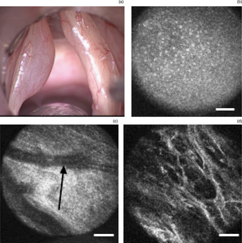
During microlaryngoscopy, the superficial layers of the epithelium needed to be identified in healthy epithelium and in benign lesions of the larynx (Figure 2). A representative image of a case of Reinke's oedema is shown in Figure 2. In this patient, confocal endomicroscopy revealed an epithelium of normal appearance, with regular cell architecture and no dysplastic cells. Manual adjustment of the focal plane enabled the surgeon to monitor the blood vessels within the lamina propria and to assess their blood flow. The depth scan showed loose fibres in the superficial layer of the lamina propria, also known as Reinke's space. The dark, fibreless areas within the lamina propria were consistent with oedema.
(a) Photomicrograph showing Reinke's oedema of the left human true vocal fold. Confocal micrographs showing: (b) regular cellular architecture at a depth of 36 µm in the left true vocal fold; (c) subepithelial vessels (arrow) at a depth of 80 µm; and (d) collagen fibres at a depth of 95 µm. Scale bar = 50 µm

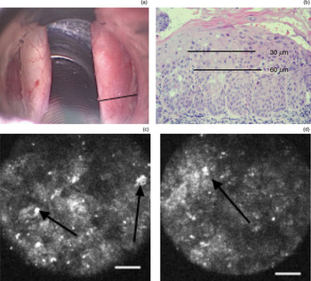
Carcinoma in situ is depicted in Figure 3(a) and 3(b). In such cases, confocal endomicroscopy demonstrated an abnormal cellular architecture and irregularly shaped nuclei (Figures 3c and 3d). These cellular abnormalities were detectable in all scan layers (two complete depth scans).
(a) Magnified endoscopic image of carcinoma in situ in the posterior part of the right true vocal fold. (b) Photomicrograph of a transverse histological section from a 120 µm thick probe, showing a thin layer of keratin on the surface. (c) & (d) Confocal endoscopic images of laryngeal epithelium, showing epithelium of irregular appearance with enlarged, irregular cell nuclei (arrows) at approximately (c) 30 µm and (d) 65 µm. Scale bar = 50 µm

Discussion
To date, the applicability of confocal endomicroscopy for imaging of experimentally induced precancerous lesions has been tested in a mouse tongue model.Reference Farahati, Stachs, Prall, Stave, Guthoff and Pau11 This initial, experimental study indicated that the penetration depth of laser light is limited in lesions with strong keratosis. Therefore, in the current, human study we used confocal endomicroscopy to study only small, suspicious, keratotic laryngeal lesions. The test outcomes for confocal endomicroscopy were comparable to those for optical coherence tomography.Reference Just, Lankenau, Prall, Hüttmann, Pau and Sommer14 It has been demonstrated that confocal endomicroscopy can describe the microanatomy of the true vocal fold.12 The border between the epithelium (basal cell layer) and the lamina propria (collagen fibres) can be easily identified. However, confocal endomicroscopy fails to visualise the basement membrane, with its two different lamina, even in healthy laryngeal tissue. Despite this, confocal endomicroscopy can still be used to monitor cellular abnormalities.
Cellular abnormalities within the epithelium are consistent with a minimally or frankly invasive carcinoma; relevant findings have already been described, in 2006.Reference Just, Stave, Boltze, Wree, Kramp and Guthoff7 In general, confocal microscopy fails to differentiate between severe dysplasia and minimally or frankly invasive carcinoma. However, we wish to emphasise the great potential of confocal endomicroscopy to detect severe cellular abnormalities during microlaryngoscopy. Carcinoma in situReference Sigston, De Mones, Babin, Hans, Hartl and Clement15 and precancerous lesions can be multifocal. Using confocal endomicroscopy, areas suspicious for epithelial dysplasia or carcinoma in situ can be detected with some confidence, and biopsies can be taken with precision. In cases with exophytic tumours or severe hyperkeratosis, confocal endomicroscopy needs to be combined with other imaging technologies that penetrate more deeply (e.g. optical coherence tomography (2.5 mm) or high frequency ultrasound (25 mm)). Further technical developments include miniaturisation of the scanner of the confocal microscope, automatic analysis of the nuclear–cytoplasmic relationship, online 3D reconstruction of the scanned volume, and increase in the penetration depth for imaging of the lamina propria.
• Intra-operative imaging of laryngeal epithelium microanatomy is not currently possible
• This study tested a combined rigid endoscope, adapter and confocal microscope, designed to achieve this aim
• The confocal laser scanning system detected pre-malignant laryngeal epithelial changes
Interpretation of the confocal endomicroscopy images depends markedly on the surgeon's experience, and scans should be compared with histopathological results in cooperation with a pathologist. The accuracy of interpretation of confocal endomicroscopy data is subject to a ‘learning curve’, similar to that for conventional histopathology, ultrasonography or other imaging modalities. In order to visualise disease-specific morphological changes of the laryngeal epithelium, we intend in future to perform fluorescent labelling studies of individual cells. Similar experiments have been performed in patients undergoing colonoscopy;Reference Hsiung, Hardy and Friedland16 the fluorescein-conjugated peptide bound more strongly to dysplastic colon cells than healthy cells.
Our future research will focus on the determination of inter-rater reliability, using kappa statistics to determine the consistency between two observers. The method presented here will be explored in a prospective clinicopathological study in patients with precancerous and cancerous lesions of the larynx (i.e. dysplasia grade I–III, carcinoma in situ and minimally invasive carcinoma) to assess the diagnostic sensitivity, specificity and accuracy.
Conclusion
Confocal endomicroscopy using a rigid endoscope in contact mode is an imaging technology that provides real-time, en-face images of the laryngeal tissue. The operating microscope can be used for visual control of the scanning site. Technical problems hindering clinical use have largely been resolved. Our imaging system enables the surgeon to scan through the laryngeal tissue automatically. The system is easy to handle intra-operatively, and no assistance is required.