I. INTRODUCTION
Perovskite manganites have attracted scientific attention since the observation of colossal magneto resistance (CMR) in La1−x A x MnO3 (A, alkaline earth element) in 1990 (review by Dagotto et al., Reference Dagotto, Hotta and Moreo2001). Cubic perovskites belong to Pm3 m space group with one formula unit per unit cell (Z = 1) where A and B site cations have 12- and sixfold oxygen coordinations (Howard and Stokes, Reference Howard and Stokes1998). Crystal structure deviates from the cubic cell depending on the ionic size of cations. Manganates of RMnO3 exhibit orthorhombic structure for larger rare earth elements (R = La–Dy) and hexagonal structure for smaller rare earth atoms (R = Ho–Lu). When La is replaced by a rare earth ion whose ionic radius is smaller than lanthanum, the coordination of A site reduces to 8 and 9 depending on the value of the ionic radius. REMnO3 belong to multi-functional materials since they exhibit a wide variety of physical properties such as CMR, multi-ferroic behavior, superconductivity, etc. Among the rare earth series, DyMnO3 (DMO) exhibits a complex magnetic structure. Low-temperature ground state of LaMnO3 is antiferromagnetic (Tokura, Reference Tokura2006), while that of analogous DMO is complex (Goto et al., Reference Goto, Kimura, Lawes, Ramirez and Tokura2004). There exist two polymorphic structures for DMO depending on the synthesis conditions. High-temperature synthesis (close to its melting point) stabilizes hexagonal structure, whereas synthesis at low temperature results in an orthorhombic phase (Harikrishnan et al., Reference Harikrishnan, Naveen Kumar, Bhat, Elizabeth, Robler, Dorr, Robler and Wirth2008). To induce a stable ferromagnetism into DMO similar to that of La1−x Sr x MnO3, we have chosen Sr and Fe substitutions at Dy and Mn sites in Dy0.55Sr0.45Mn1−x Fe x O3. So far, there is no X-ray diffraction (XRD) data of the Dy–Sr–Mn–Fe–O system in the database of ICDD-2014, so we present here our experimental powder XRD data of Dy0.55Sr0.45MnO3 and Dy0.55Sr0.45Mn0.8Fe0.2O3 to be included in the database.
II. EXPERIMENTAL
A. Synthesis
Polycrystalline compounds of Dy0.55Sr0.45Mn1−x Fe x O3 (x = 0.0 and 0.2) were prepared by high-temperature solid-state reaction method, from high-purity oxides (99.99%) of dysprosium oxide (Dy2O3), strontium carbonate (SrCO3), manganese oxide (MnO2), and iron oxide (Fe2O3). Starting oxides taken in appropriate ratio to make 2 gm of the final compounds were ground with the help of an agate mortar and pestle for several hours in order to get a homogeneous mixture. Mixed powders were calcined in air at 1573 K for 10 h with intermediate grinding until the formation of single-phase compounds.
B. XRD data acquisition
Powder XRD patterns of the compounds were collected at room temperature using STOE (Germany) X-ray powder diffractometer operated in Bragg–Brentano parafocusing geometry with fixed slits. The diffraction data were collected at room temperature with a scintillation detector, NaI (Tl) using CuKα radiation (1.5406 Å) operated at 40 kV and 30 mA. The diffractometer is equipped with a secondary monochromator, which is flat pyrolitic graphite. The angular range of the scan was from 21° to 90° with a step size of 0.05° and a count time per step was 30–40 s. The powder was loaded in a (911) Si single-crystal wafer holder, which gives a zero background intensity in the 2θ measurement range studied.
III. RESULTS
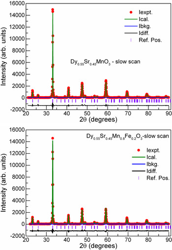
Observed XRD patterns (solid symbols) of Dy0.55Sr0.45MnO3 (upper panel) and Dy0.55Sr0.45Mn0.8Fe0.2O3 (lower panel) are shown in Figure 1. Rietveld structure refinement of the whole powder XRD data using EXPGUI version (Larson and Von Dreele, Reference Larson and Von Dreele2000) of GSAS program was undertaken. Background intensity was well fitted by a linear interpolation function. Among the four functions available in GSAS to represent the diffraction profiles, Pseudo-Voigt function (type II), which is the weighted sum of a Gaussian and Lorentzian functions was used during the refinement. All the diffraction peaks were adequately represented by this function where all the profile parameters were refined. While refining the fractional coordinates of cations, Dy, Sr and Mn, Fe ions, their x, y, and z parameters were constrained. Isotropic thermal parameters were chosen during the refinement. All the diffraction peaks were indexed using orthorhombic structure with space group Pnma. XRD patterns thus calculated matched satisfactorily with the experimental patterns and are included along with the observed patterns in Figure 1.
(Color online) Experimental XRD patterns (symbols) for Dy0.55Sr0.45MnO3 (upper panel) and Dy0.55Sr0.45Mn0.8Fe0.2O3 (lower panel) collected at room temperature. Blue color solid line corresponds to the background intensity (I bkg.). Other solid lines in the figure show calculated intensity (green color – I calc.), difference between experimental and calculated intensities (black – I diff.). The vertical lines shown at the bottom correspond to the expected Bragg reflections for Pnma space group.

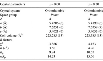
Crystal data after the final refinement are presented in Table I. Corresponding set of R parameters, which indicate the quality of the fit are also included in Table I.
Crystallographic data after refinement for Dy0.55Sr0.45Mn01−x Fe x O3.

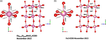
Fe substitution in the parent compound did not bring noticeable changes in lattice parameters. Figure 2 depicts ball-and-stick representation of crystal structure using Visualization for Electronic and Structural Analysis (Vesta) software (Momma and Izumi, Reference Momma and Izumi2013) of the unit cell where corner sharing Mn–O octahedra are arranged along the b-axis for both compounds. A single MnO6 along with bond distances is displayed in the same figure. It is apparent form the figure that there are significant distortions in bond distances and bond angles by nearly partial substitution of Sr at the Dy site.
(Color online) Schematic diagram of octahedral arrangement of Mn–O chains along the b-axis in (a) DSMO and (b) DSMFO, pink/yellow spheres correspond to Mn/Fe ions and oxygen ions are shown by red spheres. Deviations in the bond angles (Mn–O–Mn) and bond distances (Mn–O) from the ideal cubic perovskite structure are seen. Bond distances (Mn–O) in the MnO6 and bond angles in the plane (Mn1–O2–Mn1) and along the b-axis (Mn1–O1–Mn1) are marked in the figure.

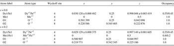
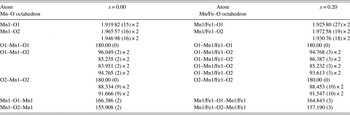
Fractional coordinates used for the refinement for both compounds are tabulated in Table II. Selected bond lengths and bond angles are given in Table III together with their estimated standard deviations. Each Mn atom is coordinated by six oxygen atoms with three pairs of different bond distances. The apical bond lengths (Mn–O1) are shorter than the in-plane Mn–O2 bond lengths. Our refinement results also indicate that Dy is ninefold coordinated with oxygen ions and the bond lengths of Dy–O vary between 2.4 and 2.9 Å.
Fractional coordinates obtained from Rietveld refinement of Dy0.55Sr0.45Mn1−x Fe x O3 for x = 0.0 and 0.2.

Selected inter-atomic distances (Å) and bond angle (°) for Dy0.55Sr0.45Mn1−x Fe x O3 obtained from the Rietveld refinement structure model used to refine whole powder diffraction patterns.

SUPPLEMENTARY MATERIAL
The supplementary material for this article can be found at http://dx.doi.org/10.1017/S0885715616000397.