Introduction
Granulomatosis with polyangiitis, also known as Wegener's granulomatosis, is a multi-systemic vasculitic process with prominent respiratory and renal involvement, characterised histologically by granuloma formation and necrotising inflammation. It is classified as an antineutrophil cytoplasmic antibody (ANCA)-associated vasculitis since ANCAs directed against proteinase 3 and, less commonly myeloperoxidase, are often present; testing for these is then a useful diagnostic tool.
Upper airway involvement in granulomatosis with polyangiitis is common, with variable clinical manifestations ranging from mild to potentially life-threatening, and may occur independently of other clinical manifestations. Subglottic laryngotracheal stenosis has been found to occur in approximately 16–23 per cent of patients with granulomatosis with polyangiitis.Reference Langford, Sneller, Hallahan, Hoffman, Kammerer and Talar-Williams1, Reference Lebovics, Hoffmann, Leavitt, Kerr, Travis and Kammerer2 It is a narrowing of the subglottic area within the cricoid cartilage; this is the narrowest area of the airway and, unlike the trachea and larynx, is a complete and non-expandable ring. Subglottic laryngotracheal stenosis may be congenital or acquired, and has been reported in granulomatosis with polyangiitis and other forms of vasculitis, such as relapsing polychondritis.Reference D'Cruz, Baguley, Asherson and Hughes3–Reference Solans-Laqué, Bosch-Gil, Canela, Lorente, Pallisa and Vilardell-Tarrés5 We describe a series of nine patients with granulomatosis with polyangiitis who developed symptomatic subglottic laryngotracheal stenosis, and discuss the medical and surgical interventions and clinical outcomes in this cohort of patients.
Methods
We retrospectively reviewed the medical and surgical notes of nine granulomatosis with polyangiitis patients with symptomatic subglottic laryngotracheal stenosis attending the systemic vasculitis clinic at St Thomas' Hospital, London, United Kingdom. This group represented 16 per cent of the total cohort of 55 granulomatosis with polyangiitis patients attending the clinic for regular review. All patients fulfilled the Chapel Hill Consensus Conference and American College of Rheumatology classification criteria.Reference Jennete, Falk, Andrassy, Bacon, Churg and Gross6, Reference Hunder, Arend, Bloch, Calabrese, Fauci and Fries7 Data were collected on the clinical manifestations related to granulomatosis with polyangiitis, and the diagnostic procedures and therapeutic management, including immunosuppression and surgical interventions. The Birmingham Vasculitis Activity Score and Vasculitis Damage Index were documented for all patients.Reference Mukhtyar, Lee, Brown, Carruthers, Dasgupta and Dubey8, Reference Exley, Bacon, Luqmani, Kitas, Carruthers and Moots9 Antineutrophil cytoplasmic antibodies were measured in all patients.
Results
The median age of patients at diagnosis was 44 years (range: 34–81 years). Seven patients were women and two were men. All patients in the cohort were white. The median Birmingham Vasculitis Activity Score was 5 (range: 2–8) and the median Vasculitis Damage Index score was also 5 (range: 2–7) at diagnosis of subglottic laryngotracheal stenosis. All nine patients presented insidiously with dyspnoea, wheezing, reduced exercise tolerance and varying degrees of stridor. One patient subsequently re-presented with acute upper airway compromise requiring emergency tracheostomy.
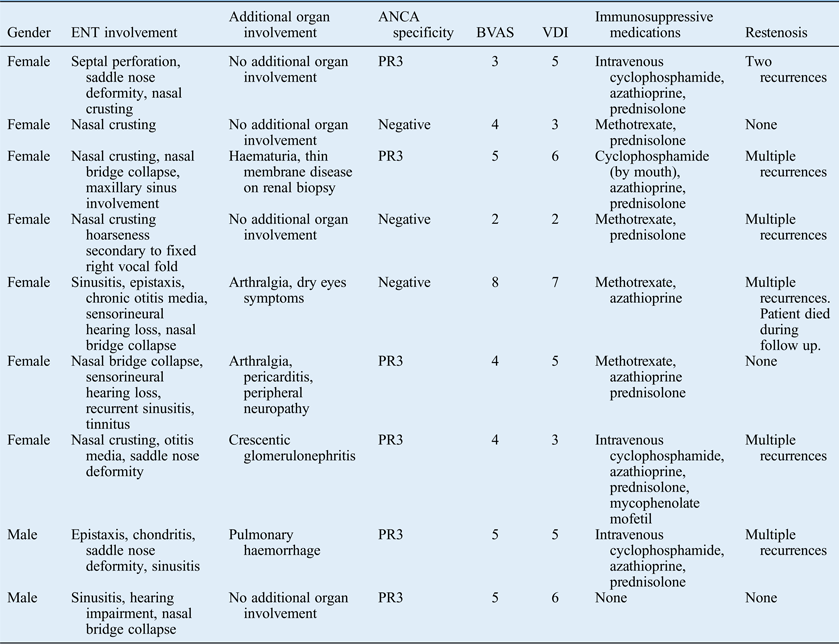
All patients in the cohort experienced longstanding ENT manifestations prior to diagnosis of subglottic laryngotracheal stenosis, including nasal crusting (n = 5), sinusitis (n = 4), recurrent epistaxis (n = 2), nasal bridge collapse (n = 4), sensorineural hearing loss (n = 3), chronic otitis media (n = 2) and nasal septum perforation (n = 1). One patient developed hoarseness and was found to have a fixed right vocal fold on flexible endoscopy. The most commonly cited symptoms at the outpatient review were fatigue, malaise and arthralgia (Table I).
Clinical features of patients with granulomatosis with polyangiitis who developed symptomatic subglottic laryngotracheal stenosis

ANCA = antineutrophil cytoplasmic antibodies; BVAS = Birmingham Vasculitis Activity Score; VDI = Vasculitis Damage Index;
PR3 = proteinase 3
One patient had experienced pulmonary haemorrhage related to granulomatosis with polyangiitis. Two patients presented with renal involvement, but activity did not correspond with episodes of subglottic laryngotracheal stenosis. One of these patients manifested clinically with persistent microscopic haematuria and normal renal function. Renal biopsy in this patient showed thin membrane disease on electron microscopy. The second patient developed chronic renal impairment secondary to crescentic glomerulonephritis related to granulomatosis with polyangiitis. There was one case of pericarditis and peripheral neuropathy related to vasculitis in this cohort. None of these vasculitic manifestations were temporally related to subglottic laryngotracheal stenosis flare-ups.
All patients except one were medically managed with immunosuppressive therapy including cyclophosphamide, azathioprine, methotrexate or mycophenolate mofetil in addition to corticosteroids. All patients with subglottic laryngotracheal stenosis received concomitant co-trimoxazole and one patient remained on co-trimoxazole therapy alone.
Six of the nine subglottic laryngotracheal stenosis patients were proteinase 3-ANCA-positive (67 per cent). Four patients were anti-cardiolipin antibody-positive and of these one had a history of recurrent miscarriages; none had experienced thrombotic events.
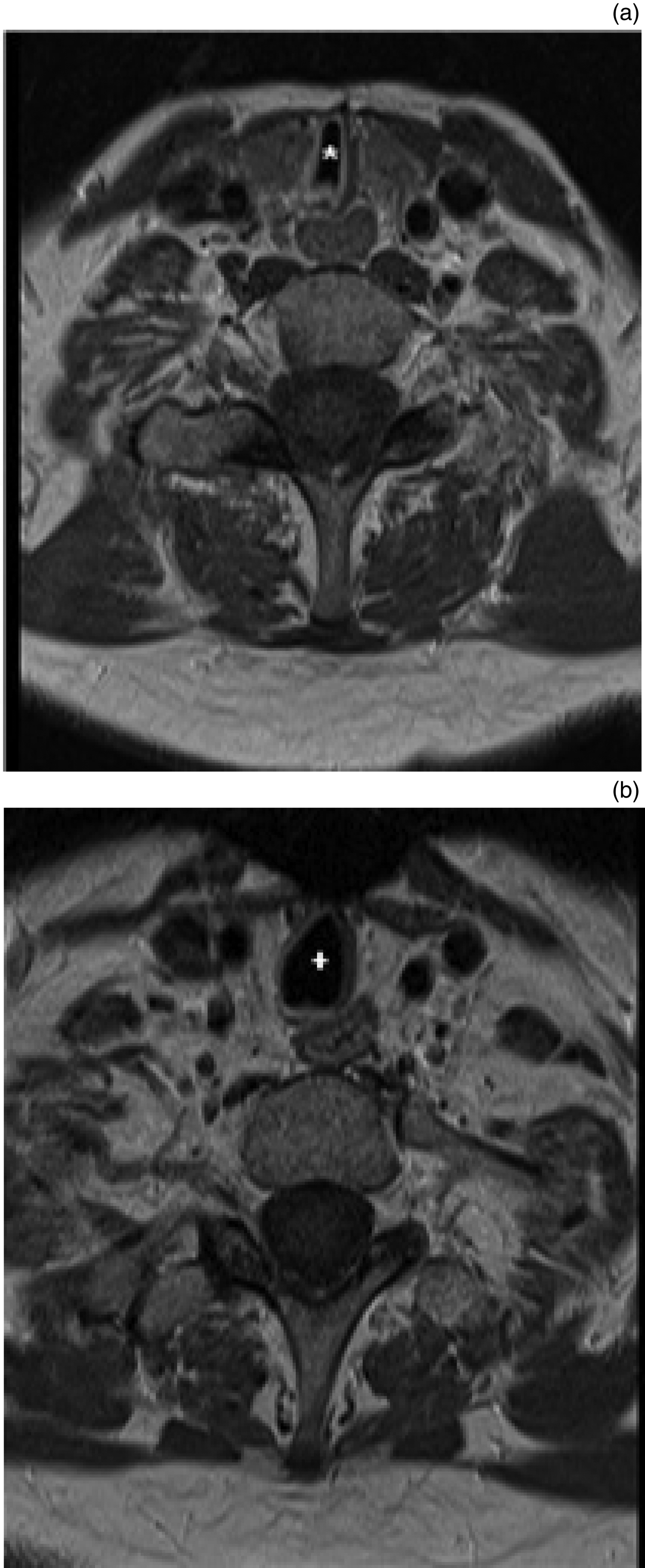
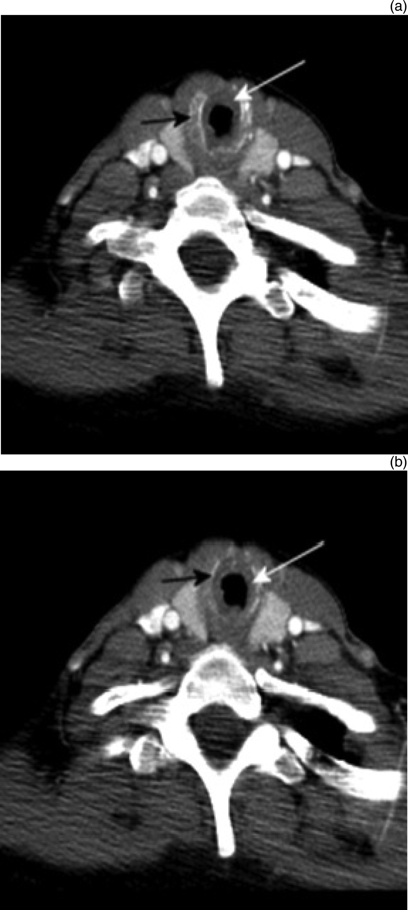
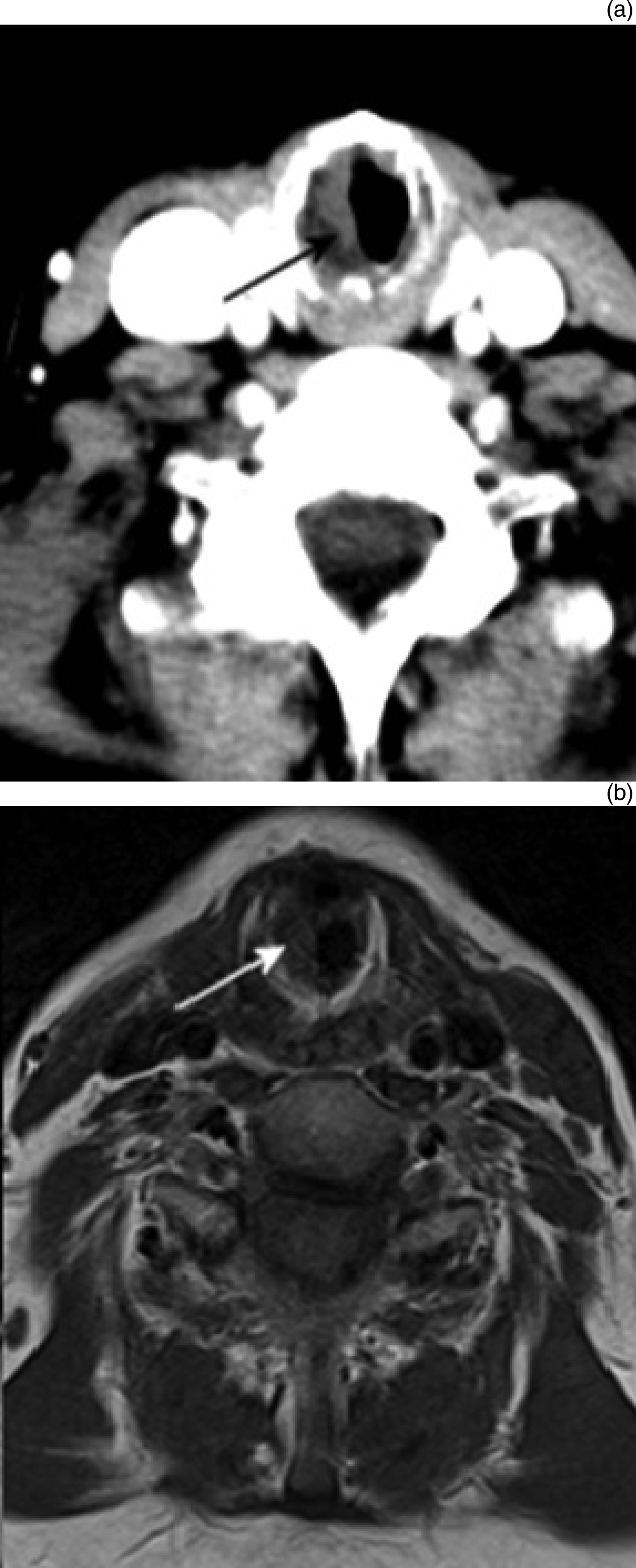
The decision to undertake surgery was made in patients who had significant stridor and limitation of exercise tolerance, following endoscopic laryngoscopy under light, spontaneous-breathing general anaesthesia. This allowed full assessment of the severity, nature and length of the stenosis. All clinical factors were weighed in the decision process. The stenosis was measured by assessing the cross-sectional diameter of the airway at the narrowest point and expressing the reduction in cross-sectional area as per the Myer–Cotton classification of subglottic laryngotracheal stenosis.Reference Myer, O'Connor and Cotton10 In all cases, the stenoses comprised mature, firm scar tissue located chiefly anteriorly or circumferentially, though posteriorly in some. As a rule of thumb, a stenosis of more than 50 per cent of the adult airway tends to be compromising and merits surgery (Figures 1–3).
Post-contrast, T1-weighted, axial magnetic resonance images of the neck in a patient with granulomatosis with polyangiitis, which demonstrate significant subglottic laryngotracheal stenosis (*): (a) at the level of C7, where it measures 3.7 mm in transverse diameter; and (b) more inferiorly, at the level of T1, where it measures 11.8 mm in transverse diameter.
Post-contrast, axial computed tomography images in a granulomatosis with polyangiitis patient: (a) and (b) at the level of the cricoid cartilage (black arrow), which demonstrates abnormal soft tissue within the subglottic larynx (white arrow), thereby resulting in subglottic laryngotracheal stenosis.
(a) Post-contrast, axial computed tomography and (b) post-contrast, T1-weighted magnetic resonance images of the granulomatosis with polyangiitis patient shown in FIG. 1 at the level of the cricoid cartilage, which demonstrate abnormal subglottic soft tissue on the right that crosses the midline (black arrow in (a) and white arrow in (b)).
Where the stenosis was deemed to require surgery, the choice was between a therapeutic endoscopic procedure or the more invasive, open laryngotracheal reconstruction surgery. The shorter and milder stenoses, even if circumferential, were treated with potassium titanyl phosphate (KTP) laser vaporisation of the organised scar tissue in the subglottis, usually in two or three radial incisions, preserving sufficient lumen mucosa, and then employing high-pressure balloon dilatation of the stenosis in addition. Endovascular balloons were used, selecting an appropriate diameter and typically with balloon pressure just below burst pressures of up to 14 atm for 2 minutes dilatation time (Table II). Steroid injections with methylprednisolone acetate were also administered into the dilated lumen submucosa in most patients. Post-operatively, all patients were managed with systemic steroids in a decreasing dosage regimen over two weeks in an attempt to avoid restenosis during the re-epithelialisation phase (Figure 4).
Bronchoscopy images of a granulomatosis with polyangiitis patient with symptomatic subglottic laryngotracheal stenosis: (a) bronchoscopy image showing airway obstruction secondary to subglottic laryngotracheal stenosis prior to intervention; (b) bronchoscopy image showing improved airway patency following endoscopic intervention with balloon dilatation and KTP laser. KTP = potassium titanyl phosphate
Frequency of open surgical interventions and endoscopic procedures in our cohort of patients with subglottic laryngotracheal stenosis
KTP = potassium titanyl phosphate
In those with more severe and longer stenoses, open surgery was selected. The laryngotracheal skeleton was fully exposed through an anterior neck incision and the stenotic region was opened with a vertical incision into the airway through the cricoid ring and adjacent lower end of the thyroid cartilage and upper tracheal rings, according to need. The anterior commissure was preserved in all cases, avoiding a full laryngofissure. Submucosal dissection and excision of scar tissue was undertaken as fully as possible and, in most cases, a non-vascularised anterior cartilage graft was inserted and sutured into the split to augment the airway diameter. The surgery was undertaken as a ‘single stage’ in which a tracheostomy was avoided; the reconstructed airway was supported for 5 days post-operatively, with intubation in the intensive care unit. Following successful extubation, the airway was typically assessed endoscopically once and in some cases twice over the following four weeks. This allowed removal of any soft granulations which can form during that stage. Once again, steroid cover was used during this period.
The most frequently performed surgical interventions in this cohort were microlaryngoscopy with balloon dilatation, local triamcinolone injection and KTP laser therapy. Five patients required laryngotracheal reconstruction surgery. One pregnant patient underwent microlaryngoscopy and bronchoscopy, KTP laser therapy and balloon dilatation at 29 weeks gestation due to significantly reduced exercise tolerance and increasing stridor. She tolerated these procedures well with significant symptomatic improvement and had a subsequent uneventful delivery by caesarean section. Her subglottic laryngotracheal stenosis remained in remission for the duration of the pregnancy and post-natal period.
Six patients had two or more recurrences of subglottic laryngotracheal stenosis, which developed independently of vasculitis activity elsewhere and required surgical intervention. Four of these six patients with relapsing subglottic laryngotracheal stenosis were proteinase 3-ANCA-positive. Peak expiratory flow rate and pulmonary function tests were found to be useful indicators of disease progression in the clinic. Two patients in our cohort had particularly aggressive disease. One of these required 18 dilatations in the course of their disease. Our most severe case was that of a female patient who had originally presented 13 years earlier with transient, bilateral hearing loss. Nine years later, she developed progressive dyspnoea and wheezing, sinusitis, recurrent epistaxis, nasal bridge collapse and arthralgia. This patient was subsequently admitted urgently to hospital with acute dyspnoea and stridor and was found to have 90 per cent subglottic laryngotracheal stenosis requiring an emergency tracheostomy. Despite immunosuppression and repeated surgical intervention, this patient died from complications of the disease. The remaining eight patients from the cohort continue to attend our clinic for routine follow up and have safely maintained their airways without the requirement for tracheostomy, albeit with some revision procedures.
Discussion
Subglottic laryngotracheal stenosis is an uncommon but significant complication of granulomatosis with polyangiitis often requiring both immunosuppressive therapy and surgical intervention. Sixty-seven per cent of patients in our series had relapsing disease requiring repeated surgical intervention. The patients reviewed in this cohort developed subglottic laryngotracheal stenosis independently of the other clinical features of vasculitis. While there was one instance of pulmonary haemorrhage and one case of crescentic glomerulonephritis, both well-recognised complications of granulomatosis with polyangiitis, these were not temporally related to the development of subglottic laryngotracheal stenosis.
All patients in our cohort received systemic co-trimoxazole prophylaxis as this has been shown to reduce the incidence of upper respiratory relapse in patients with granulomatosis with polyangiitis.Reference Stegeman, Tervaert, de Jong and Kallenberg11 However, caution should be exercised if patients are concurrently prescribed co-trimoxazole and methotrexate given the known interaction between these drugs.
Antineutrophil cytoplasmic antibodies have been found to be both a sensitive and specific marker of granulomatosis with polyangiitis in previous studies.Reference Venning, Arfeen and Bird12, Reference Lockwood, Bakes, Jones, Whitaker, Moss and Savage13 In our series, 67 per cent of subglottic laryngotracheal stenosis patients were proteinase 3-ANCA-positive, which corresponds to previously published series of subglottic laryngotracheal stenosis in granulomatosis with polyangiitis patients.Reference Solans-Laqué, Bosch-Gil, Canela, Lorente, Pallisa and Vilardell-Tarrés5, Reference Langford and Hoffman14 Our study population comprised 44 per cent anti-cardiolipin antibody-positive patients. One of these patients had suffered recurrent first- and second-trimester miscarriages, but none had developed thromboembolic phenomena. The prevalence of anti-cardiolipin antibodies was much higher than noted in our previous study, and since our unit is a referral centre for antiphospholipid syndrome, the possibility of referral bias cannot be excluded.Reference Rees, Lança, Marques, Gómez-Puerta, Moco and Oliveri15
Recurrence of subglottic laryngotracheal stenosis was common despite immunosuppressive therapy and patients should be regularly reviewed in the clinic regarding symptoms such as increasing dyspnoea, reduced exercise tolerance and stridor. The use of laser treatments within the airway for granulomatosis with polyangiitis-related subglottic stenosis is an established procedure.Reference Roediger, Orloff and Courey16 Roediger et al. however, also used topical applications of mitomycin C in an attempt to reduce re-scarring.Reference Roediger, Orloff and Courey16 We do not use this drug because of concerns about possible long-term mitogenic effects.Reference Agrawal and Morrison17
The concept of balloon dilatation as a therapeutic endoscopic procedure has gained popularity in laryngotracheal airway surgery for children and adults in recent years.Reference Schokkenbroek, Franssen and Dikkers18 In isolation, however, the balloon is unable to dilate a mature stenosis inside the fixed, complete cricoid ring. Dilatation is, however, a helpful and effective adjunct once the airway diameter has been potentially enlarged either by reduction of scarring by laser or following open surgery in which the fixed, rigid ring has been broken by an anterior, midline cricoid split. Especially in this latter situation, if the airway restenoses as a result of ongoing granulomatosis with polyangiitis activity, it has proved very successful to endoscopically redilate it since the fibrous union at the old surgical site can be stretched. This method of repeated endoscopic procedures after laryngotracheal reconstruction surgery has allowed good long-term outcomes in our patients and has typically been undertaken after two to three years in patients whose airways have deteriorated. The advantage of balloon dilatation over older methods of bougienage is that traumatic shearing forces are avoided and mucosal integrity is thus preserved. Intra-lesional steroid injection has also been previously described and may reduce the likelihood of the subglottis healing with early restenosis.Reference Hoffman, Thomas-Golbanov, Chan, Akst and Eliachar19
To our knowledge, there is little in the medical literature regarding open airway surgery for granulomatosis with polyangiitis-related subglottic laryngotracheal stenosis. Single-stage laryngotracheal reconstruction has been described by one study.Reference van den Boogert, Hans Hoeve, Struijs, Hagenouw and Bogers20 The benefit of open surgery, when required, is that it breaks the complete and closed ring of fixed cricoid cartilage and thereby allows true airway expansion and indeed subsequent airway dilatation to be more effective. Also, the open approach enables submucosal dissection and excision of the fibrous stenosis, further augmenting the airway. Open reconstruction surgery within our unit has tended to use an anterior cartilage graft to expand the lumen. Bone has been harvested from the ribcage, but it takes many months to heal and cartilage from the ala of the thyroid cartilage is sufficient.
The avoidance of complete laryngofissure at surgery preserved good voice quality for the patients in this series. One patient, however, had a pre-operative, unilateral vocal fold fixation. This was most likely caused by granulomatosis with polyangiitis with scar infiltration into the cricoarytenoid joint, but a mononeuropathy was not completely excluded as the cause. Of interest is the finding that histology from the subglottic scar tissue is usually not in itself diagnostic of granulomatosis with polyangiitis, as it is unusual to see vasculitis and the features are those of any organised fibrosis.
• This study showed a 16 per cent prevalence of symptomatic subglottic laryngotracheal stenosis in granulomatosis with polyangiitis patients
• It also showed that 67 per cent of symptomatic subglottic laryngotracheal stenosis patients were proteinase 3-antineutrophil cytoplasmic antibodies-positive; 67 per cent of subglottic laryngotracheal stenosis patients developed relapsing disease requiring repeated surgical intervention
• Subglottic laryngotracheal stenosis relapse was not associated with active systemic vasculitis elsewhere
In conclusion, in our institution, a multi-disciplinary approach, involving the careful medical management of granulomatosis with polyangiitis, together with a stepwise approach to subglottic laryngotracheal stenosis from conservative treatment of the airway, to endoscopic procedures, to open laryngotracheal reconstruction surgery using the techniques described has proved a reliable means of maintaining quality of life for these patients while almost always avoiding even a temporary tracheostomy.
Decisions regarding the combination of immunosuppression and local surgical intervention should be taken on an individual patient basis. Optimal management of subglottic laryngotracheal stenosis in granulomatosis with polyangiitis patients requires a multi-disciplinary approach involving both rheumatological and otolaryngological surgical expertise.