Introduction
Flies of the genus Drosophila (Diptera: Drosophilidae) have a wide range of hosts and species, many of which exert economically important impacts on cultivated fruits (Parshad and Paika Reference Parshad and Paika1964). For example, D. melanogaster (Meigen) prefers to lay eggs on rotten fruits (Mitsui et al. Reference Mitsui, Takahashi and Kimura2006; Atallah et al. Reference Atallah, Teixeira, Salazar, Zaragoza and Kopp2014), especially on grapes. This may cause grape sour rot and other diseases that can cause serious economic loss. Rombaut et al. (Reference Rombaut, Guilhot, Xuéreb, Benoit, Chapuis, Gibert and Fellous2017) suggested that in grapes, sour rot is attributed not only to D. melanogaster but also to previous D. suzukii (Matsumura) invasion. Drosophila suzukii originated in East Asia and spread to America and Europe (Zhai et al. Reference Zhai, Lin, Zhang, Zhang, Zheng and Yu2016). Drosophila suzukii has become an economically important invasive pest for berry and stone fruits because flies lay their eggs on fresh fruit, on which larvae feed (Schetelig et al. Reference Schetelig, Lee, Otto, Talmann, Stökl and Degenkolb2018). Moreover, D. immigrans (Sturtevant) and D. hydei (Sturtevant) are also widely found in Japan and East Asia (Katoh et al. Reference Katoh, Nakaya, Tamura and Aotsuka2007) and have been found in cherry orchards in different regions of China (Ren et al. Reference Ren, Wang, Yu and Chu2014; Wang et al. Reference Wang, Shen, Wang, Huang and Xu2017). Drosophila hydei and D. immigrans survive throughout the growing season (from grape and cherry sprouting to the falling of leaves), but their population numbers are lower than those of D. suzukii and D. melanogaster (Gao et al. Reference Gao, Zhu, Lv, Yang, Liu and Wang2018). To control these Drosophila species and to protect fruit, it is necessary to better understand the ecological strategies these important fruit pests employ.
The intestinal tract of insects is a dynamic environment involved in feeding, digestion, and excretion (Savage Reference Savage1977), and it is inhabited by many microorganisms. In insect guts, bacteria play an important role on the growth, reproduction, digestion, absorption, resistance to pathogenic bacteria, enhancement of immunity, and even resistance to pesticides of hosts (Dillon et al. Reference Dillon, Vennard, Buckling and Charnley2005; Warnecke et al. Reference Warnecke, Luginbühl, Ivanova, Ghassemian, Richardson and Stege2007; Sharon et al. Reference Sharon, Segal, Ringo, Hefetz, Zilber-Rosenberg and Rosenberg2010). As an excellent model system to study the effects of gut microbiota on host nutrition and metabolism, many studies assessed Drosophila–microbe interactions during the last decade (Douglas Reference Douglas2018). One way in which D. melanogaster adapts to the rotten-fruit environment is to feed on microbes, including yeasts and bacteria (e.g., Acetobacter and Gluconobacter) growing on rotting fruits (Becher et al. Reference Becher, Flick, Rozpędowska, Schmidt, Hagman, Lebreton and Bengtsson2012). The ethanol content is high in rotten fruits infested by D. melanogaster. The gut bacterium Acetobacter pomorum enables D. melanogaster to overcome the detrimental effects of ethanol by regulating the activity of alcohol dehydrogenase (PQ-ADH) in the insulin/insulin-like growth factor signalling pathway (Shin et al. Reference Shin, Kim, You, Kim, Kim and Lee2011). Fresh fruit is rich in sugars and other carbohydrates while also being relatively deficient in proteins and amino acids (Goodhue et al. Reference Goodhue, Rolda and Farnsworth2011; Milan et al. Reference Milan, Kacsoh and Schlenke2012). For this reason, D. suzukii must use other strategies, such as bacterial microsymbionts, to adapt to a nutrient-deficient habitat. However, very little information exists on the biological characteristics and adaptive strategies D. hydei and D. immigrans employ for their survival, especially those associated with their gut bacteria. Moreover, the gut microbiota contributes to the B vitamin requirements of Drosophila (Wong et al. Reference Wong, Dobson and Douglas2014; Douglas Reference Douglas2017). The role of the gut microbiota for Drosophila energy storage has been testified by analyses of the major macromolecular energy stores (i.e., lipid and glycogen) and two free sugars (i.e., glucose and trehalose; Shin et al. Reference Shin, Kim, You, Kim, Kim and Lee2011; Ridley et al. Reference Ridley, Wong, Westmiller and Douglas2012; Newell and Douglas Reference Newell and Douglas2014). For example, the gut microbiota of wild D. suzukii (consisting of, for example, Lactobacillus, Leuconostoc, Lactococcus, Comamonadaceae, and Enterobacteriaceae) reared on fresh fruit provides key proteins required for the development of D. suzukii (Bing et al. Reference Bing, Gerlach, Loeb and Buchon2018). In-depth knowledge on the association between Drosophila and their gut core symbionts is therefore important for understanding the adaptive ecology of Drosophila.
Over the course of evolution, the microbial diversity in insect guts and the relationship between gut microbes and insects may have been affected by both the diet and habitat of hosts (Yun et al. Reference Yun, Roh, Whon, Jung, Kim and Park2014). Many taxonomic differences have been identified between the microbiota of laboratory-reared and wild Drosophila (Winans et al. Reference Winans, Walter, Chouaia, Chaston, Douglas and Newell2017). Field-captured flies had a greater diversity of gut microbiota, which varied significantly with sampling location and season. In contrast, laboratory-reared flies possessed strikingly lower bacterial abundance and diversity (Bing et al. Reference Bing, Gerlach, Loeb and Buchon2018). To eliminate interference of other factors, in the present study, laboratory populations of D. suzukii, D. melanogaster, D. hydei, and D. immigrans were selected. The differences in core bacterial composition were analysed, and the function of intestinal bacteria was predicted. The stable and core bacteria are of the utmost importance not only from the perspective of enhancing the biological understanding of important fruit pests but also from a long-term Drosophila management perspective.
Materials and methods
Insects and diet
Drosophila suzukii, D. melanogaster, D. hydei, and D. immigrans adults were collected in May 2016 from a cherry orchard in Taian (121° 27′ E, 37° 27′ N), Shandong Province, China. Drosophila adults were attracted by semisolid bait provided in a plastic bottle with a small hole. The semisolid bait was composed of rice vinegar, red wine, sugar, berry juice, and agar. One male and one female adult of each Drosophila species were reared in a tissue-culture bottle (ø 5.5 cm × 9 cm) in the laboratory and received artificial diet (see below) for 5–6 generations. The colony was maintained in a climate-controlled growth chamber at 25 ± 0.5 °C, 70% ± 0.5% relative humidity, and a photoperiod of 16:8 hours (light:dark). These colonies of four Drosophila species formed the laboratory populations.
The artificial diet was composed of mashed bananas and apples, corn flour, sucrose, yeast extract, sorbitol, and agar, as described in Zhai et al. (Reference Zhai, Yu, Lin, Zhou, Li and Zhuang2014). Female adults (five days after eclosion) of the laboratory population were used for the isolation and identification of microorganisms. These species are common agricultural pests and are not listed on the “List of Protected Animals in China”. No specific permissions were required, as these fields are experimental plots that belong to Shandong Academy of Agricultural Sciences, Jinan, Shandong, China.
Sample collection and dissection
Adults of D. suzukii, D. melanogaster, D. hydei, and D. immigrans (five days after hatching) from the same generation were anaesthetised by chilling before dissection. Directly before dissection, to eliminate bacteria on the surface, adults were sterilised with 75% alcohol for 90 seconds and rinsed with distilled water for two minutes. The gut tissues of each individual were then dissected in phosphate-buffered saline (0.2 M, pH 7.2) under a stereomicroscope (SZX2-ILLB; Olympus, Hatagaya, Japan) in sterile conditions. Ten guts were pooled per replicate in each group and were sampled and collected in 2-mL sterile centrifuge tubes, with three biological replicates (i.e., tubes) per group. The samples were frozen at –80 °C.
DNA extraction and 16S rRNA gene Illumina MiSeq sequencing
The DNA of each sample was extracted with an insect DNA kit (OMEGA Bio-Tek, Norcross, Georgia, United States of America) and purified by the MoBio PowerSoil kit (Qiagen, Hilden, Germany) following the manufacturer’s protocol. Extracted high-quality DNA from all Drosophila species (three biological replicates per species) was high-throughput amplicon sequenced by Sangon Biotech Co., Ltd., Shanghai, China. The DNA was diluted to 1 ng/μL using DNase-free water. Polymerase chain reaction was conducted using bacterial universal primers of the V3 + V4 region: 341F (5'-CCTACACGACGCTCTTCCGA-TCTN (barcode) CCTACGGGNGGCWG CAG-3') and 805R (5'-GACTGGAGTTCCTTGGCACCCGAGAATTCCA (barcode) GACTACHVGGGTATCTAA-TCC-3') to amplify the bacterial 16S rRNA gene. In the primer sequence, the barcode was used to sort the groups in a single run. The sequencing library was generated by NEBNext Ultra DNA Library Pre-Kit (New England BioLabs, Ipswich, Massachusetts, United States of America) following the manufacturer’s protocols. The quantity and quality of the library were assessed by the Qubit 2.0 Fluorometer (ThermoFisher Scientific, Waltham, Massachusetts, United States of America) and Agilent Bioanalyzer 2100 system (Agilent, Santa Clara, California, United States of America). The library was sequenced on an Illumina Miseq sequencing platform (Hiseq 2000, Illumina, San Diego, California, United States of America) with 2 × 250 bases, and using the paired-end version. After removal of low-quality reads containing primer/adaptor sequences (identified using cutadapt, version 1.2.1 (https://pypi.org/project/cutadapt/1.2.1/)), paired-end reads were assembled using PEAR, version 0.9.6 (https://cme.h-its.org/exelixis/web/software/pear/). Individual samples then were assigned, based on unique sample-specific barcodes. Quality filtering was performed on the assembled sequences using PRINSEQ, version 0.20.4 (http://prinseq.sourceforge.net/) to obtain high-quality clean sequences. For this, the barcode, primer sequences, chimeras, low-quality read sequences, and trimming of read lengths were cut off. Then, these clean data were used for the analyses described below.
Operational taxonomic unit cluster and bacterial taxonomy analysis
Sequences of clean data were clustered into the same operational taxonomic units, applying a 97% identity threshold (3% dissimilarity level) using Mothur software, version 1.30.1. Operational taxonomic unit abundance was normalised using the sequence number corresponding to the sample with the least sequences. A Venn diagram was generated by the VennDiagram package of R software, version 1.6.16. To evaluate the phylogenetic relationships among different operational taxonomic units, multiple sequence alignment was performed between the first 50 operational taxonomic units with the largest overall abundance and the NCBI 16SrRNA database (http://ncbi.nlm.nih.gov/) using MUSCLE software, version 3.8.31 (https://www.drive5.com/muscle/).
The representative sequence for each operational taxonomic unit was screened for species annotation using the NCBI 16SrRNA database at each taxonomic rank (threshold: 0.81). The sequences that satisfied the standard (similarity > 90% and coverage > 90%) were used for the next classification, and sequences that did not satisfy the standard were categorised as “unclassified”. Then, the species and relative richness of each microorganism were analysed, and the microbial community structures were depicted as histograms. Furthermore, variation analysis of bacterial abundance based on genera among different Drosophila species was estimated using one-way analysis of variance (α = 0.05) and Student–Newman–Keuls multiple comparisons using the SPSS 20.0 statistical analysis package.
Alpha diversity analysis
According to operational taxonomic unit abundance, the Shannon and Simpson indexes were used to indicate the community diversity of these four Drosophila groups. The Chao1 and Ace indexes (which indicate community richness) and all other indexes were calculated by the software Mothur, version 1.30.1 (http://mothur.org/). Rarefaction analysis of all samples was also performed using Mothur software, version 1.30.1.
Beta diversity analysis
Beta (β) diversity analysis was used to evaluate variances in species complexity in samples of different Drosophila species. The weighted and unweighted uniFrac distances were calculated using QIIME software, version 1.8.0 (http://qiime.org/) to measure the dissimilarity coefficient between pairwise samples. Nonmetric multidimensional scaling analysis, a nonlinear model designed to improve the representation of the nonlinear biological data structure, was performed to obtain principal coordinates and to visualise complex multidimensional data.
Functional prediction
According to the microbial community structure generated from 16S rRNA sequencing, functional prediction of the metagenome was conducted based on the Kyoto Encyclopedia of Genes and Genomes (KEGG) using the software PICRUS, version 1.0.0 (http://picrust.github.io/picrust/). The histogram generated by KEGG was used to depict differences in microbial function of the four groups. Variation analysis of the top 10 gene functions contributed by relative operational taxonomic unit abundance was assessed with one-way analysis of variance (α = 0.05) and Student–Newman–Keuls multiple comparisons using the SPSS 20.0 statistical analysis package.
Results
Sequencing information
Raw data were generated by Illumina MiSeq sequencing of the 16S rRNA genes of intestinal samples of four Drosophila species (i.e., D. hydei, D. immigrans, D. suzukii, and D. melanogaster). After quality control, in which samples with a quality score of Q < 30 were discarded, filtered clean reads found in each Drosophila species (D. hydei: 58191 reads; D. immigrans: 59780 reads; D. suzukii: 58577 reads; D. melanogaster: 59776 reads) were then used for downstream bioinformatic processing (Supplemental material, Table S1). The datasets generated in the current study are available under NCBI Bio-project PRJNA719706.
Operational taxonomic unit abundance and bacterial taxonomy analysis
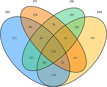
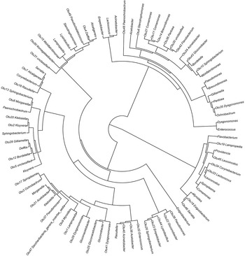
A Venn diagram of operational taxonomic units for all four groups is shown in Fig. 1. A total of 1779 operational taxonomic units was clustered in the samples of four Drosophila species. The numbers of operational taxonomic units found in D. hydei, D. immigrans, D. suzukii, and D. melanogaster were 887, 712, 832, and 799, respectively. Four Drosophila species shared 135 operational taxonomic units, including 69 bacterial genera (11 Lampropedia, eight Acetobacter, six Clostridium, and five Paenibacillus), with most of the remaining genera having more than two operational taxonomic units. Moreover, 420 operational taxonomic units were found in both D. hydei and D. suzukii groups, but only 279 were found both in D. immigrans and D. melanogaster groups. The phylogenetic tree of the first 50 abundant operational taxonomic units found in all samples is shown in Fig. 2. Among the first 50 operational taxonomic units, 32 belonged to the phylum Proteobacteria, 10 belonged to the phylum Bacteroidetes, and five belonged to the phylum Firmicutes, with one for each of Actinobacteria, Candidatus Saccharibacteria, and Tenericutes. The taxonomic information of operational taxonomic units for the four Drosophila species and the data for generating Venn plot of the four Drosophila species can be found in Supplemental material, Files S1 and S2.
Venn diagram of operational taxonomic units in four Drosophila species. DH, Drosophila hydei; DM, D. melanogaster; DY, D. immigrans; DS, D. suzukii.

Circular phylogenetic tree of the first 50 operational taxonomic units representative sequences and database sequences for the genus. Multiple sequence alignment was performed between the first 50 operational taxonomic units with the largest overall abundance and NCBI 16SrRNA database using MUSCLE software. OTU, operational taxonomic unit.

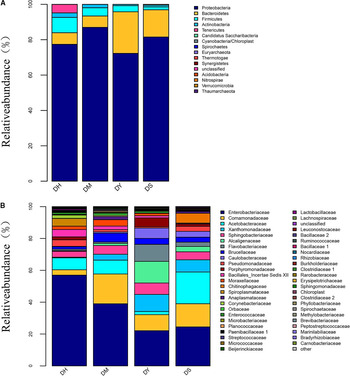
The community structure of phyla in these four Drosophila species is shown in Fig. 3A. Proteobacteria, Bacteroidetes, and Firmicutes were the dominant phyla among 16 identified phyla in each species. The sum of the percentages of the Proteobacteria, Bacteroidetes, and Firmicutes phyla was 92.48%, 98.09%, 99.21%, and 98.51% in the intestines of D. hydei, D. melanogaster, D. immigrans, and D. suzukii, respectively. Family-level community structure is shown in Fig. 3B. Enterobacteriaceae was the most abundant family in the four species. In the intestine of D. hydei, Enterobacteriaceae had a proportion of 56.99%, followed by Acetobacteraceae (7.49%), Spiroplasmataceae (4.78%), and Bacillales Incertae Sedis XII (4.58%). Besides Enterobacteriaceae (22.11%), other dominant families in D. immigrans (e.g., Alcaligenaceae, Flavobacteriaceae, Xanthomonadaceae, Comamonadaceae, and Sphingobacteriaceae) had similar relative abundances. The dominant families in both D. melanogaster and D. suzukii were Enterobacteriaceae, Comamonadaceae, and Acetobacteraceae, with total relative abundances of 66.40% and 58.96%, respectively.
A, Bar diagram of the community structure at the phylum level in four Drosophila species. B, Bar diagram of the community structure at the family level in four Drosophila species.

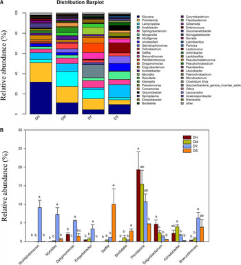
A total of 211 bacterial genera were found in the gut microbiota of the four Drosophila species, and the dominant species and amounts differed between these four species. The dominant intestinal bacteria that comprised a total percentage above 50% included four bacterial genera in D. melanogaster, including Kluyvera, Providencia, Lampropedia, and Acetobacter. The dominant bacterial genera in the intestines of D. immigrans (50.02%) were Providencia, Sphingobacterium, Alcaligenes, Wohlfahrtiimonas, and Myroides. Six bacterial genera (Kluyvera, Providencia, Acetobacter, Stenotrophomonas, Delftia, and Gluconobacter) comprised a total percentage of 54.58% and were the dominant bacteria in the intestines of D. suzukii. The dominant bacterial genera in the intestines of D. hydei were Kluyvera and Providencia, comprising a total of 50.21% (Fig. 4A).
A, Bar diagram of the community structure based on the genus level for four Drosophila species. B, One-way analysis of variance of community richness based on the genus level in four Drosophila species. Different letters in the same genus indicate significant differences (Student–Newman–Keuls multiple comparisons, P < 0.05).

According to the results of the one-way analysis of variance, a significant difference existed in the relative abundance of specific genera among the four Drosophila species (Fig. 4B). The percentages of Wohlfahrtiimonas, Myroides, Dysgonomonas, and Empedobacter in the intestines of D. immigrans were 3.39%, 7.26%, 5.57%, and 9.12%, respectively, which differed significantly from the percentages of the other three species (Wohlfahrtiimonas: F = 21.46, P < 0.01; Myroides: F = 14.60, P < 0.01; Dysgonomonas: F = 30.28, P < 0.01; Empedobacter: F = 7.12, P = 0.01). In addition, the relative abundances of the genera Delftia (10.01%) and Bordetella (2.76%) were much higher in D. suzukii than in the other three flies (Delftia: F = 5.25, P = 0.03; Bordetella: F = 7.48, P = 0.01). However, the relative abundances of Providencia and Exiguobacterium in D. suzukii were significantly lower than those among D. hydei intestinal microbes (Providencia: F = 3.83, P = 0.06; Exiguobacterium: F = 7.41, P = 0.01). No significant difference was found among other bacterial genera.
Alpha-diversity and beta-diversity analyses
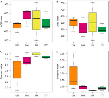
The α-diversity indexes illustrating the microbial community richness (ACE and Chao1) and diversity (Shannon and Simpson indexes) for each of the four Drosophila species are shown in Fig. 5. Although D. melanogaster had a higher ACE index (769.54 ± 56.06), no significant difference was found compared to the other three Drosophila species (ACE: F = 0.39, df = 2, P = 0.77). The Chao1 index did not differ significantly among the four Drosophila species (Chao1: F = 0.27, df = 2, P = 0.84). However, bacterial diversity also did not differ significantly among the four Drosophila species (Shannon: F = 1.81, df = 2, P = 0.22; Simpson: F = 1.77, df = 2, P = 0.23). Richness rarefaction curves of all samples tended to be flat, indicating that the amount of sequencing data was reasonable and that operational taxonomic unit coverage was sufficient (Supplemental material, Fig. S1).
Boxplot illustrating the α-diversity indexes of four Drosophila species: A, ACE index; B, Chao1 index; C, Shannon index; D, Simpson index.

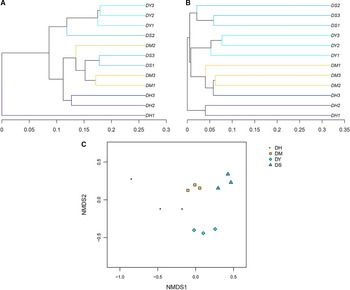
Beta (β) diversity evaluating the differences in microbial communities among different Drosophila species was represented by a Bray tree plot based on weighted uniFrac (Fig. 6A) and unweighted uniFrac (Fig. 6B) distances between samples. Similarly, according to nonmetric multidimensional scaling analysis, the clustering result of gut bacteria in the four Drosophila species suggests that the gut microbial community was affected by the host species (Fig. 6C).
A, Weighted tree plot of all samples found in four Drosophila species. B, Unweighted tree plot of all samples in four Drosophila species. Weighted and unweighted uniFrac distances were calculated using QIIME software. C, Nonmetric multidimensional scaling (NMDS) analysis reflecting the extent of variation in the gut bacterial communities among four Drosophila species.

Functional prediction
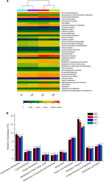
Based on the KEGG databases, the heatmap cluster illustrates the functional diversity of the bacterial communities in the four Drosophila species (Fig. 7A). The contribution of each operational taxonomic unit is associated with the function of 16S rRNA. Relative abundance was found for the top 10 gene functions in the four species: “amino acid metabolism”, “carbohydrate metabolism”, “cellular processes and signalling”, “energy metabolism”, “lipid metabolism”, “membrane transport”, “metabolism”, “metabolism of other amino acids”, “metabolism of terpenoids and polyketides”, and “replication and repair”. According to the results of the one-way analysis of variance of the top 10 functions, significant differences were found in the relative abundances of gene functions among the four Drosophila species (Fig. 7B). The relative abundances of gene functions “lipid metabolism” and “amino acid metabolism” in D. suzukii and D. immigrans were higher than those in D. hydei (F = 5.23, P = 0.03; F = 7.51, P = 0.01, respectively). However, the 16S rRNA gene of intestinal bacterial in D. hydei had the highest relative abundance of all four KEGG functions in “membrane transport”, “carbohydrate metabolism”, and “metabolism” and “cellular processes and signalling”. No significant difference was found in the relative abundance of 16S rRNA gene functions among D. melanogaster, D. immigrans, and D. suzukii.
A, Significantly enriched Kyoto Encyclopedia of Genes and Genomes (KEGG) functional analysis of bacteria in four Drosophila species. DH, Drosophila hydei; DM, D. melanogaster; DY, D. immigrans; DS, D. suzukii. B, Analysis of variance of KEGG functional analysis of the top 10 bacteria in four Drosophila species. Different letters in the same genus indicate significant differences (Student–Newman–Keuls multiple comparisons, P < 0.05).

Discussion
The gut microbiome of insects is increasingly recognised as playing an important role in shaping the ecological adaptability of insects. Drosophila has become a valuable model for microbiome research, and relevant research can increase the understanding of how the microbiome influences host traits. Research on the composition of the gut microbiota of Drosophila has principally focused on bacteria. Chandler et al. (Reference Chandler, Morgan, Bhatnagar, Eisen and Kopp2011) reported that four bacterial families contributed 90% of all sequences of the gut microbiota in 14 wild Drosophila species. These include Enterobacteriaceae, Acetobacteraceae, Lactobacillaceae, and Enterococcaceae. All wild populations are dominated by at least one of the four major clades. The gut bacterial communities of field-caught D. suzukii were dominated by two families of the phylum Proteobacteria: Acetobacteraceae and Enterobacteriaceae (Martinez-Sañudo et al. Reference Martinez-Sañudo, Simonato, Squartini, Mori, Marri and Mazzon2017). The diversity of intestinal microbes in insects may be affected by feeding conditions. Moreover, much taxonomic research has focused on the gut microbiota of laboratory-reared Drosophila (Broderick and Lemaitre Reference Broderick and Lemaitre2012; Douglas Reference Douglas2018). For example, a core member of microbiota that belongs to the genus Gluconobacter is common in wild D. melanogaster and D. simulans but is absent from laboratory-reared flies (Staubach et al. Reference Staubach, Baines, Kunzel, Bik and Petrov2013). The family Staphylococcaceae of the phylum Firmicutes mainly prevails in laboratory-reared populations (Martinez-Sañudo et al. Reference Martinez-Sañudo, Simonato, Squartini, Mori, Marri and Mazzon2017).
In the present study, the diversities of intestinal bacterial communities were analysed in four laboratory-reared Drosophila species fed by an artificial diet that includes corn, fruits, and yeasts. Comamonadaceae, of the phylum Proteobacteria, represented a high proportion of microbes in the gut of Drosophila (e.g., Delftia in D. suzukii and Lampropedia in D. melanogaster). The abundance of Enterobacteriaceae in laboratory-reared D. hydei was only 56.99% in the present study, which differs from a previous report where a single Enterobacteriaceae operational taxonomic unit represented at least 85% of bacterial microbiomes in wild D. hydei fed with fruits (Chandler et al. Reference Chandler, Morgan, Bhatnagar, Eisen and Kopp2011). Acetobacteraceae and Providencia in the family Enterobacteriaceae were dominant in the intestine of wild D. immigrans (Chandler et al. Reference Chandler, Morgan, Bhatnagar, Eisen and Kopp2011), whereas in the present study, the family Acetobacteraceae was rare in laboratory-reared D. immigrans. Another difference of D. immigrans was that the bacterial genera Alcaligenes, Wohlfahrtiimonas, Myroides, and Sphingobacterium were also abundant in laboratory-reared D. immigrans in the present study but were rare in wild flies. However, Enterobacteriaceae (Kluyvera and Providencia) and Acetobacteraceae (Acetobacter and Gluconobacter), of the phylum Proteobacteria, were still the core families in D. suzukii and D. melanogaster, which was consistent with previous studies (Broderick and Lemaitre Reference Broderick and Lemaitre2012; Chandler et al. Reference Chandler, James, Jospin and Lang2014; Martinez-Sañudo et al. Reference Martinez-Sañudo, Simonato, Squartini, Mori, Marri and Mazzon2017; Adair et al. Reference Adair, Wilson, Bost and Douglas2018). Winans et al. (Reference Winans, Walter, Chouaia, Chaston, Douglas and Newell2017) also reported that, for Enterobacteriaceae and Acetobacteraceae, laboratory-reared and wild Drosophila cannot be easily differentiated by 16S taxonomy. Therefore, taxonomic differences between the microbiota of laboratory-reared and wild Drosophila existed because of differences in diet, ecological environment, and species, whereas core bacteria (i.e., Enterobacteriaceae) remained stable and conserved (Kumar et al. Reference Kumar, Sun, Cao, Xue, Hu and Gong2019).
In the present study, significant differences were found in the relative abundance of bacterial genera among the four Drosophila species, which may be related to different ecological adaptations. For example, the relative abundance of the genus Delftia in D. suzukii was significantly higher than that in the other three species. Gut-associated bacteria are generally acquired from the environment after birth (Broderick and Lemaitre Reference Broderick and Lemaitre2012). The genus Delftia has been isolated from different environments such as fresh and marine water, soil, infected plants, clinical samples, and activated sludge. This genus has been characterised by the ability of its members to transform (or degrade) multiple organic pollutants (Ubalde et al. Reference Ubalde, Braña, Sueiro and Morel2012). Members of the genus Delftia have been described as plant growth–promoting bacteria because they promote plant growth either by providing nutrients (e.g., via nitrogen fixation, production of phytohormones, siderophores, and organic acids) or by helping with the resistance to infection by pathogens (Morel et al. Reference Morel, Ubalde, Braña and Castro-Sowinski2011). Delftia, with its high nitrogen-fixing activity, is regarded as an endophytic bacterial species that inhabits healthy plant tissue and cannot be found in rotting fruits (Di Fiore and Del Gallo Reference Di Fiore, Del Gallo, Fendrik, del Gallo, Vanderleyden and de Zamaroczy1995). Based on these results, it can be assumed that D. suzukii may obtain the bacteria of the genus Delftia from fresh fruit, and bacteria are then retained in the intestines of flies. However, the other three Drosophila species, which mainly feed on rotten fruit or other food sources, would not obtain the same amount of genus Delftia. Delftia was also abundant in wild D. suzukii, because it provides key proteins required for the development of D. suzukii reared on fresh fruit (Bing et al. Reference Bing, Gerlach, Loeb and Buchon2018). Therefore, D. suzukii has the unique ecological adaptability to consume fresh fruits, which may contribute to different gut microbes compared to those found in other Drosophila species.
Moreover, the intestine of D. immigrans had significantly higher percentages of Empedobacter, Myroides, Dysgonomonas, and Wohlfahrtiimonas compared to the microbes found in the other three Drosophila species. Thomas et al. (Reference Thomas, Hehemann, Rebuffet, Czjzek and Michel2011) had reported that the genera Myroides and Empedobacter contain pathogenic species. Wohlfahrtiimonas species were isolated from the bloodstream of a patient with septicemia and wound myiasis (Bonwitt et al. Reference Bonwitt, Tran, Dykstra, Eckmann, Bell, Leadon and Glover2018). Many findings have provided evidence that W. chitiniclastica is transmitted by larvae of house flies, Musca domestica (Diptera: Muscidae), and black soldier flies, Hermetia illucens (Diptera: Stratiomyidae), during myiasis (Gupta et al. Reference Gupta, Nayduch, Verma, Shah, Ghate and Patole2012; Lee et al. Reference Lee, Lee, Park, Sim, Choi and Lee2014). According to the results of the present study, the bacteria in these four genera comprise 25.34% of the intestinal bacteria of D. immigrans. However, whether these gut bacteria contribute to the spread of disease needs to be further evaluated.
The gut microbiota plays a very important role in many aspects of host physiology, especially in digestive, nutritional, metabolism-related, and immune functions (Fraune and Bosch Reference Fraune and Bosch2010; Douglas Reference Douglas2018). Warnecke et al. (Reference Warnecke, Luginbühl, Ivanova, Ghassemian, Richardson and Stege2007) reported that the gut microbes of Nasutitermes ephratae (Blattodea: Termitidae) can help the host degrade lignocellulose to compensate for the shortage of lignocellulose-degradation genes in termites. The herbivorous turtle ants of the genus Cephalotes (Hymenoptera: Formicidae) possess a conserved gut microbiome that enriches the nutrient composition by recycling nitrogen-rich metabolic waste to increase the production of amino acids (Duplais et al. Reference Duplais, Sarou-Kanian, Massiot, Hassan and Moreau2021). The results of the present study identified the metabolism of carbohydrates, amino acids, inorganic ions, lipids, and secondary metabolites as important functions of gut bacteria in Drosophila. These results show that gut microbes were probably related to the metabolism of fruit flies. However, the species and distribution of these microbes differed between the four Drosophila species. The differences during metabolism of carbohydrates, amino acids, inorganic ions, lipids, and secondary metabolites may be related to differences of the living habits and ecological niches among the four Drosophila species. However, the specific bacteria related to the adaptation to a specific ecological niche need to be identified in further studies.
In conclusion, taxonomic differences in microbiota were found among four laboratory-reared Drosophila species. The predicted function of bacteria can serve as targets for future downstream functional studies of core gut bacterial communities, which would expand the available knowledge about the fruit flies’ adaptations for survival in hostile environments. Breaking the relationship between gut bacteria and Drosophila species is one of the strategies to control them.
Acknowledgements
This work was financially supported through a grant from National Natural Science Foundation of China (31972273), Natural Science Foundation of Shandong Province, China (ZR2019MC034). The funders had no role in study design, data collection and analysis, decision to publish, or preparation of the manuscript.
Supplementary material
To view supplementary material for this article, please visit https://doi.org/10.4039/tce.2021.45.
Data availability
The datasets generated in the current study are available under NCBI Bio-project PRJNA719706 (Biosample of SAMN18614805, SAMN18614806, SAMN18614807, and SAMN18614808).
Author contributions
Qingcai Lin, Yifan Zhai, and Dongyun Qin conceived and designed the experiments, performed the experiments, analysed the data, contributed reagents/materials/analysis tools, authored or reviewed drafts of the paper, and approved the final draft. Hao Chen and Li Zheng conceived and designed the experiments, contributed reagents/materials/analysis tools, authored or reviewed drafts of the paper, and approved the final draft. Yifan Zhai and Huanhuan Gao analysed the data, prepared figures and/or tables, authored or reviewed drafts of the paper, and approved the final draft.
Grant disclosures
The following grant information was disclosed by the authors: National Natural Science Foundation of China: 31972273, 31801750; Natural Science Foundation of Shandong Province, China: ZR2019MC034; Innovation Project of Shandong Academy of Agricultural Sciences: CXGC2021A48.
Competing interests
The authors declare no competing interests.