





Introduction
Palpitations are a common reason for presentation to the paediatric emergency department and frequently generate stress for children and their families due to concerns about underlying heart disease. Reference Dhillon1,Reference Türe, Akın and Özbek2 Although palpitations are often perceived as a marker of serious cardiac pathology, the majority of paediatric cases are ultimately benign. Population-based and hospital studies consistently demonstrate that palpitations account for approximately 1–3% of paediatric emergency department visits, yet fewer than 10% of these presentations are due to a true cardiac arrhythmia, and less than 1% represent life threatening conditions. Reference Türe, Akın and Özbek2–Reference Macinnes, Martin, Fulton and McLeod4 Despite this low prevalence of serious pathology, intensive investigations are frequently undertaken, unnecessarily raising healthcare expenditures and amplifying family anxiety and distress. The primary clinical challenge is the need for rapid, critical decision-making. Emergency clinicians must differentiate benign causes such as sinus tachycardia which is related to anxiety, dehydration, fever, or pain, from potentially life-threatening conditions, including supraventricular tachycardia, Wolff–Parkinson–White syndrome, long QT syndrome, myocarditis, and ventricular arrhythmias. Reference Dhillon1,Reference Kang, Kumar and Hayes5 This differentiation must often be made within minutes of the patient’s presentation, sometimes without recording the symptomatic rhythm on electrocardiography; while over-investigation of low-risk children exposes them to unnecessary testing, prolonged monitoring, and expert referrals, delays in diagnosis or misclassification of high-risk patients may result in missed emergencies. Reference Türe, Akın and Özbek2,Reference Yakut3 Retrospective analyses of children presenting with palpitations show that although electrocardiograms are performed in nearly all cases, the diagnostic yield for clinically significant arrhythmia remains relatively low. Holter monitoring, event recorders, cardiac enzymes, echocardiography, and cardiology consultations are commonly used, even in children with reassuring histories and normal initial examinations. Conversely, rare serious disorders such as inherited channelopathies or accessory pathway mediated tachycardias may be overlooked if clinicians rely solely on symptom description without structured risk stratification. This creates a decision-making gap in the emergency department that closely mirrors challenges previously seen in paediatric chest pain and anaphylaxis, where low incidence but high consequence conditions demand systematic assessment pathways.
Evidence suggests that specific historical and clinical features can substantially increase the likelihood of true arrhythmia. Reference Türe, Akın and Özbek2,Reference Yakut3,Reference Kang, Kumar and Hayes5 Palpitations that are sudden in onset and offset, last longer than one hour, occur at a rate too rapid to count, are associated with syncope or near syncope, or occur during exertion are associated with higher arrhythmia rates. Family history of sudden cardiac death in a first-degree relative under 40 years old, known structural heart disease, or abnormal baseline electrocardiogram findings significantly increase the risk. Surveys in paediatric cardiology clinics and emergency departments have reported arrhythmia detection rates of 18% to 26% under these conditions. This highlights the need for detailed history and specific clinical examination. At the same time, new diagnostic methods are being developed to cut down on unnecessary testing and improve diagnosis accuracy. Innovations in ambulatory electrocardiogram technology such as smartphone-based event recorders/apps, have proven to be more effective in diagnostic yield and patient satisfaction than traditional short-term cardiac rhythm recording devices. These instruments underscore the fundamental principle that the acquisition of rhythm during symptomatic episodes is paramount to accurate diagnosis, rather than depending on extensive evaluations during asymptomatic intervals. Reference Macinnes, Martin, Fulton and McLeod4
This narrative review was conducted through a structured search of the PubMed and Google Scholar databases, as well as relevant paediatric cardiology and emergency medicine journals, for articles published between 2010 and 2026. Key search terms included “pediatric palpitations,” “arrhythmia,” “emergency department,” “risk stratification,” and “syncope.” Inclusion criteria prioritised observational studies, retrospective and prospective cohort studies, and clinical reviews focusing on paediatric patients presenting with palpitations in an emergency setting. Studies describing epidemiology, diagnostic yield of investigations, risk stratification, and management outcomes were selected. Non-English articles, isolated adult-only studies, and single case reports were excluded. A narrative review approach was chosen over a systematic review due to the heterogeneity of study designs and the primary aim of synthesising evidence to formulate a practical, emergency department-focused clinical framework. The aim of this review is to identify presenting red flags to emergency department decision-making and final diagnoses in children with palpitations. By aligning clinical features with evidence-based risk stratification, this paper seeks to support consistent, safe, and cost-effective emergency department practice ensuring that high risk patients are promptly identified while avoiding unnecessary investigations in the large proportion of children with features associated with their chief complaint of palpitations. Reference Dhillon1–Reference Kang, Kumar and Hayes5
Epidemiology and pathophysiology of paediatric palpitations
Palpitations may indicate a child has a cardiac rhythm abnormality/arrhythmia, which may be benign or life-threatening. Hence, it is crucial to identify the patterns in age, sex, and recurrence rates as early as possible to allow the right assessment, triage, and treatment. Palpitations in children and adolescents arise from a spectrum of mechanisms, including physiological, ectopic, supraventricular, ventricular, channelopathy-related, and structural causes. Understanding these mechanisms is essential for accurate diagnosis, risk assessment, and management.
Epidemiology and Age–sex Patterns
While palpitations frequently cause concern among caregivers, the complaint is among the least frequent ones in paediatric emergency departments. In fact, in a study that looked back at data from more than 140,000 paediatric emergency visits, only 0.07% of the patients complained of palpitations, and just 13.5% of these patients had arrhythmic aetiologies verified that necessitated intervention. Reference Bobbo, Amoroso and Tamaro6 Supraventricular tachycardia is the most common type of arrhythmia in children, and it is found in 0.1% to 0.4% of individuals who are otherwise healthy. Reference Balsam, Lodziński and Tymińska7 The incidence of supraventricular tachycardia increases steadily from the time of birth to adolescence when it reaches approximately 1.3 cases per 1,000 patients per year at the age of 15. Reference Balsam, Lodziński and Tymińska7 The age distribution is commonly bimodal with peaks between 6 and 9 years and again in adolescence. This is explained by the changes in autonomic tone, the development of the cardiac conduction system, and the fact that older children become more aware of palpitations and can verbalise symptoms to caregivers. Reference Balsam, Lodziński and Tymińska7
Paediatric arrhythmias vary greatly between the sexes at different ages. For instance, males tend to have more first episode supraventricular tachycardia due to an accessory pathway, Brugada syndrome, and arrhythmogenic right ventricular cardiomyopathy, whereas females experience atrioventricular nodal reentrant tachycardia and arrhythmias related to a long QT interval more often. Reference Clausen, Theophilos, Jackno and Babl8 It is very important to understand these patterns when selecting the diagnostic tests and determining the risk. Repeated palpitations have a major impact on the choices of long-term care and treatment. It is estimated that about 35% of children with atrioventricular nodal reentrant tachycardia who are treated with supraventricular tachycardia ablation will experience recurrent palpitations. Reference Çınar, Kafalı, Şahin and Ergül9 Previous arrhythmia attacks, younger age at first presentation, and the intensity of the first tachycardia are the factors associated with the recurrence. Reference Bobbo, Amoroso and Tamaro6,Reference Çınar, Kafalı, Şahin and Ergül9 It was pointed out that continuous monitoring by wearable electrocardiogram devices or external event recorders allows the correlation of symptoms with arrhythmias, thus leading to targeted follow-up care and reducing unnecessary hospitalisations. Reference Çınar, Kafalı, Şahin and Ergül9
Physiologic and benign causes
The most frequent benign cause of palpitations in children is sinus tachycardia. Fever, dehydration, pain, anxiety, or strenuous activity are the usual triggers. Reference Crosson and Hanash10 True emergencies are uncommon; most cases are well-tolerated. Palpitations are also caused by paroxysmal atrial contractions and premature ventricular contractions, which, while frequently frightening, usually have positive results and spontaneously resolve in follow-up. Reference Mohsin, Sangi and Sheikh11 Transient factors such as emotional stress, caffeine consumption, or acute illness can aggravate these benign arrhythmias. In order to avoid needless investigations and parental anxiety, it is critical to identify physiological triggers. Ectopic beats that originate outside of the typical conduction pathway are represented by premature atrial contractions and premature ventricular contractions. They can be found in perfectly healthy children or be the consequence of easily treatable conditions such as an electrolyte imbalance or drug side effects. Usually, premature ventricular contractions and premature atrial contractions are without symptoms; however, in some cases, they may cause palpitations or a brief sensation of lightheadedness. When there is no structural heart disease, the outlook is excellent and spontaneously occurring cases in children are many. Reference Mohsin, Sangi and Sheikh11
Supraventricular arrhythmias
Among the most common pathologic arrhythmias in paediatric emergency settings are supraventricular arrhythmias, such as atrioventricular reentrant tachycardia (from concealed accessory pathway or manifest ventricular preexcitation/Wolff–Parkinson–White syndrome) and atrial tachycardia. Between 1 in 1,000 and 1 in 250 children are thought to be affected. Reference Balsam, Lodziński and Tymińska7,Reference Taylor, Thomas and Lawrence12 Because of its quick conduction through accessory pathways, atrioventricular reentrant tachycardia can be more dangerous than other supraventricular tachycardias because it can cause syncope or, in rare cases, ventricular fibrillation. Reference Taylor, Thomas and Lawrence12 Acute stabilisation, medication therapy, and increasingly catheter ablation are all part of the management. Due to its high success rates and low risk of complications, catheter ablation has become a reasonable option for children who are deemed appropriate for the procedure. Reference Hafez, Abu-Elkheir and Shokier13,Reference Smith and Clark14 Early detection of ablation-suitable patients can lower long-term morbidity and enhance quality of life.
Ventricular arrhythmias
Children with or without underlying structural heart disease may experience ventricular arrhythmias, such as idiopathic ventricular tachycardia. Prevalence may reach 17% in certain cohorts, such as those with left ventricular noncompaction. Reference Brescia, Rossano and Pignatelli15 Idiopathic ventricular tachycardia in otherwise healthy children usually has a benign course and responds well to medical or interventional therapy. However, children with documented ventricular tachycardia require additional diagnostic evaluation and risk stratification by a paediatric cardiologist, as ventricular tachycardia is much more likely to be life-threatening in the setting of structural heart disease or a channelopathy, and may require close monitoring after first presentation. Reference Mohsin, Sangi and Sheikh11 Since children with underlying cardiomyopathy may have higher morbidity and need close monitoring, the identification of structural abnormalities through echocardiography or MRI is essential to stratify risk and direct treatment. Reference Brescia, Rossano and Pignatelli15
Channelopathies
Abrupt syncope/loss of consciousness or an aborted sudden cardiac death event can be the first manifestation of a cardiac channelopathy such as Brugada syndrome, long QT syndrome, and catecholaminergic polymorphic ventricular tachycardia. Consequently, a typical presentation of long QT syndrome is the occurrence of torsades de pointes, syncope, or sudden cardiac death. In children with the family history of sudden cardiac death or unexplained syncope, it is vital that early detection is done by means of electrocardiogram screening and genetic testing. Reference Clausen, Theophilos, Jackno and Babl8 The management of patients includes the use of medications, lifestyle modifications and when indicated implantation of a cardioverter-defibrillator.
Structural and inflammatory causes
Structural and inflammatory issues of the heart, such as myocarditis and different types of cardiomyopathies, may manifest as palpitation symptoms or documented rhythm abnormalities. Myocarditis, which is commonly caused by viral infections, can abruptly lead to rhythm changes and may at times necessitate hospital admission for close monitoring and supportive therapy. Reference Brescia, Rossano and Pignatelli15 Certain cardiomyopathies, e.g., dilated, hypertrophic, and left ventricular noncompaction, can severely affect a child’s health and typically require the collaborative management of multiple healthcare professionals. Reference Brescia, Rossano and Pignatelli15 Children with CHDs, those who have recently undergone cardiac surgery, and those with genetic syndromes or a family history of early sudden cardiac death are at increased risk of serious complications. Early identification, thorough evaluation, and close monitoring are the key steps in reducing this risk.
Risk stratification in paediatric palpitations
Palpitations can cause anxiety to children and caregivers when there are concerns about an underlying arrhythmia. However, many patients will be describing a benign awareness of heartbeats. Reference Kang, Kumar and Hayes5 Palpitations are considered serious when associated with cardiac red flags (syncope, exertional chest pain or poor exercise tolerance, a family history of sudden death, and structural heart disease) as well as the presence of electrocardiographic abnormalities (Figure 1).
Pathophysiological mechanisms leading to paediatric palpitations and associated red-flag groups. (Conceptual and derived from the current narrative).

Red-flag features (“never-miss” list)
Syncope is a transient loss of consciousness and postural tone with spontaneous recovery. It is a common paediatric presentation and most cases are benign. A small proportion are due to life threatening conditions. Reference Stewart, van Dijk, Balaji and Sutton16 Cardiac syncope is associated with high morbidity and substantial mortality. Any syncope due to a confirmed cardiac cause is classified as severe. Arrhythmias are the most common cardiac cause of syncope. Reference Brignole, Moya and de Lange17 Arrhythmic syncope in children is more likely to occur in genetic conditions associated with cardiac disease. Certain triggers may suggest a specific inherited disease; startle such as by loud sound or cold water on the face (long QT syndrome), sudden onset syncope in fever (Brugada syndrome), and syncope during exercise (or similar catecholamine states; catecholaminergic polymorphic VT or arrhythmogenic right ventricular cardiomyopathy). Also, very rapid ventricular tachycardia, as seen in many channelopathies, may lead to syncope when sustained for more than 3–5 s.
Exertional palpitations: Sudden cardiac death remains a leading cause of death in young athletes. Exercise-related sudden cardiac death is commonly caused by hereditary conditions: channelopathies and cardiomyopathies. In particular, long QT syndrome, catecholaminergic polymorphic ventricular tachycardia, hypertrophic cardiomyopathy, and arrhythmogenic cardiomyopathy have an increased risk of triggered ventricular arrhythmias with exertion/catecholamines. Reference Katyal, Li, Franciosi and Sanatani18
Structural heart disease: A retrospective review of 5,307 paediatric exercise stress tests performed between 2013 and 2015 showed that, although self-terminating arrhythmias were common (46%), clinically significant arrhythmias were rare (0.6%) in a high-volume paediatric cardiology criterion. The absence of any high-risk criterion was associated with a negative predictive value of 99.7% (95% CI: 99.5–99.8%) for arrhythmias requiring test termination and 99.96% (95% CI: 99.85–99.99%) for arrhythmias requiring intervention beyond test termination. Reference Barry, Gauvreau and Rhodes19
Family history of sudden death: In a study of 419 paediatric relatives of 256 decedents, 27% of children were diagnosed with a cardiac disease or had findings of uncertain significance. An inherited cardiac disease was identified in 39 patients (9.3%), while non-heritable cardiac disease was diagnosed with an additional 5.5%. Findings of uncertain significance were present in 52 patients (12.4%), including abnormal electrophysiological findings (41 of 52) or imaging abnormalities (11 of 52). Among patients diagnosed with inherited cardiac disease, the nearest affected relative was almost always a first-degree relative (95%). The strongest predictors of a diagnosis were an abnormal electrocardiogram and a first-degree relationship to the nearest affected relative, with odds ratios of 24.2 and 18.8, respectively. Reference Webster, Olson and Schoppen20
Abnormal electrocardiogram: Electrocardiogram findings associated with arrhythmic risk include narrow or wide QRS complex tachycardia. In most cases, supraventricular tachycardia produces a narrow QRS complex; however, a wide QRS tachycardia can be the result of a baseline bundle branch block or can be transient due to rate-related aberrant conduction in supraventricular tachycardia. In atrioventricular reentrant tachycardia, P waves may not be visible when they overlap with the QRS complex. In atrioventricular reentrant tachycardia, ventricular preexcitation—manifesting as a delta wave, short PR interval, and wide QRS complex (Wolff–Parkinson–White syndrome pattern)—is typically appreciated in sinus rhythm, not during tachycardia. Importantly, a patient can have atrioventricular reentrant tachycardia from an accessory pathway that conducts only in the retrograde direction (concealed pathway) and thus may not exhibit ventricular preexcitation on a resting electrocardiogram.
Risk tiers and clinical meaning
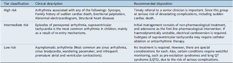
In a review focused on studies between 2019 and 2024, included patients aged 2–18 years with different types of arrhythmias, a symptom-based guide designed to help non-specialist physicians in the management of paediatric arrhythmias (Table 1). Reference Cepeda-Nieto, Roblero-Aguilar, Martínez-López and Balderrábano-Saucedo21
Risk stratification tool. The table summarises risk stratification of palpitations in paediatric patients, along with the proper disposition for each category Reference Cepeda-Nieto, Roblero-Aguilar, Martínez-López and Balderrábano-Saucedo21

Table 1. Long description
A table categorizing arrhythmias in pediatric patients by risk level and recommended management strategies. The table has three rows and three columns. The columns are labeled ‘Tier classification’, ‘Clinical description’, and ‘Recommended disposition’. The rows are labeled ‘High risk’, ‘Intermediate risk’, and ‘Low risk’. Row 1: High risk, Arrhythmia associated with any of the followings: Syncope, Family history of sudden cardiac death, Exertional palpitation, Abnormal electrocardiogram, Structural heart diseases, Timely referral to a senior clinician is important. Since this group at serious risk of devastating complications, including sudden cardiac death. Row 2: Intermediate risk, Episodes of paroxysmal arrhythmia, supraventricular tachycardia is the most common arrhythmia in children, mainly as a result of re-entry mechanisms, Initial management consists of non-pharmacological treatment, and adenosine as the first-line pharmacological intervention. If haemodynamically unstable, electrical cardioversion is required. Subtypes of supraventricular tachycardia may require catheter ablation or antiarrhythmic therapy. Row 3: Low risk, Asymptomatic arrhythmia (Most common are sinus arrhythmia, sinus bradycardia, wandering pacemaker, and infrequent premature atrial and ventricular contractions), No treatment is required. However, there are special considerations for each. Also, certain conditions require watchful monitoring, such as pre-excitation syndromes and long QT syndrome (LQTS), due to the risk of serious complications.
The first group is asymptomatic arrhythmias, which can be considered a low-risk group. The most common asymptomatic arrhythmias are sinus arrhythmia, sinus bradycardia, wandering pacemaker, and infrequent premature atrial and ventricular contractions. Many asymptomatic cases are benign and require no treatment; however, there are special considerations for each. Also, certain conditions, such as pre-excitation syndromes and long QT syndrome, require watchful monitoring due to the risk of serious complications.
The second group is patients with episodes of paroxysmal tachycardia, which can be considered as an intermediate-risk. Supraventricular tachycardia is commonly encountered in children. Initial management consists of non-pharmacological, and adenosine as the first-line pharmacological intervention. If haemodynamically unstable, electrical cardioversion is required. Subtypes of supraventricular tachycardia may require catheter ablation or antiarrhythmic therapy. Timely referral involves identifying patients at high risk of serious complications, including sudden cardiac death, while managing benign arrhythmias conservatively. Reference Cepeda-Nieto, Roblero-Aguilar, Martínez-López and Balderrábano-Saucedo21
The high-risk group includes any arrhythmia associated with red-flag features such as syncope, exertional symptoms, a family history of sudden cardiac death, known structural heart disease, or a significantly abnormal electrocardiogram. These patients are at serious risk of devastating complications and require immediate, thorough evaluation.
Common pitfalls in risk assessment
Diagnosing arrhythmias in children can be challenging. Precise histories can be difficult to elicit from many patients, particularly children in addition the physical examination can be normal during the initial assessment. Also, normal rhythm variants may be seen in healthy paediatric patients, including sinus arrhythmia, wandering atrial pacemaker, ectopic atrial rhythm, and intermittent nodal rhythm. Reference Goto, Witkowska, Slusarczyk, Grotek, Dziubinski and Clark22 A study included 1,884 healthy newborns, infants, children, and adolescents showed that the values of 15 key electrocardiogram parameters change characteristically during growth and development. The study also demonstrated that sex impacts electrocardiogram parameters, accordingly, participants aged 6–9 years, 9–13 years, and 13–18 years were divided into male and female groups. Additionally, some electrocardiogram parameters may change with race, ethnicity, and socioeconomic and nutritional status. Reference Lue, Wu and Wang23 A significant pitfall is over-relying on a single, asymptomatic electrocardiogram tracing while downplaying a concerning history. Another is dismissing a family history of “fainting spells” or “seizures” without considering an underlying channelopathy.
When a “normal” electrocardiogram Is not reassuring
A critical aspect of senior-level cardiology judgement is recognising that a single “normal” electrocardiogram does not definitively exclude a high-risk condition. The primacy of the clinical history, particularly exertional syncope or a strong family history of sudden death, must always be respected, even in the face of a non-diagnostic tracing. Clinicians should maintain a high index of suspicion for subtle abnormalities, such as borderline QTc values that may vary with age and sex, or subtle pre-excitation (delta waves) that can be intermittent or difficult to discern. The absence of overt pathology on one electrocardiogram does not negate the risk conferred by compelling red-flag features, which should always prompt further investigation or specialist consultation.
Limitations of electrocardiogram interpretation in the emergency department setting
An important limitation that must be acknowledged in the context of this framework is the challenge of accurate electrocardiogram interpretation for paediatric patients in the emergency department. The diagnostic pathways outlined in this review presuppose proficiency in recognising the subtle and age-dependent electrocardiogram abnormalities described herein. However, many emergency departments—particularly those without dedicated paediatric services—do not have continuous access to a trained paediatric cardiologist who can confirm that the electrocardiogram abnormalities referenced in this manuscript are accurately identified and appropriately interpreted. Emergency physicians who are not formally trained in paediatric cardiology may find electrocardiogram interpretation in this age group challenging, given the evolving normal ranges across different developmental stages and the subtlety of findings such as intermittent delta waves, borderline QTc prolongation, or low-amplitude changes in atrioventricular nodal reentrant tachycardia. Accordingly, a low threshold for remote consultation with a paediatric cardiologist—via telemedicine or formal referral—is strongly recommended whenever any electrocardiogram abnormality is identified or when clinical suspicion of an arrhythmia is high despite a seemingly normal tracing.
Diagnostic testing
The goal in paediatric palpitations is not to diagnose every arrhythmia, but to safely exclude dangerous ones. This principle guides a tiered approach to diagnostic testing, emphasising the high negative predictive value of a normal electrocardiogram combined with an absence of red-flag features. Reflexive testing without clear indication should be avoided (Figure 2).
Diagnostic algorithm flowchart for paediatric palpitations in the emergency department. This flowchart presents a standardised clinical pathway for the diagnostic assessment of paediatric patients presenting to the emergency department with palpitations. The pathway distinguishes typical low-risk presentations from atypical or episodic histories that warrant ambulatory rhythm monitoring despite a normal or non-diagnostic resting ECG, and from high-risk presentations requiring selective testing, advanced cardiac imaging, telemetry, or urgent cardiology evaluation. Abbreviations: ECG = electrocardiogram. (Conceptual and derived from the current narrative).

Figure 2. Long description
The flowchart begins with the initial assessment, which includes history and physical examination, 12-lead ECG, orthostatic vitals, and blood glucose. The next step is to classify the presentation using history and ECG. If the patient has a typical low-risk history with a clear physiologic trigger and normal ECG, the pathway leads to reassurance, outpatient follow-up if symptoms recur, and discharge. If the patient has an atypical or episodic history with a normal or non-diagnostic ECG and no classic red flags, the pathway suggests ambulatory rhythm monitoring, such as a Holter or event monitor, to correlate symptoms with rhythm. If there are red flags, an abnormal ECG, or persistent concern for cardiac disease, selective testing is indicated, which may include laboratory tests, imaging, telemetry, or urgent cardiology review. If the standard workup is complete but the etiology remains unclear, structured follow-up is arranged, and monitoring is extended if symptoms recur or concern persists. The final step is disposition based on documented risk and monitoring findings.
Mandatory testing
In addition to a thorough history and physical exam, there are several investigations that are essential to leading clinicians towards finding the root cause of palpitations in the paediatric population presenting to the emergency department. A 12-lead electrocardiogram is mandatory and considered to be the gold standard in all children with palpitations, as proposed by paediatric cardiology experts. Electrocardiograms serve as an important initial screening tool, capable of identifying patterns suggestive of dangerous arrhythmias or structural heart disease that may predispose childrense to sudden cardiac death; Reference Sebudandi, Brockmeier, Atmatzidou, Khalil and Wendt24 however, as emphasised in section When a “normal” electrocardiogram is not reassuring, a single normal electrocardiogram does not definitively exclude all high-risk conditions when compelling clinical red-flag features are present. Effective interpretation of electrocardiograms in the paediatric emergency department requires foundational knowledge of high yield pathologies. A short PR interval with a delta wave should prompt suspicion for Wolff–Parkinson–White syndrome. A prolonged correct QT interval should be concerning for congenital long QT syndromes or acquired including electrolyte disturbances, medications, myocarditis, and head injuries. ST segment elevations or depressions may be pathological in myocarditis, while convexity of the ST segment indicates pericarditis. Hyperkalemia may lead to peaked T waves, though hypokalaemia displays flattened T waves. Reference Lambrechts and Fourie25 Electrocardiograms can help steer further testing if indicated or support a timely and safe discharge.
Orthostatic vitals, including blood pressure and heart rate assessment, are simple measurements that should not be overlooked when working up palpitations in a child. Once the patient has been supine for at least 5–10 minutes to allow for fluid equilibration, blood pressure and heart rate should be measured. Repeat both of these measurements after standing for 1, 3, 5, 8, and 10 minutes. Positive orthostatic hypotension is defined as a decrease in systolic blood pressure of ≥20 mmHg or diastolic blood pressure of ≥10 mmHg within 3 minutes of standing. Orthostatic tachycardia consistent with postural orthostatic tachycardia syndrome in adolescents is defined as a heart rate elevation of ≥40 bpm within 10 minutes of standing from the supine position, in the absence of orthostatic hypotension. A criterion of ≥10 bpm at the 3-minute mark was referenced in the original text; however, in keeping with established clinical practice and current guidelines, the ≥40 bpm/10-minute threshold is recommended for diagnosing orthostatic intolerance in adolescents. Reference Raj, Fedorowski and Sheldon26 Orthostatic vitals combined with electrocardiogram findings help provide guidance and reassurance to the patient, parent, and clinician towards a specific diagnosis.
Research studies have suggested a correlation between increased heart rates and hypoglycaemia in children. Reference Calcaterra, Bosoni and Sacchi27 Because of this, assessing blood glucose levels is necessary to unmask secondary causes of tachycardia, which may manifest as palpitations in children. A point of care blood glucose is a quick and noninvasive screening tool; therefore, emergency care clinicians should not hesitate to obtain it before moving further down the diagnostic testing chain.
Selective testing
If the diagnosis for palpitations remains unclear following mandatory investigations mentioned above, workup may be continued with exploring the use of selective tests. These studies (e.g. laboratory testing and imaging) should be intentional and driven by the complete clinical picture, rather than routine or reflex. For example, a complete blood count may be ordered if there is speculation of anaemia or infection, and electrolytes may be analysed if their disturbance appears to be the underlying cause of the palpitations. Reference Govender, Nashed, Rangiah, Okeke and Maphasha28 Troponin, a cardiac marker, has been commonly used in the paediatric emergency department to detect myocarditis or pericarditis. However, this marker should be avoided in children unless there is strong clinical suspicion for cardiac aetiology of palpitations. Although high sensitivity for myocardial injury, studies specifically in children have demonstrated troponin elevation to also occur in pathologies of noncardiac origin, such as sepsis or asthma. Even then, research has shown cardiac troponin to be at normal levels in patients already diagnosed with myocarditis. In accordance with experts, blood work for troponin in children with palpitations should be limited to those with concurrent alarming symptoms (e.g. chest pain), an abnormal electrocardiogram, or abnormal chest radiography. Similarly, the use of chest radiography adds limited benefit to children with isolated palpitation symptoms. Chest radiography may uncover suspected diseases like cardiomegaly and pneumopericardium; however, this may be unnecessary given that such cases can also be discovered with a thorough history and physical exam. Reference Huang and Liu29 This is consistent with evidence from standardised clinical pathways in the paediatric emergency department, which support selective use of chest radiography based on clinical indication rather than routine ordering. Reference Mohan, Nandi, Stephens, M’Farrej, Vogel and Bonafide30
Cardiac monitoring and advanced testing in children have diagnostic significance, since children often have greater difficulty expressing their symptoms. Outlined by the American College of Cardiology, the use of transthoracic echocardiogram is warranted in children with palpitations along with a family history of early cardiac arrest, pacemaker or defibrillator placement, or cardiomyopathy. Guidelines also suggest that a transthoracic echocardiogram may be appropriate in those with an atypical electrocardiogram or known channelopathy. And although rare, transthoracic echocardiogram may be used in cases with a normal electrocardiogram but with strong indication of structural heart disease. Reference Puchalski, Lui and Miller-Hance31
Cardiac monitoring devices, such as Holter monitors, are investigations of value since palpitations may be transient and not present at the time of initial consultation in the emergency department. Ambulatory Holter or event monitoring should also be considered when the clinical history is atypical or episodic, even without classic red-flag symptoms and when the resting electrocardiogram is unremarkable or non-diagnostic. This circumstance differs from an unclear aetiology after a completed standard workup: an atypical history indicates that the diagnostic evaluation remains open and requires symptom–rhythm correlation. Reference Goto, Witkowska, Slusarczyk, Grotek, Dziubinski and Clark22,Reference Bieganowska, Miszczak-Knecht and Brzezińska32,Reference Leng, Aldalati and Homme33 Holter monitors have been shown to detect arrhythmias as well as normal sinus rhythm during a symptomatic episode. Having both positive and negative results during episodes of palpitations is especially important in the paediatric population. Patients with negative symptomatic episodes are more likely to be discharged from paediatric cardiology care. For the child and family, this implies less invasive testing, financial strain, and mental stress. Reference Goto, Witkowska, Slusarczyk, Grotek, Dziubinski and Clark22 Those with recorded arrhythmias on a Holter monitor, abnormal electrocardiogram, or coexisting high-risk symptoms (e.g. syncope) require further analysis with telemetry. Studies have shown that long-term telemetry monitoring considerably increases arrhythmia detection compared to a short-term Holter monitor in paediatric patients. Long-term telemetry of this calibre should be reserved for children who have first undergone mandatory testing and whose evaluation remains non-diagnostic or raises persistent concern. Reference Bieganowska, Miszczak-Knecht and Brzezińska32
Cost-effectiveness discussion
Electrocardiogram, orthostatic vitals, and point of care glucose serve as dual benefit investigations in many countries as they hold significant diagnostic value while also providing a low cost. These tests are easily accessible, less invasive, and conducted quickly in most acute care facilities and hence they may be considered the first line investigations when evaluating symptomatic children. Studies have shown that including an electrocardiogram in initial evaluation, especially in symptomatic patients, substantially increases the likelihood of detection of serious cardiac disease whilst providing cost advantages compared to pathways without electrocardiograms. Reference Dhutia, Malhotra and Finocchiaro34 In addition to their direct diagnostic value, such baseline investigations assist in standardising clinical practices, early stratification of risk, as well as facilitate transparent communication with families on possible causes and future course of treatment. The use of standardised baseline testing also provides objective data which may be combined with the findings from the patient’s history and physical examination to narrow down clinical judgement pertaining to both diagnosis and treatment.
After cost-effective mandatory testing, diagnostic testing must be hypothesis-driven to reduce false positive results and mitigate the financial burden of healthcare investigations, without sacrificing efficacy in diagnostic testing, patient safety, and family reassurance. An analytical and progressive strategy to using additional studies is useful in balancing between the vigilance of over-testing, minimisation of redundant tests, incidental results, and the length of stay in the hospital associated with unnecessary costs in patient care.
Emergency department management pathways and disposition
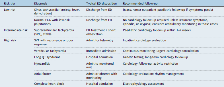
The main challenge for emergency physicians is the accurate identification of low-risk patients who can be safely discharged, in contrast to patients who require further and more specialised evaluation. The disposition logic hinges on the risk stratification performed through history, physical exam, and a 12-lead electrocardiogram (Table 2). Children with benign findings, such as sinus tachycardia with a clear physiologic trigger (e.g. fever, anxiety) and a normal electrocardiogram, can be safely discharged with reassurance and instructions for outpatient follow-up if symptoms persist. Reference Dhillon1
Final disposition decision matrix integrating diagnosis, risk tier, and recommended follow-up timeline. (Conceptual and derived from the current narrative)

Table 2. Long description
A table categorizing diagnoses by risk tier and recommended follow-up. The table has four rows and four columns. Column headers are Risk tier, Diagnosis, Typical ED disposition, and Recommended follow-up. Row 1: Low risk, Sinus tachycardia (anxiety, fever, dehydration), Discharge from ED, Reassurance; outpatient paediatric follow-up if symptoms persist. Row 2: Low risk, Normal ECG with low-risk palpitations, Discharge from ED, No cardiology follow-up required unless recurrent symptoms, episodic, or atypical; consider ambulatory monitoring in those cases. Row 3: Intermediate risk, Supraventricular tachycardia (SVT), stable, ED treatment short observation, Paediatric cardiology follow-up within 1-2 weeks. Row 4: High risk, SVT with recurrence or poor response, Admit for telemetry, Inpatient cardiology evaluation. Row 5: High risk, Ventricular tachycardia, Immediate admission, Continuous monitoring; urgent cardiology consultation. Row 6: High risk, Long QT syndrome, Hospital admission, Genetic testing; long-term cardiology follow-up. Row 7: High risk, Myocarditis, Admit to monitored unit, Cardiology follow-up; activity restriction. Row 8: High risk, Atrial flutter, Admit or observe with monitoring, Cardiology evaluation; rhythm management. Row 9: High risk, Complete heart block, Hospital admission, Electrophysiology assessment.
ED = emergency department; ECG = electrocardiogram; QT = QT interval in electrocardiogram; SVT = supraventricular tachycardia.
Patients with intermediate-risk features, such as a first episode of haemodynamically stable supraventricular tachycardia that resolves with or without intervention, may be suitable for discharge after a period of observation, with prompt outpatient cardiology follow-up arranged. Reference Abbasi, Vijayashankar and Goldman35,Reference Przybylski, Michelson, Neuman, Porter, Alexander and Lyons36 However, those with recurrent supraventricular tachycardia, poor response to initial treatment, or any concerning features may require admission for telemetry monitoring and inpatient cardiology evaluation. Reference Przybylski, Michelson, Neuman, Porter, Alexander and Lyons36
High-risk patients, identified by red-flag features (e.g. syncope, exertional symptoms, abnormal electrocardiogram suggesting channelopathy or ventricular tachycardia), require immediate admission for continuous monitoring and urgent cardiology consultation. Reference Cepeda-Nieto, Roblero-Aguilar, Martínez-López and Balderrábano-Saucedo21 This group includes those with suspected myocarditis, new-onset complete heart block, or atrial flutter, which, while rare, necessitates inpatient management. Reference De Nigris, Arenella and Di Nardo37 The key is to use the initial risk assessment to guide disposition, ensuring that high-risk children receive immediate, specialised care while avoiding unnecessary hospitalisation for the large majority with benign symptoms. Reference Goto, Witkowska, Slusarczyk, Grotek, Dziubinski and Clark22,Reference Li, Kallas, LePoidevin, Hart, Franciosi and Sanatani38 While the gold standard for diagnosing arrhythmia is performing an electrocardiogram at the time of symptom onset, many patients present after symptoms have resolved. In such cases, ambulatory cardiac monitoring with electrocardiogram or Holter monitoring can be helpful for diagnosis post-discharge for select intermediate-risk patients. Reference Leng, Aldalati and Homme33
Future directions
The development in wearable electrocardiogram technology, artificial intelligence-enabled electrocardiogram interpretation, and standardised emergency department protocols can manage paediatric arrhythmias and syncope. Current limitations in validation, risk stratification, and economic evaluation reveal research gaps that future studies must address to improve emergency department decision-making and reduce unnecessary testing. This chapter covers important aspects related to paediatric-specific validation, prospective registries, and integrated care system. Reference Littell, Roelle and Dalal39,Reference Winder, Marietta, Kerr, Puchalski and Zhang40
Wearable electrocardiograms in paediatrics
Wearable electrocardiogram devices, including smartwatches, offer noninvasive, ambulatory monitoring suitable for detecting intermittent arrhythmias missed during brief emergency department evaluations. While studies on devices like the Apple Watch Series 6 show feasibility in paediatric populations, significant validation gaps remain, particularly across diverse age groups from neonates to adolescents. Reference Littell, Roelle and Dalal39 Younger patients present unique challenges due to smaller size, higher heart rates, and motion artefacts, highlighting the need for paediatric-specific algorithms. Post-emergency department use of wearables could enable prolonged monitoring for intermediate-risk patients, but research is needed to define optimal protocols and address barriers to clinical integration, such as data management and false alarms. Reference Zahedivash, Chubb, Giacone, Boramanand and Dubin41 Future randomised controlled trials should compare wearable-enabled discharge strategies against standard admission to evaluate safety, diagnostic yield, and cost-effectiveness.
Artificial intelligence-electrocardiogram advancements
AI-electrocardiogram algorithms show promise in identifying subtle high-risk features better than human interpretation. Expert-level deep neural networks have demonstrated high accuracy in paediatric electrocardiogram diagnosis in research settings. Reference Mayourian, La Cava, de Ferranti and Mah42 However, their translation to the chaotic emergency department environment is a major hurdle. Emergency department-specific validation is lacking, and models must be tested against the noisy tracings and confounding comorbidities common in acute care. While artificial intelligence could potentially reduce interpretation variability, significant barriers to adoption exist, including the need for explainable artificial intelligence models to build clinician trust, seamless integration into electronic health records, and rigorous prospective testing to ensure they do not increase false positives or miss low-prevalence conditions like long QT syndrome. Reference Pantelidis, Ruipérez-Campillo, Vogt and Antonopoulos43,Reference Follansbee, Landstrom and Aziz44
Emergency department arrhythmia registries
Prospective registries are the cornerstone for defining true arrhythmia incidence, validating red-flag criteria (such as exertional syncope and family history), and connect emergency department dispositions to outcomes such as sudden cardiac death or recurrent events. Existing quality improvement efforts lowered paediatric syncope testing by 50% without safety signals achieved by current quality improvement initiatives. Reference Winder, Marietta, Kerr, Puchalski and Zhang40 When combined with inpatient and ambulatory cohorts, multicentre emergency department registries could serve as a benchmark for national practices and reveal areas that could be improved.
Standardised phenotyping for inherited arrhythmias, including genetic correlations such as those in long QT or catecholaminergic polymorphic ventricular tachycardia, is one of the main deliverables. In order to create comprehensive risk models, registries should incorporate wearables and artificial intelligence outputs, track patients over time (for example, one to five years), and record social determinants. Funding from bodies like the Pediatric Heart Network could facilitate this, which would produce predictive scores similar to adult CHADS2-VASc for customised follow-up. Reference Winder, Marietta, Kerr, Puchalski and Zhang40,Reference Mariani, Pierucci, Fanisio and Laviola45 Such infrastructure would empower meta-analyses, refining guidelines from bodies like the American Academy of Pediatrics and informing public health campaigns on arrhythmia awareness.
Standardised emergency department pathways
Risk-based algorithms can help reduce unnecessary testing, such as echocardiograms, Holters, and event monitors, while also protecting high-risk cases. Minimal (observation), intermediate (wearables), or comprehensive (admission) pathways could result from algorithms based on patient history, electrocardiogram results, and artificial intelligence scores. This is similar to syncope quality initiative successes. Reference Winder, Marietta, Kerr, Puchalski and Zhang40 Testing through stepped-wedge cluster trials in emergency rooms would confirm that safety metrics, such as missed diagnoses, are unaffected.
Adding channelopathy, red flags, and speckle-tracking insights for premature ventricular contractions-induced cardiomyopathy improves the accuracy of resource allocation. Future protocols must address implementation challenges, including getting clinicians on board through simulation training and tools integrated into electronic health records. Patient-centred designs, developed in collaboration with families, could improve compliance and fairness. Reference Winder, Marietta, Kerr, Puchalski and Zhang40,Reference Follansbee, Landstrom and Aziz44
Cost-effectiveness evaluations
Formal health economic assessments are essential but currently lacking for minimal-testing paradigms in paediatric palpitations. While such approaches promise savings by avoiding admissions and tests, the upfront costs of technologies such as wearables and artificial intelligence must be considered. Robust Markov models using registry-derived data are needed to evaluate the long-term cost-effectiveness and quality-adjusted life years compared to standard care. Reference Winder, Marietta, Kerr, Puchalski and Zhang40 Paediatric-specific valuations, which account for the long-term impact of arrhythmias, remain an underexplored but critical area for future research to inform payers and policymakers. Reference Winder, Marietta, Kerr, Puchalski and Zhang40
Conclusion
A structured, patient-centred, risk-based approach enables the safe discharge of most children with palpitations while ensuring the early identification of inherited and acquired arrhythmias with life-threatening potential. The evaluation of paediatric palpitations in the emergency department requires balancing vigilance for rare but serious conditions against the harms of over-investigation in a predominantly benign population. This review affirms that a meticulous clinical history focused on red-flag features, a thorough physical examination, and a standard 12-lead electrocardiogram form the cornerstone of a safe and effective evaluation. The absence of red flags in a child with a normal electrocardiogram is a powerful predictor of a benign outcome, allowing for confident discharge and reassurance. Conversely, the presence of any red flag mandates a higher level of investigation and specialist consultation. Looking forward, the integration of validated technologies such as wearable monitors and artificial intelligence-driven electrocardiogram analysis into standardised clinical pathways holds promise. However, their implementation must be guided by rigorous, paediatric-specific research to address current gaps in validation and cost-effectiveness. Ultimately, refining these evidence-based, risk-stratified pathways will optimise resource utilisation, reduce iatrogenic harm, and ensure that the small number of children with dangerous underlying conditions receive the timely, life-saving care they require.
Acknowledgements
Not applicable.
Author contributions
Khaled Abouelmagd served as the lead author, taking primary responsibility for study design, data acquisition, project coordination, facilitating communication among co-authors, and drafting and revising the initial manuscript. Temitomi Jane Oyedele, Gabriela Berger, Sarah Fakhiraldeen, Habiba Ali, Nouran Hishmat, and Atiqullah Sadaqat contributed equally to data collection, analysis, interpretation, and drafting and revising specific sections of the manuscript. Lauren A. Carr, revise the final draft of the manuscript. Mohammed Alsabri acted as the senior and corresponding author, conceptualised the research idea, oversaw the project, participated in manuscript drafting, and provided critical revisions to ensure that the final version met high intellectual and scientific standards. All authors reviewed, approved the final manuscript, and accepted responsibility for the integrity and accuracy of the work.
Financial support
Not applicable
Competing interests
We confirm that there is no conflict of interest, and this manuscript has not been published elsewhere and is not under consideration by another journal.
Ethical standard
Not applicable.
Consent for publication
All authors have approved the manuscript and agree with its submission and publication to the journal.
Availability of data and materials
All data generated or analysed during this study are included in this published article.
Clinical trial number
Not applicable.
Checklist usage
Not applicable.
Consent to participate
Not applicable.



 Open access

