Systemic right ventricular function in congenitally corrected transposition of the great arteries is partly influenced by interventricular septal and ventricular geometry. A leftward shift of the ventricular septum after physiological repair pulls the tricuspid leaflets apart, leads to tricuspid incompetence and right ventricular failure. Right ventricular function is preserved after pulmonary artery banding.Reference Kral Kollars, Gelehrter, Bove and Ensing 1 In cyanosed patients with congenitally corrected transposition and pulmonary stenosis, Fontan-type surgery is sometimes preferred as the chamber geometry is unaltered. Ventricular function is an important prerequisite for Fontan candidacy. An intact ventricular septum after Fontan surgery leads to a left ventricle with suprasystemic pressure.
Case report
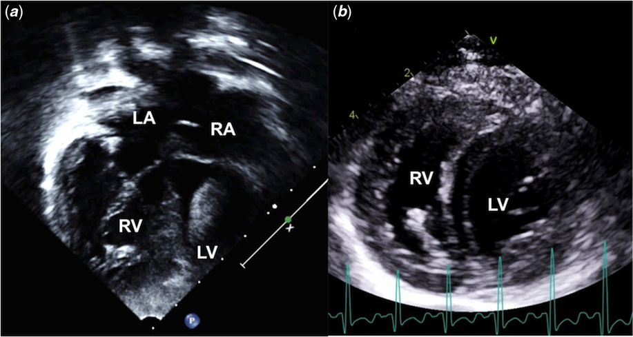
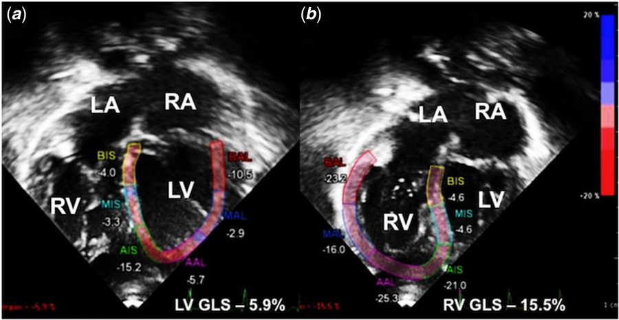
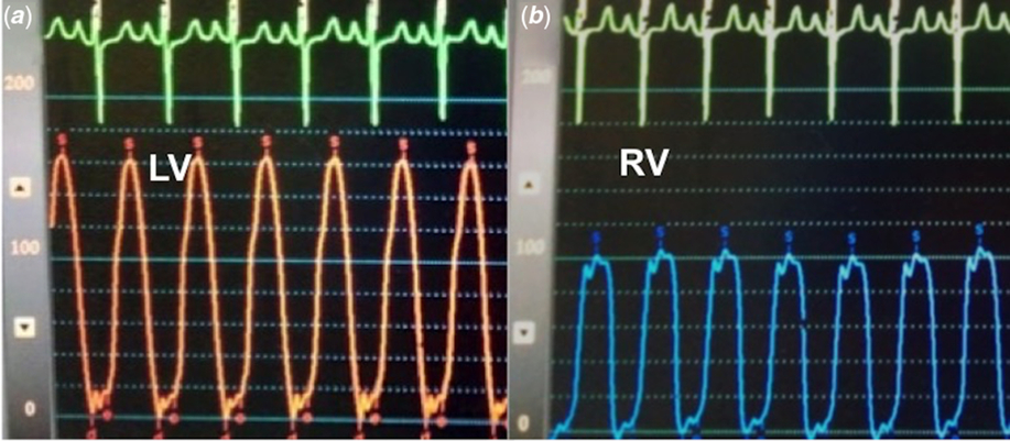
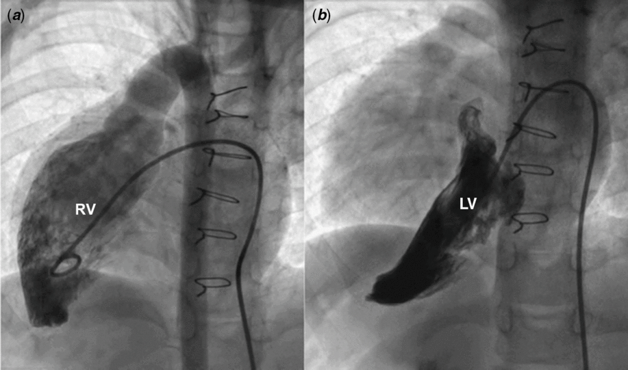
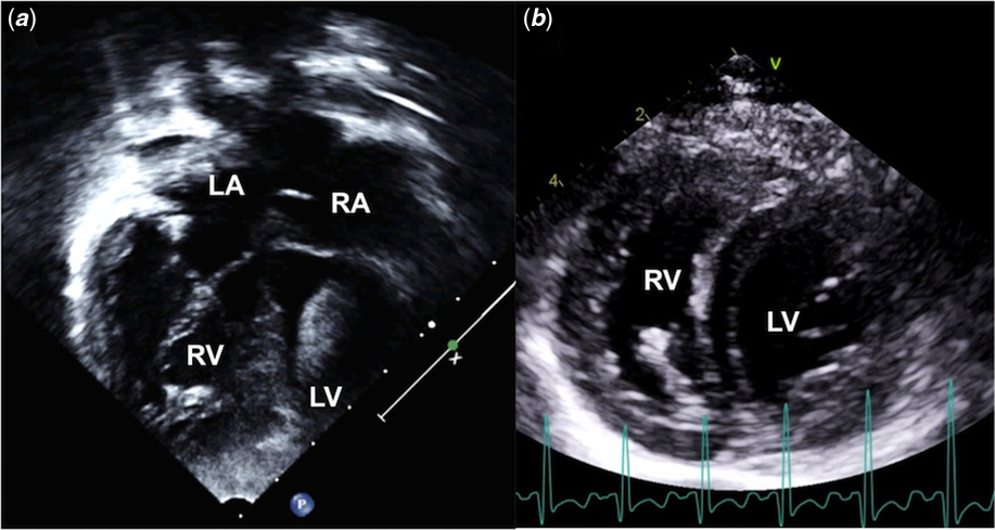
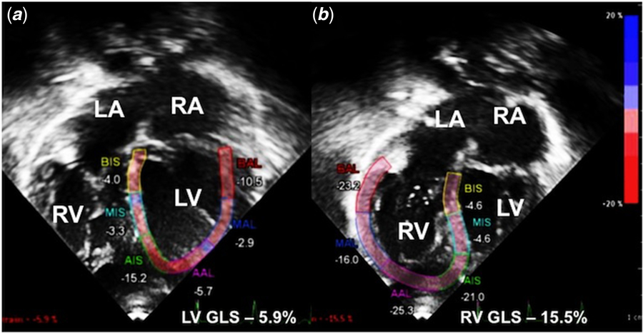
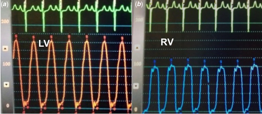
Left ventricular hypoplasia in a 4-year-old girl with congenitally corrected transposition of the great arteries, a restrictive ventricular septal defect and severe pulmonary stenosis precluded biventricular repair and warranted a bidirectional superior cavopulmonary Glenn shunt. After 3 years, spontaneous closure of the ventricular septal defect led to a left ventricle with suprasystemic pressure, altering the septal geometry and favourably preserving the crescentic right ventricular shape and function (Figs 1 and 2, Supplementary video 1a, 1b). As left ventricular decompression by a conduit was not desirable, she underwent Fontan-type completion uneventfully. Even though a suprasystemic left ventricle with high end-diastolic pressures is undesirable in a Fontan circulation, congenitally corrected transposition offers a paradox (Fig 3). The septal shift normalised the systemic right ventricular and tricuspid valve function and geometry on follow-up (Fig 4, Supplementary video 2a, 2b). Thus corrected transposition offers a paradox in candidacy for Fontan-type surgery.
Apical view (a) shows situs inversus with atrioventricular discordance, there is dense spontaneous echo contrast in the LV. A parasternal short axis view (b) shows preservation of the crescentic shape of the RV. LA = left atrium; LV = left ventricle; RA = right atrium; RV = right ventricle.
Speckle tracking shows global longitudinal strain of the LV (a) of −6%. Even though the global right ventricular longitudinal strain is −16%, the right ventricular free wall segmental strain values (b) of −23, −16, −25 and −21% indicate preserved right ventricular function. LA = left atrium; LV = left ventricle; RA = right atrium; RV = right ventricle.
LV (a) showing systolic pressures of 165 mmHg, triangular contour, elevated end-diastolic pressures of 22 mmHg. RV (b) showing systolic pressures of 100 mmHg, similar to aortic systolic pressures, normal end-diastolic pressures of 6 mmHg and a rectangular contour. LV = left ventricle; RV = right ventricle.
Right ventriculogram (a) shows situs inversus, dextrocardia, d-looped discordant ventricles, good contractility, absence of dilatation and tricuspid regurgitation, and d-malposed aorta; Left ventriculogram (b) shows complete spontaneous closure of the ventricular septal defect, surgically closed pulmonary artery and the mitral valve as the only egress for the ventricle. LV = left ventricle; RV = right ventricle.

Acknowledgements
None.
Financial Support
This research received no specific grant from any funding agency, commercial or not-for-profit sectors.
Conflicts of Interest
None.
Ethical Standards
The authors assert that all procedures contributing to this work comply with the ethical standards of the Indian Council of Medical Research and with the Helsinki Declaration of 1975, as revised in 2008, and has been approved by the institutional committees of Madras Medical Mission, Chennai, India.
Supplementary material
To view supplementary material for this article, please visit https://doi.org/10.1017/S1047951119002324