Introduction
There exist numerous preantral (primordial, primary and secondary) follicles in the ovaries, but in one's life only less than 1% typically develop into the Graafian follicles that could release mature oocytes into the fertilization site (Moore & Persaud, Reference Moore and Persaud1998). The rest remain ‘developmentally dormant’ in ovarian tissue and finally became degenerated via apoptosis. In the field of animal biotechnology, efforts have been made over the last decade to utilize preantral follicles for increasing reproductivity and further application of the preantral follicle culture has been subsequently suggested. As a result, follicular oocytes derived from in vitro-cultured secondary follicles have been developed (Cortvrindt et al., Reference Cortvrindt, Smitz and Van Steirteghem1996; Eppig & Schroeder, Reference Eppig and Schroeder1989) and a marvellous success to generate preimplantation embryos by in vitro manipulation of embryonic stem (ES) cells has also been reported (Hubner et al., Reference Hubner, Fuhrmann, Christenson, Kehler, Reinbold, Fuente, Wood, Strauss, Boiani and Scholer2003). Nevertheless, basic information on preantral follicle culture has not yet been reported and a standard protocol of follicle manipulation has not been established. Furthermore, the feasibility of the immature follicle culture technique should be confirmed in other strains and species.
In this study, a comparison of the efficiency between enzymatic and mechanical retrieval was made in order to select the optimal method. Subsequently, in vitro growth of the follicles retrieved by the optimal method and subsequent maturation of intrafollicular oocytes were monitored. We employed C57BL/6 × DBA2 F1 mice as the follicle donors, which were conventionally employed for cloning by somatic cell nuclear transfer and embryo culture models.
Materials and Methods
Experimental animals
Female F1 hybrid (C57BL/6 × DBA2) mice bred in the Laboratory of Embryology and Gamete Biotechnology, Seoul National University were maintained under controlled lighting (14 h light/10 h dark), temperature (20–22 °C) and humidity (40–60%) and 2-week-old sexually immature (prepubertal) females were subsequently provided for this study. All procedures for animal management, breeding and surgery followed the standard operation protocols of Seoul National University. Appropriate management of experimental samples and quality control of the laboratory facility and equipment were also conducted.
Isolation of preantral follicles
Females were sacrificed by cervical dislocation and their ovaries were removed aseptically. For mechanical isolation of follicles, the ovaries were placed in 2 ml Leibovitz L-15 medium (Sigma–Aldrich) supplemented with 10% (v/v) heat-inactivated fetal bovine serum (FBS) and 1% (v/v) lyophilized penicillin–streptomycin solution at 37 °C. Two types of retrieval methods were employed for this study. Preantral follicles were retrieved mechanically by using a 30 gauge needle (Lenie et al., Reference Lenie, Cortvrindt, Adriaenssens and Smitz2004). In the other example, for enzymatic retrieval, the collected ovaries were placed in ribonucleoside and deoxyribonucleoside-containing α-MEM–glutamax medium supplemented with 0.1% (v/w) collagenase type I (198 U/mg; Sigma–Aldrich), 0.02% (v/w) DNase I (11.2 U/mg; Sigma–Aldrich) and 0.03% (v/v) fetal bovine serum (FBS) for 1 h at 37 °C. To facilitate proteolytic digestion, the ovaries were mixed every 30 min by gentle pipetting (Pangas et al., Reference Pangas, Saudye, Shea and Woodruff2003).
Culture of preantral follicles
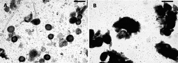
Preantral follicles, isolated either mechanically or enzymatically, were washed three times in 10 μ1 droplets of Leibovitz L-15 medium and subsequently classified into three categories by measuring the diameter with the ocular micrometer of an inverted microscope (TE-2000; Nikon, Tokyo, Japan) at ×40 magnification. The selection criteria were as follows: primary follicle 75–99 μm, early secondary follicle 100–125 μm and late secondary follicle 126–180 μm in diameter. In addition to the size of the follicles, the typical morphology of the preantral follicles was employed for classification (Fig. 1): primary follicles had a round follicular structure consisting of a single compact layer of granulosa cells and a follicular oocyte. Early and late secondary follicles also had a round structure consisting of multiple layers of granulosa cells and a follicular oocyte. All categorized follicles were subsequently cultured at 37 °C, 5% CO2 in an air atmosphere.
Morphology of preantral follicles at retrieval (×120). The preantral follicles were collected either singly (A) or in groups (B). The follicles collected in groups were difficult to separate from each other and were not suitable for single preantral culture using microdroplet (scale bar 250 μm).

In vitro growth of primary and secondary follicles
The primary follicles were placed singly in 10 μl culture droplets overlaid with washed mineral oil (Sigma-Aldrich) in 60 × 15 mm Falcon plastic Petri dishes (Becton Dickinson). The medium used for the culture of primary follicles is ribonucleoside- and deoxyribonucleoside-free α-MEM–glutamax medium, to which 1% (v/v) heat-inactivated fetal bovine serum (FBS), 5 μg/ml insulin, 5 μg/ml transferrin, 5 ng/ml selenium, 100 mIU/ml recombinant human FSH (Organon), 10 mIU/ml LH (Sigma–Aldrich) and 1% (v/v) penicillin and streptomycin were added. On day 1 of culture, an additional 10 μl fresh medium was added to each droplet and half of a medium was changed everyday from day 3 of culture (Lenie et al., Reference Lenie, Cortvrindt, Adriaenssens and Smitz2004). Cultured follicles were frequently detached from the bottom of culture dishes by mechanical pipetting. When the diameter of the follicles reached 100 μm (approximately on day 5 of culture), they were placed into 10 μl droplets of ribonucleoside and deoxyribonucleoside-containing α-MEM–glutamax medium supplemented with FBS, insulin, transferrin, selenium, FSH and antibiotics. On the following day, 10 μl of fresh medium was added to each droplet and, from the third day after replacement, half the medium was replaced every other day (Lenie et al., Reference Lenie, Cortvrindt, Adriaenssens and Smitz2004).
The secondary follicles were also cultured individually and the culture protocol was similar to that for primary follicles except for the use of ribonucleoside and deoxyribonucleoside-containing α-MEM–glutamax medium. The morphological change in preantral follicles was monitored everyday throughout their culture.
Assessment of the maturation of follicular oocytes
To induce maturation of follicular oocytes in preantral follicles, 2.5 IU/ml hCG (Pregnyl™) and 5 ng/ml epidermal growth factor (Sigma–Aldrich) were added to the culture medium 16–18 h prior to culturing for oocyte maturation. Progress of meiotic maturation was monitored by staining oocytes with Lacmoid solution and the occurrence of germinal vesicle (GV) and GV breakdown (GVBD) in oocytes that did not have a first polar body was examined under a phase-contrast microscope. Oocyte maturation, developed to the metaphase II (MII) stage, was evaluated by the extrusion of the first polar body and by mucification and expansion of cumulus cells enclosing the oocyte. To monitor the extrusion of the first polar body, oocytes retrieved from cultured follicles were freed from cumulus cells by mechanical pipetting in M2 medium supplemented with 200 IU/ml hyaluronidase.
The capacity of matured oocytes to form a pronucleus in order to indirectly confirm cytoplasmic maturation was monitored after parthenogenetic activation using Ca2+-free KSOM medium supplemented with 10 mM SrCl2 and 5 μg/ml cytochalasin B. The formation in activated oocytes was assessed by Hoechst staining under an inverted fluorescence microscope. Conversely, the size (diameter) and zona thickness of MII stage oocytes derived from the cultured preantral follicles were also monitored under an inverted microscope equipped with an ocular micrometer.
Statistical analysis
A generalized linear model (PROC–GLM) in a Statistical Analysis System (SAS) program was employed and significant differences among treatments were determined where the p-value was less than 0.05.
Results
Comparison of retrieval efficiency
Total 2432 preantral follicles were retrieved from the ovaries by two different methods. When cell population was compared at retrieval, the number of early secondary follicles was larger (p < 0.0001) than that of primary and late secondary follicles (1249 cells vs. 485–698 cells), regardless of the retrieval methods (realloted data of Table 1). As shown in Fig. 1, the preantral follicles collected from the ovaries were present singly or in groups. In the case of follicles being collected in groups, it is difficult to separate single follicles from the complexes, thus single culture of follicles collected in groups was not possible.
Retrieval of preantral follicles of different stages (primary, early secondary and late secondary) by either a mechanical or an enzymatic (use of collagenase and DNase) method

Total 16 female F1 mice were sacrificed and each treatment replicated eight times.
Model effects in the total number of preantral follicles retrieved, subtotal number of the follicles retrieved singly and in groups was less than 0.0001 (p-values).
aMean ± SD.
b, cDifferent superscripts within a column indicate values that are significantly different, p < 0.05.
As shown in Table 1, the total number of preantral follicles retrieved per mouse was larger (p < 0.0001) when using the mechanical method than when using the enzymatic method (339 ± 48 cells vs. 202 ± 28 cells). Due to the enzyme treatment, the degree to which preantral follicles aggregated to each other was very low. The number of primary, early secondary and late secondary follicles retrieved in groups by the mechanical method was 84±14, 97±12 and 56 ± 17 cells, respectively. The enzymatic method yielded more (p < 0.0001) preantral follicles collected as a single complex than the mechanical method (202 ± 28 cells vs. 102 ± 26 cells). An increased number of primary (52 ± 12 cells vs. 35 ± 9 cells), early secondary (110 ± 18 cells vs. 46 ± 13 cells) and late secondary (39 ± 12 cells vs. 21 ± 7 cells) follicles in the enzymatic retrieval was detected.
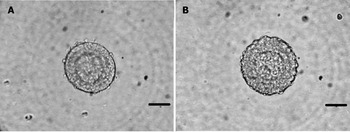
As shown in Fig. 2, the preantral follicles retrieved by the mechanical method had spherical shapes and their basement membranes remained intact. Only a few theca cells were still attached to the basement membrane. The preantral follicles retrieved by the enzymatic method lost the basement membrane partly or wholly and the theca cells no longer attached to the follicles. The cytoplasm, especially in the marginal region, of the preantral follicles retrieved by the enzymatic method became coarse compared with that of the follicles collected by the mechanical method.
Morphological difference of preantral follicles and follicular oocytes retrieved from mouse (C57BL/6 × DBA2) ovaries by different methods. Either a mechanical method using a syringe needle or an enzymatic method using collagenase and DNase was employed. (A) The follicle retrieved by the mechanical method (day 0 of culture): basement membrane was intact and several theca cells were still attached to the membrane (×600). (B) The follicle retrieved by the enzymatic method (day 0 of culture): the basement membrane was not visible and the theca cells were completely detached from the membrane (×600), (scale bar, 50 μm).

In vitro growth of preantral follicles
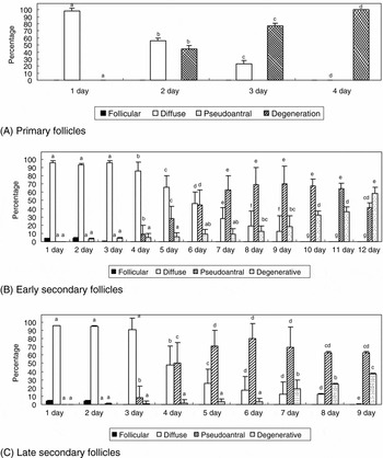
Only the enzymatic method was used for follicle retrieval based on our previous results (Table 1). Regardless of the types of preantal follicles, all follicles cultured in vitro went through step-by-step growth from the follicular to degenerative stages via diffuse and pseudoantral stages (Fig. 3). In the case of primary follicles, the major proportion (97%) entered into the diffuse stage on day 1 of culture (97%). However, no follicles developed to the pseudoantral stage and all of the follicles become degenerated by day 4 of culture. In the case of early secondary follicles, the pseudoantral stage was firstly detected on day 4 of culture. The incidence of follicles that had developed to the diffuse and pseudoantral stages peaked on day 4 (86%) and 9 (70%) of culture. For late secondary follicles, the incidence of cells at the diffuse stage peaked on day 3 (91%) of culture. Follicles that had developed to the pseudoantral stage first appeared on day 3 (8%) of culture and the incidence peaked on day 7 (63%) and day 6 (80%) of culture, respectively.
In vitro growth of preantral follicles retrieved by an enzymatic method. Primary, early secondary and late secondary follicles were cultured in α-MEM–glutamax medium supplemented with fetal bovine serum, insulin, transferrin, selenium, FSH and antibiotics and grown in vitro to reach follicular (black bar), diffuse (white), pseudoantral (diagonal) and degenerative (hatched) stages and these were monitored daily under an inverted microscope. The values indicate the mean percentage ± SD. (A) Growth of primary follicles: all follicles retrieved ceased their growth at the diffuse stage after day 4 of culture. (B) Growth of early secondary follicle: the incidence of the pseudoantral stage peaked on day 9 (70%) of culture. (C) Growth of late secondary follicles: the peak of pseudoantrum formation was on day 6 (80%) of culture. Different letters in the same stage of follicle development demonstrated a significant (p < 0.05) difference among observation times.

Maturation and pronuclear formation of intrafollicular oocytes
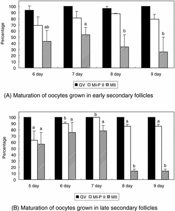
No primary follicles retrieved by the enzymatic method developed to the pseudoantral stage (Fig. 4). Oocytes that were present in early secondary follicles reached the MII stage after day 6 (43%) of culture and the optimal time to retrieve MII stage oocytes was on day 7 (54%) of culture. In the case of late secondary follicles, oocytes reached the MII stage from day 5 (57%) of culture and the peak time for oocyte maturation was day 7 (78%) of culture.
Meiotic maturation of oocytes derived from the pseudoantral stage of primary, early secondary or late secondary follicles retrieved by an enzymatic method. Maturational status was monitored daily and hCG and epidermal growth factor was added into the culture medium 16 h prior to the oocyte maturation culture. The values indicate the mean percentage ± SD and the percentage of oocytes that developed to germinal vesicle (GV), germinal vesicle breakdown (GVBD) and metaphase II (MII) stages were monitored at each time of observation. (A) Maturation of oocytes grown in early secondary follicles, significant increase in MII oocytes was detected on day 7 (54%). (B) Maturation of oocytes grown in late secondary follicles, significant increase in MII oocytes was detected from day 7 (78%). Different letters in the same category of follicle development demonstrated a significant (p < 0.05) difference between observation times.

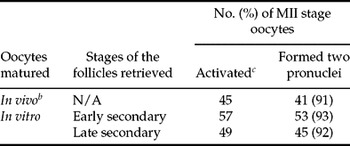
As shown in Table 2, oocyte diameter generally decreased in all oocytes derived from in vitro-cultured follicles compared with oocytes derived in vivo (65.2–65.53 μm vs. 75 μm). Thickness was significantly lower in the follicle-derived oocytes (5.41–5.74 μm vs. 7.76 μm). As shown in Table 3, the rate of pronuclear formation after parthenogenetic activation was within the range 86–94% and 91% of in vivo-derived oocytes formed pronuclei after activation. No significant difference was detected between treatments.
Effects of follicle retrieval methods on the thickness of zona pellucida and the diameter of metaphase II (MII) stage oocytes grown in in vitro-cultured primary, early secondary or late secondary follicles

Model effects in the thickness of zona pellucida and the diameter of oocytes were less than 0.0001 (p-values).
a, b, cDifferent superscripts within a column indicate values that are significantly different, p < 0.05.
Formation of pronucleus after the parthenogenetic activation of mature oocytes derived from primary, early secondary or late secondary follicles a cultured in vitro

Model effect in the number of MII oocytes formed pronuclei was 0.972 (p-values).
aPreantral follicles cultured were retrieved from the ovaries by two different methods.
bOocytes were collected from the oviduct flushing after natural ovulation.
cParthenogenetic activation was conducted by the treatment with SrCl2 and cytochalasin B.
Discussion
This is the first report of the culture of preantral follicles derived from C57BL/6 × DBA2 F1 mice, one of major hybrid strains used for nuclear transfer and embryo culture. Intrafollicular oocytes in early or late secondary follicles developed to MII stage (mature) by subsequent culture and formed pronuclei after artificial activation. The efficiency of follicle retrieval could be improved by using an enzymatic method with collagenase and DNase and more than 200 follicles per ovary could be provided for culture when using the optimized method.
In rodents, several methods for culturing preantral follicles from another F1 hybrid strain (C57BL/6 × CBA/Ca) have been developed. It has been reported that oocytes derived from in vitro-cultured secondary follicles acquire full maturity and develop further into viable offspring following in vitro fertilization and embryo transfer. (Cortvrindt et al., Reference Cortvrindt, Smitz and Van Steirteghem1996; Eppig & Schroeder, Reference Eppig and Schroeder1989; Nayudu & Osborn, Reference Nayudu and Osborn1992; Roy & Greenwald, Reference Roy and Greenwald1985). Nevertheless, it has not been confirmed whether this established protocol could be used for the culture of preantral follicles from other strains and even detailed protocols for follicle manipulation have not been standardized. The results of this study confirmed that, at least to ensure that oocytes have the capacity to mature and to be parthenogenetically activated, the basic follicle culture protocol for C57BL/6 × CBA/Ca mice could be applied when culturing follicles of other F1 mice (such as C57BL6 × DBA2).
A single cell culture system (Cortvrindt et al., Reference Cortvrindt, Smitz and Van Steirteghem1996) was employed for culturing preantral follicles at different stages, which were retrieved from 2-week-old prepubertal mice by mechanical or enzymatic (Roy & Greenwald, Reference Roy and Greenwald1996) treatment. This system is beneficial for scrutinizing the various effects of exogenous substrates on the development of single follicles, without the interference from adjacent follicles and for monitoring the capacity of individual follicles to grow in vitro. The results suggest that the basic composition of α-MEM medium was at least suitable for supporting in vitro growth of preantral follicles and the maturation of intrafollicular oocytes. However, it could not be determined if α-MEM medium was the optimal culture medium for supporting embryogenesis of intrafollicular oocytes after fertilization or parthenogenetic activation. In fact, intrafollicular oocytes from primary follicles were impossible to mature and it was apparent that critical factors for the growth of primary follicles and their intrafollicular oocytes were not present in the culture system employed. Probably, different culture regimes were required for optimizing the yield of developmentally competent oocytes from in vitro-cultured preantral follicles. This hypothesis is supported by our results, which showed that the capacity of the follicles to grow in vitro was increased as the follicle stage at retrieval was advanced: the period to reach pseudoantral stage was shortened and required duration for maturing intrafollicular oocytes also decreased.
When compared with previous data obtained from other (C57BL/6 × CBA/Ca) F1 mice (Cortvrindt & Smitz, Reference Cortvrindt and Smitz1998; Cortvrindt et al., Reference Cortvrindt, Smitz and Van Steirteghem1996, Reference Cortvrindt, Hu, Liu and Smitz1998a; Demeestere et al., Reference Demeestere, Delbaere, Gervy, Van Den Bergh, Devreker and Englert2002; Smitz & Cortvrindt, Reference Smitz and Cortvrindt2002), the results of this study showed strain specificity for follicle growth and oocyte maturation. The secondary follicles derived from C57BL/6 × DBA mice required 5–7 days to reach the pseudoantral stage, while the follicles derived from C57BL/6 × CBA mice required 12–14 days (Cortvrindt & Smitz, Reference Cortvrindt and Smitz1998; Cortvrindt et al., Reference Cortvrindt, Smitz and Van Steirteghem1996, Reference Cortvrindt, Hu, Liu and Smitz1998a; Demeestere et al., Reference Demeestere, Delbaere, Gervy, Van Den Bergh, Devreker and Englert2002). It has been reported that a number of either exogenous or endogenous substrates greatly influence follicle growth: various gene families such as growth differentiation factor and bone morphogenic proteins (Dong et al., Reference Dong, Albertini, Nishimori, Kumar, Lu and Matzuk1996; Elvin et al., Reference Elvin, Yan and Matzuk2000) critically stimulate follicular growth, while exogenous growth factors, gonadotrophins and inorganic substrates affect development (Cortvrindt et al., Reference Cortvrindt, Hu, Liu and Smitz1998a, Reference Cortvrindt, Hu and Smitzb; Eppig et al., Reference Eppig, O'Brien, Pendola and Watanabe1998).
Enzymatic retrieval increased the number of singly isolated preantral follicles that were provided for culture in vitro (Table 1), so further increasing the number of preantral follicles singly isolated from the ovaries, which improved the efficiency of the single culture system used in this study. However, the follicle retrieval method also influenced the morphology of preantral follicles. From observations using microscopy, collagenase and DNase induced breakdown of the basement membrane and detachment of granulosa cells from the membrane (Demeestere et al., Reference Demeestere, Delbaere, Gervy, Van Den Bergh, Devreker and Englert2002; Roy & Treacy, Reference Roy and Treacy1993). Furthermore, the enzymes used for retrieval profoundly affected oocyte structures such as the zona thickness and oocyte diameter (Table 2). Other reports (Epigg & O'Brien, Reference Epigg and O'Brien1998; Liu et al., Reference Liu, Van Der Elst, Van Den Broecke, Dumortier and Dhont2000) also demonstrated that a decrease in diameter of approximately 60–70% of original length was detected in mature oocytes derived from in vitro-cultured follicles.
The results from parthenogenetic activation clearly show that follicular oocytes retrieved from cultured follicles acquired maturity during in vitro culture. There was no difference in the capacity to form pronucleus among oocytes derived from primary, early secondary and late secondary follicles (Table 3). However, detailed analysis on developmental competence of oocytes derived from in vitro-cultured preantral follicles is required. Further studies are now being conducted to evaluate the developmental competence of oocytes grown in cultured follicles and efforts have been made to produce viable blastocysts; the information from these further studies might confirm the applicability of the optimal retrieval and culture regime developed in this study for human medicine and medical biotechnology.
Acknowledgements
We greatly appreciate Dr Jeffery A. Hubbell for his critical review of this manuscript. The authors also acknowledge Mrs Jong E. Ihm for her technical assistance in follicle culture. This research was supported by a grant from the Stem Cell Research Center of the 21st Century Frontier Research Program funded by the Ministry of Science and Technology, Republic of Korea. The authors also acknowledge a graduate fellowship provided by the Korean Ministry of Education through the Brain Korea 21 project.