




No CrossRef data available.
Current issues and perspectives in hypoplasia of the left heart
Published online by Cambridge University Press: 14 April 2005
Abstract
Hypoplastic left heart syndrome is a rare but serious form of congenital cardiac disease, characterized by underdevelopment of the components of the left heart, rendering the left ventricle non-functional. Its aetiology is largely unknown, but there is certainly a genetic component. Prenatal diagnosis nowadays uncovers about half of cases. Postnatal options for treatment include comfort care, 3-stage palliative surgery, or cardiac transplantation. In this review, we discuss the morphology, possible pathogenetic mechanisms, clinical management, and perspectives of prenatal intervention based on work in animal models.
Information
- Type
- Continuing Medical Education
- Information
- Copyright
- 2005 Cambridge University Press
The term “hypoplastic left heart syndrome” encompasses a morphologically diverse spectrum of malformations, characterized by underdevelopment of the components of the left heart. Functionally, the most important is presence of a diminutive left ventricle, leading to a functionally univentricular circulatory arrangement, with the morphologically right ventricle serving as the pump to both systemic and pulmonary circulations, with systemic perfusion being duct-dependent. Historically, pathologists have described individual cases since the nineteenth century (see Reference #1 for review of older literature). The definition of the condition stems from Lev,2 who used the term “hypoplasia of the aortic arch complex”. The following half-century witnessed a shift in perception of left heart hypoplasia, from a natural oddity that belongs in the autopsy room, to a complex but treatable, although not yet curable, disease. In this review, we will describe its morphology, discuss the possible aetiopathogenesis, describe experimental models, assess the current options for diagnosis and management, and finish with our perspectives concerning prenatal treatment.
Pathologic anatomy
The spectrum of hypoplasia of the left heart includes stenosis of the mitral and aortic valve, although either valve can be atretic, as well as the presence of other associated anomalies, such as anomalous pulmonary venous connections, atrial septal defects, hypoplasia of the aortic arch, anomalies of coronary arteries, and so on.3, 4 Some of these anomalies may influence the options for surgical treatment and its outcome, so detailed diagnostic evaluation of the anatomical features is essential. Left ventricular endocardial fibroelastosis is a crucial feature, found in the setting of a patent mitral and severely stenotic or atretic aortic valve, but not seen in the absence of flow of blood into the left ventricle, in other words, mitral atresia.4 From a clinical standpoint, the common denominator of the entire spectrum is a small, non-functional left ventricle (Fig. 1), hypoplasia of the ascending aorta, and a resulting univentricular circulatory physiology.

Figure 1. Morphology of left heart hypoplasia. Four-chamber views of fetal and newborn hearts showing small size and thick wall of the hypoplastic chamber. Note the whitish appearance of endocardial fibroelastosis in these cases with a patent mitral valve. Matching ultrasound pictures show the resolution available for diagnosis. The postnatal scan shows heart with atretic mitral valve and no demonstrable flow in the left ventricle. Inset shows hyperechogenic left ventricle with endocardial fibroelastosis (arrowheads) in another fetus with mitral and aortic stenosis. LA: left atrium; LV: left ventricle; RA: right atrium; RV: right ventricle.
From the anatomic standpoint, however, hypoplasia of the left heart is far from being a uniform disease. The morphology of individual cases will be dictated by the presence or absence of an inlet to the left ventricle, the presence or absence of a patent outflow tract, and any associated septal or other cardiac defects. It is possible, nevertheless, to divide the syndrome into broad sub-groups (Table 1). Any of these forms can be associated with any type of atrial arrangement, although more often than not the arrangement will be usual, or so-called “situs solitus”. Other, much more complex, types of malformation can occur, in which the “systemic” ventricle is hypoplastic, but is of morphologically right pattern, albeit that these are rare.5
Table 1. The morphology of left heart hypoplasia is variable, but can be divided into 5 broad categories. Although it is possible that there is a single common mechanism, evidence so far suggests it is more likely that there are multiple pathways leading to hypoplasia of the left heart.

The likelihood of encountering the differing forms of morphology will depend, in part, on the age-group being studied. For instance, hypoplasia of the left ventricle associated with marked hypoplasia of the aortic arch and coarctation is much more frequently seen in fetal life than postnatally. In contrast, hypoplasia associated with aortic valvar dysplasia and stenosis can progress with age, and so will be more obvious towards the end of fetal gestation and postnatally than earlier during fetal life.6 Within each sub-group, nonetheless, there are some consistent features that give some clues to the processes involved in the disease, and suggesting avenues for research.
Mitral and aortic atresia
Absence of a left atrioventricular connection or severe hypoplasia of the mitral valve results in a thin-walled and virtual left ventricle. This is most easily appreciated in hearts with both mitral and aortic atresia, which, in the majority of cases has resulted from muscular separation both between the left atrium and ventricle, and also between the left ventricle and aortic root. Thus, both the atrioventricular and ventriculo-arterial connections on the left side of the heart are effectively absent from the outset. As a result, the left ventricle is slit-like, and its posterior wall is much thinner than expected for age. In essence, the left ventricular cavity takes the appearance of a slit within the postero-inferior wall of the ventricular mass (Fig. 2, upper panel), but can be proven to be the remnant of the left ventricle histologically. If there is a ventricular septal defect present in this setting, then the left ventricle will be slightly wider, and the wall of the left ventricle will be proportionally thicker. The larger the ventricular septal defect (Fig. 2, lower panel), the more the dimensions of the left ventricle will approach normality. Similarly, in the presence of patent but severely hypoplastic mitral and aortic valves, as can be seen in the group with severe hypoplasia of the aortic arch, the thickness of the left ventricular free wall is proportional to its inlet. This implies that the thickness of the left ventricle is dependent, in part, on the flow of blood through it, whether the blood enters the left ventricle from the left atrium, via a patent, hypoplastic mitral valve, or from the right ventricle via a ventricular septal defect. In the presence of a large ventricular septal defect or multiple smaller defects and mitral atresia, the aortic valve will more often be patent and severely hypoplastic, rather than atretic. The aortic valvar leaflets are still usually abnormal, being fused together to varying degrees in a complete spectrum from mild stenosis to severe stenosis or atresia. Again, this situation is reflected in hearts with patent but severely hypoplastic mitral and aortic valves, indicating that aortic valvar patency and structure can be affected by the flow of blood, even if the blood is not entering the left ventricle by conventional means through the mitral valve. Of note, there is an important distinction to be made between the morphology of the fused aortic valvar leaflets in these settings, which are fused but thin and pliable, as compared to those in aortic valvar stenosis, which are fused but distinctly nodular, thickened, and dysplastic.

Figure 2. Phenotype of mitral and aortic atresia in the mid-trimester fetus. In the majority of fetuses, the left atrioventricular junction (yellow lines) has joined the muscular base of the atrial septum (*), walling the left atrium from the left ventricle. The presence of a ventricular septal defect (VSD) allows some blood flow to the left ventricle, resulting in a significant mitigation of the left ventricular hypoplasia.
Aortic atresia or stenosis with patent mitral valve
The second common theme is that, in the presence of an adequate inlet and an obstructed outlet, the left ventricle becomes thicker-walled. This also requires, however, that the ventricular septum is intact, since patients with aortic atresia and a large ventricular septal defect can have normally formed left ventricles. In the absence of such defects, the left ventricular free wall and ventricular septum become thickened, as does the endocardium. The endocardium becomes visible to the naked eye as a pearly white lining composed of fibroelastotic tissue (Fig. 1). In fetal life, this lining is highly cellular, containing numerous smooth muscle cells and fibroblasts, but following birth this cellular characteristic has all but disappeared, leaving interleaving layers of collagen and elastic fibres. This suggests that proliferation of the endocardium is a more active process prenatally, with the potential for alteration, than it is following birth. Overall, these features give the characteristic morphology of hearts with aortic atresia or severe stenosis and a patent mitral valve. The left ventricle, when seen in four-chamber section, appears as a globular mass embedded within the posterior ventricular wall, containing a small, fibrotic core (Fig. 1).
The presence of a thick-walled yet hypoplastic left ventricle can give rise to some confusion. For us, this is resolved by reserving the term ventricle for the cavity, and describing the surrounding ventricular walls independently. From an anatomic stance, it would be incorrect to use the term “left ventricular hypertrophy” to describe the thickening of the ventricular wall since, in essence, the thickening is due to myocytic hyperplasia rather than hypertrophy. Again, this can be seen more easily in fetuses. It indicates that much, if not all, of the thickening has occurred during fetal life, at a time when mitotic division is still prevalent. It is also important to remember that the ventricular hypoplasia is not only due to encroachment on the cavity by the thickened walls. In most hearts with aortic valvar atresia encountered in the mid-trimester of pregnancy, the left ventricular apex does not form the cardiac apex. Indeed, there is often a characteristic dimple at the distal extent of the left ventricle. Towards term, the distance between the apical extent of the left ventricle and apex of the heart is even greater (Fig. 1). It appears, therefore, that following an initial period of cellular hyperplasia, the left ventricle reaches a point of equilibrium, and then ceases to grow.
So what leads to atresia of the aortic valve in the first place? Several studies have illustrated that there can be considerable variation in size of the aorta in the setting of aortic atresia when the mitral valve is patent, albeit that in most the aortic root and ascending aorta will be patent but minute.7 There is some evidence from study of fetal hearts that the pathway between the left ventricle and ascending aorta was initially patent, since in the majority of fetal specimens obtained at mid-trimester, the pathway between the left ventricle and aortic root is blocked not by muscle but by an imperforate aortic valve.8 Thus, the atresia is at valvar rather than sub-valvar level, in contrast to hearts with both mitral and aortic atresia. More detailed examination of the imperforate aortic valve shows that its hinge points are often asymmetric, in a similar manner to those seen in patent aortic valves in hearts with severe aortic valvar stenosis. Taken with evidence that arterial valvar stenosis can progress to atresia in fetal life, it seems probable that, in most fetuses with aortic valvar atresia, there was a patent connection between the ventricular mass and aortic root at some stage during embryonic development. Against this hypothesis is the observation that, in hearts with aortic valvar stenosis rather than atresia, the aortic valve has a characteristic thickened and dysplastic appearance with nodularity on the ventricular surface of the leaflets as well as fusion between leaflets. This thickening is not initially seen in any other form of left heart disease, even in those with aortic valvar atresia.
Left heart hypoplasia in association with hypoplasia of the aortic arch
In most patients with hypoplasia of the left ventricle, hypoplasia of the aortic root will continue into the aortic arch, and there will be coarctation of the aorta. There is a characteristic sub-group of patients with left heart hypoplasia, however, in which the hypoplasia of the aortic arch is out-of-step with that of the remainder of the left heart. In this group, there is a variable degree of hypoplasia of the mitral and aortic valves, together with proportional hypoplasia of the left ventricle and severe hypoplasia of the transverse aortic arch. This group undoubtedly overlaps morphologically with patients with coarctation of the aorta and no evident hypoplasia of the left ventricle. During fetal life, the thickness and width of the left ventricle will be in proportion to the size of its inlet, and there is no evidence of endocardial fibrosis. Postnatally, however, both hypertrophy and fibrosis can develop. Frequently, the mitral and aortic valves are abnormal, with fusion between leaflets, but they are not thickened and dysplastic.
Aetiology and pathogenesis
Congenital cardiac disease affects nearly 1 percent of all newborns. Specific aetiologies for most of these cases are unknown. Genetic causes are clearly present in a substantial proportion of cases, but the clinical identification of specific genetic anomalies remains in the rarified province of research laboratories. Congenital cardiac disease has remained the most common cause of premature death from congenital malformations in the United States of America over the past decades, despite heroic advances in surgical techniques and markedly increased survival of patients. Hypoplasia of the left heart is the paradigm of this situation. The syndrome affects over 2000 infants a year in the United States of America alone, and continues to carry a significant mortality in the period from birth to 3 to 5 years of age, when the series of palliative surgeries employed has been completed. Although it accounts for only approximately less than one-twentieth of human congenital cardiac defects, the syndrome contributes up to one quarter of current mortality.9 As with the other malformations, the aetiology of hypoplasia of the left heart in humans is most often unknown. There is obviously a genetic component in some families. In the 1970s, Shokeir published two reports documenting probable autosomal recessive inheritance in specific ethnic groups.10, 11 Others have noted the high incidence of cardiac defects in first-degree relatives,12 as well as the frequent co-occurrence in up to three-tenths of cases with well defined genetic and chromosomal anomalies,13 including Turner's syndrome,14 mosaic deletions of chromosome 22,15 trisomy 9,16 and terminal deletions of chromosome 11.17 Biochemically, decreased levels of platelet-derived growth factor have been reported in surgical samples from hypoplastic hearts.18 These changes may more likely reflect changes secondary to altered hemodynamic patterns19 rather than the primary cause.
Haemodynamic aetiologies for some cases have long been discussed, especially mechanical obstructions influencing the flow of blood, such as left atrial myxoma, mitral atresia, or premature closure of the oval foramen.2, 20 There is little evidence so far, however, at least from human specimens, to suggest a common aetiology. In the past, it has been suggested that abnormalities of the atrial septum (Fig. 3) could alter flow into the left side of the heart, thus leading to hypoplasia. More recent evidence from the fetal age-group suggests it is more likely that abnormalities of the atrial septum are secondary to the left heart hypoplasia itself.8 Of course, it is possible that, once set in place, acquired abnormalities of the atrial septum then lead to a worsening of the hypoplasia. Despite this possibility, we now believe, on current evidence, that the initial insult in the majority of cases is not at the level of the atrial septum. When the size of the interatrial communication is mapped out against the size of left heart structures in fetuses, there is a very poor correlation between the two. In contrast, there is relatively good correlation between the size of the components of the left heart downstream of the atrial septum, in particular between the size of the left atrioventricular junction and left ventricle or aortic root.8 This indicates that factors controlling the initial formation of the left atrioventricular junction, or its subsequent expansion, may be more important in the development of left heart hypoplasia than the size or flow across the atrial septum. Experimental left atrial clipping21 or ligation22 in the chick embryo may lead to a restrictive interatrial communication and a secondary mitral stenosis. Analysis of embryonic inbred minipigs23 suggested enlargement of the atrioventricular cushions as another possibility leading to haemodynamic alterations. No known cardiac teratogens have been shown to be specific to left heart hypoplasia.24 An association with maternal infection of the upper respiratory tract, and exposure to organic solvents, was suggested, but the small size of the sample precluded definite conclusions. The hypothesis that fetal inflammation such as endocarditis could lead to endocardial fibroelastosis and hypoplasia has been out of favour for sometime.3 There is, however, some evidence that prenatal infection could account for some cases of left ventricular hypoplasia with endocardial fibroelastosis.25, 26 Additional support to this hypothesis comes from observations of non-uniform occurrence of left heart hypoplasia in time, namely its tendency to come in mini-epidemics (Bartelings, personal communication). Echocardiographically, the striking feature is of a poorly functioning and echogenic left ventricle. Although it is easy, at first, to attribute the poor function and echogenicity to the obstructed outlet, the pattern of echogenicity would also fit with a left ventricular cardiomyopathy such as is seen postnatally with infection by enteroviruses such as coxsackie B. Indeed, the consistency and severity of left ventricular abnormality as compared to more variable nature of aortic valvar pathology suggests to us that this is the case. In our laboratory in London, we have begun to investigate a possible link between enteroviral infection and aortic valvar stenosis and left heart hypoplasia. Initial studies indicate that sequences consistent with enteroviral RNA are indeed present in myocardial samples from fetal hearts with aortic valvar stenosis, and not in controls. As yet, our number of cases is small, but similar results have been established in other studies, warranting further investigation.25, 26 In any event, independent of the primary cause, the alteration in the flow of blood and loading of the ventricles results in progressive worsening of the phenotype.20, 27

Figure 3. Morphology of the prematurely closed interatrial septum. The top panel shows an example of mildly thickened flap valve, which is completely fused to the rims of the oval foramen, in a fetus with aortic valvar stenosis. The bottom panel shows thickening of the left atrial walls, the interatrial septum, and the pulmonary veins. The atretic mitral valve is overgrown by left atrial myocardium.
Left ventricular hypoplasia can also apparently develop in the later fetal period.28 This explains why it might easily be missed on routine antenatal second-trimester ultrasound examination,29 where the left ventricle can be of normal size in standard view.30 The cause of this second distinct group of hypoplastic left ventricles is most likely critical aortic stenosis.31, 32
Animal models of hypoplasia of the left heart
Left heart hypoplasia occurs sporadically in different animal species such as cattle and lambs,33 minipigs,23 or domestic cats,34 which suggests that the cause may lie in perturbation of a conserved developmental mechanism. Many different aetiologies can lead to altered haemodynamics, which is the most likely ultimate causative agent, raising the possibility of phenocopies similar to so-called “thin compact myocardium syndrome”, which has been linked to deficiencies in many different genes (see review in Reference #35).
Left heart hypoplasia was created surgically in mid-gestation lambs36 by either producing obstruction to the inflow using a rubber-filled balloon in the left atrium, or creating tight aortic stenosis; but none of the fetuses with obstructed inlets survived more than 7 days, showing the inability of the fetal heart to cope with such sudden haemodynamic alterations. Because of the lack of long-term survival, this model is thus of limited utility.
The chick embryo can provide another model of the hypoplastic left heart syndrome.22, 37–39 Essentially identical phenotypes (Fig. 4) are produced by clipping or ligation of the developing left atrial appendage, or by temporary occlusion of the left atrioventricular junction by a nylon thread. The chick embryo has often been used in studies of cardiovascular physiology,40 and the development of its heart is well described.41–45 In the model of left atrial ligation, the flow of blood is redistributed from the developing left ventricle towards the right ventricle.22, 37, 38 Profound remodelling of ventricular myocardial architecture can be observed as early as 2 days after ligation, and left ventricular myocardial volumes are significantly reduced after 4 days without adequate compensation by the right ventricle.22 These changes in myocardial architecture were linked to alterations in proliferative structure of the embryonic ventricle, and changes in expression of growth factors19 (Fig. 5). These results correspond well with acute physiological measurements46 showing decreased pressure loading of the left ventricle resulting in decreased stress and strain and increased myocardial stiffness.

Figure 4. Left heart hypoplasia in the chick model. By the end of cardiac septation (incubation day 8 of 21), the four-chamber view shows that the left ventricle normally forms apex of the heart. In the heart subjected to left atrial ligation (right panel), the interatrial septum (ias) is deviated to the left, and the left ventricle is underdeveloped. As in the human situation, whole range of degrees of hypoplasia of the left heart structures is produced in this model, and the phenotype becomes progressively more severe. LA: left atrium; LV: left ventricle; RA: right atrium; RV: right ventricle. Left panel originally appeared in Reference #22.

Figure 5. Growth factor expression in left heart hypoplasia. In two-chamber views of the chick heart reacted with anti-fibroblast growth factor-2 antibody, the hypoplastic left ventricle shows a clear decrease in staining intensity. A similar situation can be observed in the fetal human heart immunostained for fibroblast growth factor-1. Pseudocolour display of fluorescent staining intensity is shown on the right. LV: left ventricle; RV: right ventricle. Top panels originally appeared in Reference #19.
No mouse transgenic models described to date show a phenotype similar to hypoplasia of the left heart as seen in the human. Nkx2.5 null embryos were reportedly missing the left ventricle,47 but they also lack ventricular trabeculations, and have died by the eleventh embryonic day. Extensive mutagenesis screens with ethylnitrosourea reportedly resulted in production of phenotypes similar to human left heart hypoplasia, but the genetics are still not clearly defined (Lo, personal communication). A neonatal pig model was developed for purposes of studying the physiology of univentricular circulation,48 but as far as we know has not been used beyond this purpose.
Diagnosis
Prenatal diagnosis is possible as early as 14 weeks of gestation31, 49 (Fig. 1), and the severity usually progresses with time.20 Sadly, the only option currently available subsequent to prenatal diagnosis is termination of pregnancy, although balloon catheter intervention is now offered in some centres for critical aortic stenosis,50, 51 and creation of an atrial septal defect in those rare cases with an intact interatrial septum was recently reported by the Boston group.52 Accuracy of diagnosis is of paramount importance, since it has a dramatic influence on surgical approach. In less severe cases of left heart hypoplasia, which represent the milder end of the spectrum, postnatal biventricular repair is a viable and preferred approach.53 Defining the group of patients that are candidates for biventricular repair is difficult in borderline cases, and remains subject to clinical judgement. Clearly, developing a set of criterions that could help in predicting the outcome would be of great benefit.
Diagnosis of left heart hypoplasia is nowadays readily made with prenatal ultrasonography, but approximately half of cases are still only recognized postnatally, possibly reflecting the different aetiologies.31, 32 Association with various genetic syndromes was reported,12 and some authors report congenital neurologic anomalies in as many as three-tenths of neonates with left heart hypoplasia.54 Although this is higher than typically seen in practice, a search for other malformations, especially neurological and renal, is reasonable once hypoplastic left heart is discovered. One particularly striking, although rare, syndromic association is Meacham's syndrome, manifested by double vagina and other genital anomalies, XY karyotype, pulmonary anomalies, and hypoplasia of the left heart.55
In the absence of prenatal diagnosis, presentation is most common within the first 2 weeks of life, and is most often due to closure of the arterial duct, resulting in global systemic hypoperfusion and acidosis. This results in symptoms from poor systemic oxygenation and heart failure,4 such as jaundice, hepatomegaly, ashen skin colour, cyanosis, weak pulse, poor feeding, hypothermia, dyspnoea, tachycardia, and tachypnea, and clinical findings of acidosis and failure of multiple organs.
The primary modality nowadays for diagnosis is transthoracic echocardiography.56 Once the patient is stable, a complete echocardiogram is performed, employing the segmental approach to cardiac diagnosis. The subcostal views of the heart are of great value. As the subcostal long axis sweep progresses antero-superiorly, abnormal vascular structures should be sought crossing the diaphragm. These include abnormal courses and connections of the pulmonary veins, particularly the right lower pulmonary vein to the inferior caval vein. This pattern of abnormal connection may be seen in association with an arterial collateral off the descending aorta that supplies the lower lobe of the right lung, the so-called Scimitar syndrome. The atrial septum may be thick, or even intact (Fig. 3). The latter can be associated with the presence of a decompressing vein, the levoatriocardinal vein, from either the left atrium or the pulmonary venous confluence to the superior caval vein or its tributaries (Fig. 6). The left atrium may be small and thick-walled. The interatrial communication may be restrictive, with elevated left atrial pressure as evidenced by Doppler flow gradients. A combination of cross-sectional and colour flow Doppler imaging helps to evaluate for fenestrations and multiple atrial septal defects. The flap valve of the primary interatrial septum may be deviated posteriorly and leftward, attaching to the left atrial free wall to the left of the superior interatrial fold. This can lead to abnormal drainage of the right, and sometimes even the left, pulmonary veins to the right of the plane of the atrial septum. The subcostal views also provide for estimation of the presence and severity of pulmonary regurgitation or stenosis, which is of importance if the Norwood procedure is being considered.
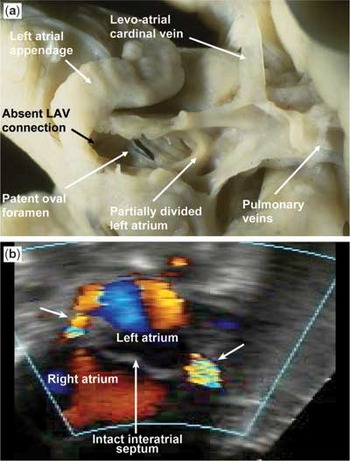
Figure 6. The levo-atrial cardinal vein in a fetus with mitral and aortic atresia. Such veins allow pulmonary venous return to the right atrium in absence of interatrial communication (closed interatrial septum, seen on the bottom postnatal subcostal long-axis echocardiogram). In that patient, there were three decompressing veins draining to the right atrium, two of which are visible in this picture and indicated by arrows.
The apical four-chamber view shows obvious disparity between the dilated chambers of the right heart and the diminutive left-sided structures (Fig. 1). The interatrial septum may bulge from left-to-right (Fig. 3), particularly in the setting of a small or absent interatrial communication. The left ventricle may be too small to identify, or it may be a small slit (Fig. 2), best seen posteriorly and leftwards in the left atrioventricular groove. The mitral valvar annulus, supravalvar obstructive rings, and subvalvar apparatus, specifically the tendinous cords, intercordal spaces and papillary muscles, are best assessed from the apical view. Prograde flow across the mitral valve may be difficult to identify due to a combination of elevated left ventricular diastolic pressure and a narrow flow jet. This might necessitate a decrease in the Nyquist limit, and/or an increase in the colour gain. The left ventricular outflow tract and the aortic valve are also well seen from the apical view. The latter is amenable to assessment for stenosis versus atresia. It is usually the right ventricle that forms the cardiac apex. Right ventricular systolic function can be assessed qualitatively. Dilation of the tricuspid annulus may contribute to tricuspid regurgitation.
The parasternal views demonstrate the discrepancy between the sizes of the two ventricles. The parasternal long axis view demonstrates the diminutive aorta arising from the small left ventricle. The left ventricle exhibits little contractility, and may exhibit hyperechogenic areas of endocardial fibroelastosis (Fig. 1 inset). The mitral valvar annulus, along with supra- and subvalvar levels of obstruction, may be evaluated from parasternal views. These views also enable identification of a dilated coronary sinus. Drainage of a left superior caval vein to the coronary sinus is a well-known associated finding in hypoplastic left heart syndrome, and is of surgical importance, given its implications for cannulation during cardiopulmonary bypass.
Views from the suprasternal notch provide for clear definition of the aortic arch. The entire aortic arch, including the ascending aorta, the proximal and distal arch, and the isthmus, is amenable to echocardiographic visualization from this view. Retrograde flow in the aortic arch and ascending aorta (Fig. 7) is a marker of the inability of the left ventricle to provide adequate prograde flow, typically in the setting of aortic atresia or severe aortic stenosis. Ascending aortic diameter is important for prognosis. A diameter less than 2 millimetres has been associated with increased risk of mortality following the Norwood procedure. The arrangement of the aortic arch and its pattern of branching can also be determined from this view. The latter allows the surgeon to plan the orientation of the aortopulmonary shunt that is integral to the Norwood procedure. The lumen of a widely patent arterial duct may mask the anterior wall of a coarcted aorta. A high index of suspicion, combined with careful scrutiny of the posterior wall of the aortic isthmus for a posterior shelf, helps avoid this diagnostic pitfall. In the presence of ductal flow, measurements of the diameters of the aortic arch are more meaningful than are Doppler gradients. Suprasternal views also help identify patterns of pulmonary venous drainage and connection. Decompressing veins that connect from the pulmonary to the systemic veins, typically the superior caval vein or its tributaries, can be identified from subcostal and suprasternal views.

Figure 7. Aortic arch hypoplasia and coarctation in hypoplastic left heart syndrome. Various degrees of severity of the coarctation shelf are illustrated in the pathology specimens. In (a) a fetus with mitral and aortic atresia, the shelf is large, there is infolding of the aortic wall and the isthmus joins the side of the arterial duct. In (b), a fetus with aortic atresia and patent mitral valve, there is only slight infolding and the obstruction comprises mainly of a large intimal shelf continuous with the mouth of the arterial duct. In (c), a suprasternal long-axis view of a neonate, one can appreciate a diminutive ascending aorta. The dilated pulmonary trunk is seen in cross-section. Retrograde flow is seen on colour Doppler in the aortic arch (*) and ascending aorta. The coarctation is indicated by white arrow. The descending aorta is of normal size.
In hypoplastic left heart syndrome, the arterial duct typically exhibits systolic flow from the pulmonary arteries to the aorta, with diastolic reversal of flow reflecting pulmonary vascular resistance that is lower than systemic vascular resistance. Pure right-to-left flow, with absence of diastolic reversal in the arterial duct, is consistent with elevated diastolic pressure in the pulmonary arteries, and implies severe elevation of pulmonary vascular resistance. This scene is typically encountered in the setting of persistent fetal circulation, in cases with an intact atrial septum, or in infants who have been awaiting transplantation for extended periods.
Preoperative care
The baby diagnosed prenatally to have hypoplasia of the left heart should be delivered in an appropriate specialized centre to avoid the stress of neonatal transfer. In cases of intact interatrial septum, emergency neonatal transcatheter atrial septostomy might be performed, but the mortality remains high in this sub-group.57 Prostaglandin E1 is started to prevent closure of, or to open, the arterial duct. In the presence of non-restrictive ductal flow, the usual state while in the hospital awaiting surgical care, the clinical course is determined by the possible complications of prostaglandin infusion, principally apnoea, in combination with the anatomic and physiologic features that determine the balance between pulmonary and systemic blood flows. When the resistance in the pulmonary circulation is low, flow of blood to the lungs is typically excessive, with symptoms of pulmonary overcirculation and systemic hypoperfusion. The anatomic predictor of this physiologic state is a large and unrestrictive shunt at atrial level. In these patients, mechanical ventilation, and/or reduced oxygen or increased carbon dioxide environments, can be used to increase pulmonary vascular resistance. Mild-to-moderate elevations of pulmonary vascular resistance due to a moderately restrictive atrial septal or oval foramen are well tolerated preoperatively due to the “balancing” effects on the ratio of systemic to pulmonary flows. Excessive restriction to pulmonary venous return, as in premature closure of the oval foramen, is poorly tolerated, and constitutes an urgent indication for interventional catheterization or surgical decompression of the left atrium, although even in the most skilled hands the outcome is likely to be poor.
Clinical management
In the initial descriptions of infants with hypoplasia of the left heart, the prognosis was invariably noted as uniformly poor, with surgery considered risky and experimental, and medical treatment being of temporary and limited value.4 Improvements in diagnostic techniques and treatments have changed the outlook for many patients with congenital cardiac disease, but have also introduced new challenges in decision-making for medical professionals.49 Hypoplastic left heart syndrome was considered a universally fatal disease a mere two decades ago. Today, staged surgical palliation or cardiac transplantation can provide survival of up to 70 percent at 5 years in patients deemed to be at “standard risk”.58 The staged palliative surgical approach consists of three operations (Figs 8 and 9). In the first stage, the Norwood procedure, performed in the first week after birth, either a modified Blalock–Taussig shunt is constructed, usually between the brachiocephalic and pulmonary arteries, or else a shunt is placed between the right ventricle and the pulmonary arteries.59 The aortic arch is reconstructed and connected to the pulmonary trunk, which is disconnected from the pulmonary arteries. Adequate mixing at the atrial level is assured by septectomy (Fig. 8). Any associated anomaly, such as venous obstructions, is corrected. The goal is to provide balanced and adequate blood flow to both the lungs and the body. Saturations of oxygen are still lower than normal, and the right ventricle is volume-overloaded, since it must perfuse both lungs and body in parallel circuits. Survival after this until the next surgery is difficult, and survival is typically in the range of 40 to 60 percent,9 although some centres recently report survival in the range of 80 to 90 percent.60, 61 The second stage, the so-called hemi-Fontan procedure, or a bidirectional Glenn operation, is usually performed between 6 and 8 months of age, and consists of removal of the shunt and connection of the superior caval vein to the pulmonary arteries (Fig. 9). This results in normalization of the volume load for the right ventricle, because the lungs are now connected in series, but mixing still occurs since both pulmonary venous blood and inferior caval venous return reach the right ventricle. As the venous return to the lungs is unassisted by ventricular pulsatility, pulmonary vascular resistance must be low for this stage to be successful. The normal elevation of pulmonary vascular resistance during infancy prevents reliance on the hemi-Fontan and bidirectional Glenn shunts before 2 to 3 months of age.62 The third stage, completion of the Fontan circulation, is performed on survivors at between 1.5 and 3 years of age. It consists of connection of the inferior caval vein to the pulmonary arteries, resulting in complete separation of the oxygenated and deoxygenated blood (Fig. 9). If the Fontan completion is extracardiac, a body weight of 15 kilograms is sought to maximize the dimensions of the extracardiac inferior caval vein to the pulmonary arterial conduit. The conversion from a shunt-dependent circulation to the full Fontan circulation is performed in two steps to avoid sudden and dramatic changes in haemodynamic loading, giving the heart time to adapt gradually.

Figure 8. Staged surgical approach to left heart hypoplasia. The first stage, or the Norwood procedure, seeks to enlarge the oval foramen (1) to allow unrestricted interatrial blood flow, reconstructs the ascending aorta (2), resects completely the ductal tissues (3), and places a shunt to provide a controlled blood flow to the pulmonary circulation (4). Note that pulmonary trunk is disconnected from the right ventricle. Colours from blue to red indicate relative oxygen saturation. AAo: ascending aorta; AoA: aortic arch; ICV: inferior caval vein; LA: left atrium; LPu: left pulmonary artery; LV: left ventricle; Pu: pulmonary trunk; RA: right atrium; RPu: right pulmonary artery; RV: right ventricle; SCV: superior caval vein; DA: arterial duct.

Figure 9. Separation of systemic and pulmonary circulation completes the palliation. The second stage, or hemi-Fontan procedure, removes the shunt, and connects the superior caval vein from the right atrium to the right pulmonary artery. The last stage, completing the Fontan circulation, connects also the inferior caval vein to the pulmonary circulation, effectively separating systemic and pulmonary circulation. Colours from blue to red approximate increasing oxygen saturation. ICV: inferior caval vein; LPu: left pulmonary artery; RPu: right pulmonary artery; SCV: superior caval vein.
Meticulous follow-up with serial echocardiography between the surgeries is essential,56 since late recoarctation attributed to unresected ductal tissue occur in up to one-quarter of patients,63 a complication that can be lethal if not recognized and dealt with appropriately.
Long-term prognosis is still highly uncertain, as there is few data for patients more than 10 years following completion of the Fontan circulation for patients with hypoplasia of the left heart. Although this circulatory arrangement is clearly competent for most children in the 5 to 10 years encompassed thus far postoperatively, the generic complications of Fontan palliation, such as protein losing enteropathy, arrhythmias, and thrombosis, are still applicable to this population. In addition, concerns remain that the morphologically right ventricle is ill-suited to perform as a systemic pump over multiple decades of life, potentially leading to premature heart failure. When detection of hypoplastic left heart syndrome occurs early in gestation, some families choose to terminate pregnancy. Although there is considerable commitment and success in the current care of these children on the part of medical providers, due to the high stress and uncertain outcome of palliation, especially when significant risk factors are present, as many as one-tenth of families still select compassionate care as a postnatal option.64
Cardiac transplantation is also an option for management that is vigorously pursued at some centres.64–66 It has the obvious benefit of resulting in a biventricular circulation, but shortage of suitable donors and the shortcomings of currently available long-term immunosuppression present substantial drawbacks.
Perspective of prenatal repair
As we already noted, if the left ventricle is small but potentially functional, postnatal biventricular repair is possible, and carries much better prognosis than current palliation.53 A prenatal intervention that would salvage the left ventricle for possible restoration of two-ventricular circulation physiology67 would thus dramatically change the management, significantly improving the potential to achieve a near-normal lifestyle. Despite the growing success rate of the Norwood procedure,49 a less heroic surgical intervention would conserve resources, the Norwood protocol requiring three major surgical procedures in the first 3 years of life, and decrease the stress put on both patients and their families. In addition, it leaves the patient with functionally univentricular physiology, in which perfusion of lungs is maintained by venous pressure alone. Frequent postoperative complications, such as arrhythmias, are also a persisting problem.68
Although most prenatally diagnosed anatomic malformations are best managed after birth, prenatal therapy can be offered to an increasing number of fetuses with simple anatomical defects that have predictably devastating developmental consequences. Current indications for fetal surgery include obstructive uropathy, diaphragmatic hernia, cystic adenomatoid malformation of the lung, sacro-coccygeal teratoma, severe neurological defects, twin-to-twin transfusion syndrome, and tracheal obstruction (see Review in Reference #69). A condition amenable to prenatal intervention must, however, meet a number of conditions, most important being the possibility of reliable prenatal diagnosis, and sufficient severity to warrant treatment. Hypoplasia of the left heart would fulfil such conditions for both diagnosis27 and severity.9, 49 Since prenatal surgery is still considered risky and experimental, it needs sound scientific justification. With increasing experience of fetal endoscopic procedures, cardiac procedures such as implantation of pacemakers for heart block and extreme bradycardia, and balloon dilation of obstructed pulmonary or aortic valves, or prematurely closed atrial septums, have also been attempted.52, 70 Although definitely of experimental nature,71 transcatheter repair during fetal life is a viable approach, and first reports of successful outcome were already published.50 It can be anticipated that the rates of success for such procedures will improve with increasing experience, and resolution of technical issues such as tocolytic control. We are trying in our lab at Charleston to provide scientific rationale for such procedures, using fetal surgical manipulations of the embryonic chick (Fig. 10). Currently, the biggest obstacles to successful prenatal interventions identified by the clinicians attempting those procedures are insufficient knowledge of the natural history, and lack of reliable criterions for selection of patients,6, 51, 72 coupled with the need for finer dedicated instruments for interventions at earlier fetal stages.52

Figure 10. Prenatal interventions in the chick embryo. The left panel shows a silver microclip that was used to reduce the volume of the right atrium (RA). This resulted in distension of the left atrium (LA). In experimental left ventricular hypoplasia, such a procedure would restore the flow of blood and the loading of left heart structures, possibly reversing the already established hypoplasia in the remaining two-thirds of incubation period. The right panel shows targeted injection of green fluorescent protein-expressing adenovirus to the left ventricle (LV). Such an approach could be used to deliver growth factors that may restore decreased myocytic proliferation in the hypoplastic left ventricle. RV: right ventricle.
Why should prenatal repair of left heart hypoplasia work? During prenatal development, the myocardial mass increases by addition of new cells, that is hyperplasia, while soon after birth, the mechanism switches to an increase in the size of the cell, in other words, hypertrophy. It was demonstrated previously that the same adaptive mechanisms are employed under artificially modified conditions of loading.19, 73, 74
The pattern of growth of the ventricular myocardium is by apposition, with a decreasing gradient of proliferative activity from the outer compact layer toward the inside of the heart.75–79 Isolated embryonic cardiac myocytes are capable of self-regulating their proliferative rate in response to mechanical stress, as was shown using systems of cell culture.80 The rapid response of embryonic myocytes to haemodynamic alterations has also been demonstrated in the whole animal models.22, 73, 81, 82 On the basis of these studies, it is reasonable to believe that prenatal intervention that would normalize hemo-dynamic conditions in carefully selected cases of congenital cardiac disease can be more beneficial in the long term, since such early intervention holds greater potential to restore normal heart structure and function.
Regulation of cardiomyocytic proliferation by growth factors. The cascade of events translating the mechanical stimulus to cell division, or hypertrophy in postnatal stages, is not yet fully understood. Nevertheless, autocrine or paracrine regulation by various growth factors is presumably an important factor.83, 84 It was emphasized that the results of studies on isolated cells can not be automatically implicated for situation in the whole organism.85 Below, we will review the available data that suggest that growth factors might be a potential therapeutic strategy for improving prenatal myocardial growth. Since most of these studies employed the chick embryo due to its easy amenability to experimental mechanical manipulations, we will focus on this model.
In the chick embryo, platelet-derived growth factor86, 87 and fibroblast growth factor-2, previously known as basic fibroblast growth factor,85, 88–91 are associated with the regulation of cardiomyocytic proliferation. Platelet-derived growth factor is a growth factor stimulating proliferation in various cell types. Three isoforms, A, B, and C, have been described, forming homo- or heterodimers that bind to specific α and β receptors, and trigger autophosphorylation on the tyrosine residues. The downstream targets are mitogen-activated protein kinase, protein kinase C, and signal transducers and activators of transcription. In the heart, expression of platelet-derived growth factor is widespread throughout the myocardium.87 The levels of protein were almost doubled by experimentally increased pressure loading in the embryonic heart,86 indicating its induction by increased cell stretching. Defects of its α and β receptors have deleterious effects on cardiac development, presenting with phenotype of myocardial hypoplasia.92, 93 While platelet-derived growth factor-AA and -CC isoforms influence mostly fibroblasts,94, 95 the -BB isoform was shown to be involved in control of cardiomyocytic proliferation.96 We have recently shown significant down-regulation of platelet-derived growth factor -BB in chick embryonic hearts after left atrial ligation.19
The role of fibroblast growth factor-2 in control of myocytic proliferation is well established. Beads soaked in fibroblast growth factor-2 increased myocytic proliferation in chick embryonic heart.91 Elegant studies of Mima et al.85, 97 demonstrated that these effects are mediated through fibroblast growth factor receptor-1 receptors, and showed also decreasing dependence on this pathway at later developmental stages. Some of the effects are, however, receptor-independent, implicating direct action of the produced protein on the cell nucleus.98 Physiological regulation of the number of receptors on cell surface plays an important role, since over-expression of receptors results in fibroblast growth factor-2 dependent proliferation in neonatal myocytic cultures.99 These results are particularly encouraging for potential success of treatment with this growth factor in rescuing the hypoplastic phenotype, since there was a report of massive up-regulation of receptors, together with decreased amount of ligand in the chick model of left heart hypoplasia.19 Interestingly, a similar decrease in the factor is found in fetal human hearts with hypoplasia of the left heart (Fig. 5). Furthermore, both platelet-derived growth factor and fibroblast growth factor have also positive effects on differentiation of the contractile apparatus,100 offering the possibility of rescuing the decreased amounts of contractile protein,19 and thus possibly improving function. Analysis of phenotypic differentiation of human cardiomyocytes isolated from hypoplastic left ventricles showed the immature, or “heart failure” patterns of expression of genes,101 validating the relevance of results obtained from animal models.
In mammals, insulin-like growth factor signalling102, 103 was also implicated in control of cardiomyocytic proliferation. Mice lacking both type 1 and type 2 receptors, as well as insulin-like growth factor-2, are dwarfs and die at birth, while up-regulation of insulin-like growth factor-2 leads to an increase in the size of the animals. In the postnatal development of the rat, decrease of proliferative capacity was correlated with down-regulation of insulin-like growth factor-1 signalling. For proliferation of cultured human fetal ventricular myocytes, epidermal growth factor signalling, as well as beta-1 integrin-mediated interaction with extracellular matrix, was found to be crucial.104
Possibly the most successful strategy for long-term delivery of growth factors to the diseased myocardium would be via adenoviral vector. Adenovirus-mediated gene transfer has already successfully been used to treat experimental anomalies of conduction in a pig model,105 and high-efficiency cardiac transfer was reported in mouse fetuses.106 We are currently using targeted delivery of growth factors through this vector to the chick embryonic left ventricle to alleviate experimentally produced hypoplasia of the left heart (Fig. 10). This modality of treatment could be used either alone, or as an adjuvant to surgical repair.
Stem cells for myocardial repair. Stem cells are cells that are self-renewing, or give rise to at least two daughter cells at the same stage as the parent, and have the ability to differentiate into other cell types. The haematopoietic stem cells are pluripotent cells in the bone marrow that give rise to all blood cells including erythrocytes, granulocytes, monocytes, mast cells, lymphocytes, and megakaryocytes. Mesenchymal stem cells, also found in bone marrow, give rise to other cell types, including fibroblasts and muscle cells. Various studies have attempted to transplant stem cells into the myocardium to treat cardiac disease, and restore myocardial function following damage such as a myocardial infarction.107–112 It was demonstrated that adult mesenchymal stem cells isolated from fatty tissue could be chemically induced by 5-azacytidine to transform into cardiomyocytes in cell culture.113, 114 The potential of transplanted bone marrow stem cells to differentiate into skeletal myocytes and cardiomyocytes was also demonstrated in the dystrophic mdx mouse.115, 116 All these studies were performed in adult animals, and little information is available on the potential of using stem cells in the fetus.69 Our current studies using quail-chick chimeras (Fig. 11) try to fill this gap. Avian fetal bone marrow cells have the potential to form cardiomyocytes in cell culture,117 but some optimisation may be needed to make it occur also in the whole animal setting. Although there might be significant difference in behaviour of stem cells between adults and fetuses, this avenue might provide a new therapeutic modality, using either direct targeted delivery to enhance mass of the hypoplastic left ventricle, or, if practical, by mobilization treatment with injections of cytokines.112

Figure 11. Bone marrow injection in the chick embryonic heart. The left panel shows donor cells of quail origin detected by quail-specific QH1 antibody in the chest wall and subepicardium (arrows). The right panel shows that these cells (green, confocal microscopy) continue to proliferate 48 hours after injection (2 hour pulse-labelling with 5-bromodeoxyuridine, BrdU, red). Our current goals are to optimise the conditions for differentiation of these cells into cardiomyocytes in an attempt to restore decreased myocardial mass in the experimental left heart hypoplasia. Scale bars 500 and 100 microns, respectively.
Conclusions
We have come a long way from recognizing the hypoplastic left heart syndrome as a pathological entity to prenatal diagnosis, and we are now fairly efficient, at least in the medium-term, at postnatal operative management. With recent advances in basic research, as well as more experience in fetal procedures, prenatal therapeutic modalities may change the way we approach this and other forms of congenital cardiac disease.
Supported by NIH HL067135 (TCM), RR16434 (DS), March of Dimes 5-FY02-269 (DS), and British Heart Foundation (AC). Technical assistance of Ms. Angela de Almeida and Deanna Mustin is gratefully acknowledged.
References
Gittenberger-de Groot AC. Het links hypoplasticsch hart als aangeboren afwijking. In: Anatomy and Embryology. Leiden: Rijksuniversiteit te Leiden, 1972, p 134.
Lev M. Pathologic anatomy and interrelationship of hypoplasia of the aortic tract complexes. Lab Invest 1952; 1: 61–70.Google Scholar
Friedman S, Murphy L, Ash R. Aortic atresia with hypoplasia of the left heart and aortic arch. J Pediatr 1951; 38: 354–368.Google Scholar
Noonan JA, Nadas AS. The hypoplastic left heart syndrome. An analysis of 101 cases. Pediatr Clin North Am 1958; 5: 1029–1056.Google Scholar
Restivo A, Ho SY, Anderson RH, Cameron H, Wilkinson JL. Absent left atrioventricular connection with right atrium connected to morphologically left ventricular chamber, rudimentary right ventricular chamber, and ventriculoarterial discordance. Problem of mitral versus tricuspid atresia. Br Heart J 1982; 48: 240–248.Google Scholar
Simpson JM, Sharland GK. Natural history and outcome of aortic stenosis diagnosed prenatally. Heart 1997; 77: 205–210.Google Scholar
Freedom RM, Culham JA, Moes CA, Harrington DP. Selective aortic root angiography in the hypoplastic left heart syndrome. Eur J Cardiol 1976; 4: 25–29.Google Scholar
Cook AC. The anatomy of left heart hypoplasia in the human fetus. Thesis, University of London, 2003.Google Scholar
Bradley SM. Neonatal repair and dealing with a single ventricle. J S C Med Assoc 1999; 95: 335–338.Google Scholar
Shokeir MH. Hypoplastic left heart. Evidence for possible autosomal recessive inheritance. Birth Defects Orig Artic Ser 1974; 10: 223–227.Google Scholar
Shokeir MH. Hypoplastic left heart syndrome: an autosomal recessive disorder. Clin Genet 1971; 2: 7–14.Google Scholar
Brenner JI, Berg KA, Schneider DS, Clark EB, Boughman JA. Cardiac malformations in relatives of infants with hypoplastic left-heart syndrome. Am J Dis Child 1989; 143: 1492–1494.Google Scholar
Natowicz M, Chatten J, Clancy R, et al. Genetic disorders and major extracardiac anomalies associated with the hypoplastic left heart syndrome. Pediatrics 1988; 82: 698–706.Google Scholar
Natowicz M, Kelley RI. Association of Turner syndrome with hypoplastic left-heart syndrome. Am J Dis Child 1987; 141: 218–220.Google Scholar
Consevage MW, Seip JR, Belchis DA, et al. Association of a mosaic chromosomal 22q11 deletion with hypoplastic left heart syndrome. Am J Cardiol 1996; 77: 1023–1025.Google Scholar
Saneto RP, Applegate KE, Frankel DG. Atypical manifestations of two cases of trisomy 9 syndrome: rethinking development delay. Am J Med Genet 1998; 80: 42–45.Google Scholar
Phillips HM, Renforth GL, Spalluto C, et al. Narrowing the critical region within 11q24-qter for hypoplastic left heart and identification of a candidate gene, JAM3, expressed during cardiogenesis. Genomics 2002; 79: 475–478.Google Scholar
Burton PB, Hauck A, Nehlsen-Cannarella SL, et al. Hypoplastic left heart syndrome: some clues to its aetiology. Lancet 1991; 338: 1148.Google Scholar
Sedmera D, Hu N, Weiss KM, et al. Cellular changes in experimental left heart hypoplasia. Anat Rec 2002; 267: 137–145.Google Scholar
Towbin JA, Casey B, Belmont J. The molecular basis of vascular disorders. Am J Hum Genet 1999; 64: 678–684.Google Scholar
Rychter Z, Rychterova V. Angio- and myoarchitecture of the heart wall under normal and experimentally changed morphogenesis. In: Pexieder T (ed.). Perspectives in Cardiovascular Research. Raven Press, New York, NY, USA, 1981, pp 431–452.
Sedmera D, Pexieder T, Rychterova V, Hu N, Clark EB. Remodeling of chick embryonic ventricular myoarchitecture under experimentally changed loading conditions. Anat Rec 1999; 254: 238–252.Google Scholar
van der Linde-Sipman JS, Wensing CJ. The left hypoplastic heart syndrome in the minipig. Birth Defects Orig Artic Ser 1978; 14: 295–314.Google Scholar
Tikkanen J, Heinonen OP. Risk factors for hypoplastic left heart syndrome. Teratology 1994; 50: 112–117.Google Scholar
Oyer CE, Ongcapin EH, Ni J, Bowles NE, Towbin JA. Fatal intrauterine adenoviral endomyocarditis with aortic and pulmonary valve stenosis: diagnosis by polymerase chain reaction. Hum Pathol 2000; 31: 1433–1435.Google Scholar
Ni J, Bowles NE, Kim YH, et al. Viral infection of the myocardium in endocardial fibroelastosis. Molecular evidence for the role of mumps virus as an etiologic agent. Circulation 1997; 95: 133–139.Google Scholar
Kohl T. Fetal echocardiography: new grounds to explore during fetal cardiac intervention. Pediatr Cardiol 2002; 23: 334–346.Google Scholar
Carvalho JS. Early prenatal diagnosis of major congenital heart defects. Curr Opin Obstet Gynecol 2001; 13: 155–159.Google Scholar
Allan LD, Sharland G, Tynan MJ. The natural history of the hypoplastic left heart syndrome. Int J Cardiol 1989; 25: 341–343.Google Scholar
Anderson NG, Brown J. Normal size left ventricle on antenatal scan in lethal hypoplastic left heart syndrome. Pediatr Radiol 1991; 21: 436–437.Google Scholar
Sharland GK, Chita SK, Fagg NL, et al. Left ventricular dysfunction in the fetus: relation to aortic valve anomalies and endocardial fibroelastosis. Br Heart J 1991; 66: 419–424.Google Scholar
McCaffrey FM, Sherman FS. Prenatal diagnosis of severe aortic stenosis. Pediatr Cardiol 1997; 18: 276–281.Google Scholar
van der Linde-Sipman JS. Hypoplasia of the left ventricle in four ruminants. Vet Pathol 1978; 15: 474–480.Google Scholar
van Nie CJ, van Messel MA, Straatman TJ. Congenital bicuspid stenosis with left ventricular hypoplasia in a kitten. Tijdschr Diergeneeskd 1980; 105: 58–62.Google Scholar
Sedmera D, Pexieder T, Vuillemin M, Thompson RP, Anderson RH. Developmental patterning of the myocardium. Anat Rec 2000; 258: 319–337.Google Scholar
Fishman NH, Hof RB, Rudolph AM, Heymann MA. Models of congenital heart disease in fetal lambs. Circulation 1978; 58: 354–364.Google Scholar
Rychter Z, Rychterova V, Lemez L. Formation of the heart loop and proliferation structure of its wall as a base for ventricular septation. Herz 1979; 4: 86–90.Google Scholar
Tobita K, Keller BB. Right and left ventricular wall deformation patterns in normal and left heart hypoplasia chick embryos. Am J Physiol Heart Circ Physiol 2000; 279: H959–H969.Google Scholar
Harh JY, Paul MH, Gallen WJ, Friedberg DZ, Kaplan S. Experimental production of hypoplastic left heart syndrome in the chick embryo. Am J Cardiol 1973; 31: 51–56.Google Scholar
Clark EB, Hu N, Dummett JL, et al. Ventricular function and morphology in chick embryo from stages 18 to 29. Am J Physiol 1986; 250: H407–H413.Google Scholar
Harh JY, Paul MH. Experimental cardiac morphogenesis. I. Development of the ventricular septum in the chick. J Embryol Exp Morphol 1975; 33: 13–28.Google Scholar
Ben-Shachar G, Arcilla RA, Lucas RV, Manasek FJ. Ventricular trabeculations in the chick embryo heart and their contribution to ventricular and muscular septal development. Circ Res 1985; 57: 759–766.Google Scholar
Icardo JM, Fernandez-Teran A. Morphologic study of ventricular trabeculation in the embryonic chick heart. Acta Anat 1987; 130: 264–274.Google Scholar
Pexieder T. Development of the outflow tract of the embryonic heart. Birth Defects Orig Artic Ser 1978; 14: 29–68.Google Scholar
Sedmera D, Pexieder T, Hu N, Clark EB. Developmental changes in the myocardial architecture of the chick. Anat Rec 1997; 248: 421–432.Google Scholar
Tobita K, Schroder EA, Tinney JP, Garrison JB, Keller BB. Regional passive ventricular stress-strain relations during development of altered loads in chick embryo. Am J Physiol Heart Circ Physiol 2002; 282: H2386–H2396.Google Scholar
Jay PJ, Berul CI, Tanaka M, et al. Cardiac conduction and arrhythmia: insights from Nkx2.5 mutations in mouse and humans. In: Chadwick DJ, Goode J (eds). Development of the Cardiac Conduction System. Wiley, Chichester, 2003, pp 227–241.
Riordan CJ, Randsbaek F, Storey JH, et al. Inotropes in the hypoplastic left heart syndrome: effects in an animal model. Ann Thorac Surg 1996; 62: 83–90.Google Scholar
Halnon NJ, Wetzel GT. Implications of improving survival in the management of life-threatening congenital heart disease: new challenges in pediatric cardiology. Curr Opin Cardiol 2002; 17: 271–273.Google Scholar
Allan LD, Maxwell DJ, Carminati M, Tynan MJ. Survival after fetal aortic balloon valvoplasty. Ultrasound Obstet Gynecol 1995; 5: 90–91.Google Scholar
Kohl T, Sharland G, Allan LD, et al. World experience of percutaneous ultrasound-guided balloon valvuloplasty in human fetuses with severe aortic valve obstruction. Am J Cardiol 2000; 85: 1230–1233.Google Scholar
Marshall AC, van der Velde ME, Tworetzky W, et al. Creation of an atrial septal defect in utero for fetuses with hypoplastic left heart syndrome and intact or highly restrictive atrial septum. Circulation 2004; 110: 253–258.Google Scholar
Tchervenkov CI, Jacobs ML, Tahta SA. Congenital Heart Surgery Nomenclature and Database Project: hypoplastic left heart syndrome. Ann Thorac Surg 2000; 69: S170–S179.Google Scholar
Glauser TA, Rorke LB, Weinberg PM, Clancy RR. Congenital brain anomalies associated with the hypoplastic left heart syndrome. Pediatrics 1990; 85: 984–990.Google Scholar
Meacham LR, Winn KJ, Culler FL, Parks JS. Double vagina, cardiac, pulmonary, and other genital malformations with 46,XY karyotype. Am J Med Genet 1991; 41: 478–481.Google Scholar
Scheurer M, Shirali GS. Hypoplastic left heart syndrome: native and post-norwood. Pediatric Ultrasound Today 2003; 8: 65–84.Google Scholar
Vlahos AP, Lock JE, McElhinney DB, van der Velde ME. Hypoplastic left heart syndrome with intact or highly restrictive atrial septum: outcome after neonatal transcatheter atrial septostomy. Circulation 2004; 109: 2326–2330.Google Scholar
Bove EL, Lloyd TR. Staged reconstruction for hypoplastic left heart syndrome. Contemporary results. Ann Surg 1996; 224: 387–394; discussion 394–385.Google Scholar
Malec E, Januszewska K, Kolcz J, Mroczek T. Right ventricle-to-pulmonary artery shunt versus modified Blalock-Taussig shunt in the Norwood procedure for hypoplastic left heart syndrome – influence on early and late haemodynamic status. Eur J Cardiothorac Surg 2003; 23: 728–734.Google Scholar
Tweddell JS, Hoffman GM, Mussatto KA, et al. Improved survival of patients undergoing palliation of hypoplastic left heart syndrome: lessons learned from 115 consecutive patients. Circulation 2002; 106: I82–I89.Google Scholar
Poirier NC, Drummond-Webb JJ, Hisamochi K, et al. Modified Norwood procedure with a high-flow cardiopulmonary bypass strategy results in low mortality without late arch obstruction. J Thorac Cardiovasc Surg 2000; 120: 875–884.Google Scholar
Reddy VM, McElhinney DB, Moore P, Haas GS, Hanley FL. Outcomes after bidirectional cavopulmonary shunt in infants less than 6 months old. J Am Coll Cardiol 1997; 29: 1365–1370.Google Scholar
Fraisse A, Colan SD, Jonas RA, Gauvreau K, Geva T. Accuracy of echocardiography for detection of aortic arch obstruction after stage I Norwood procedure. Am Heart J 1998; 135: 230–236.Google Scholar
Gutgesell HP, Gibson J. Management of hypoplastic left heart syndrome in the 1990s. Am J Cardiol 2002; 89: 842–846.Google Scholar
Chang RK, Chen AY, Klitzner TS. Clinical management of infants with hypoplastic left heart syndrome in the United States, 1988–1997. Pediatrics 2002; 110: 292–298.Google Scholar
Morrow WR, Naftel D, Chinnock R, et al. Outcome of listing for heart transplantation in infants younger than six months: predictors of death and interval to transplantation. The Pediatric Heart Transplantation Study Group. J Heart Lung Transplant 1997; 16: 1255–1266.Google Scholar
Silverman NH, McElhinney DB. Which two ventricles cannot be used for a biventricular repair? Echocardiographic assessment. Ann Thorac Surg 1998; 66: 634–640.Google Scholar
Mahle WT, Gaynor JW, Spray TL. Atrioventricular valve replacement in patients with a single ventricle. Ann Thorac Surg 2001; 72: 182–186.Google Scholar
Sydorak RM, Nijagal A, Albanese CT. Endoscopic techniques in fetal surgery. Yonsei Med J 2001; 42: 695–710.Google Scholar
Maxwell D, Allan L, Tynan MJ. Balloon dilatation of the aortic valve in the fetus: a report of two cases. Br Heart J 1991; 65: 256–258.Google Scholar
Hornberger LK, Sanders SP, Rein AJ, et al. Left heart obstructive lesions and left ventricular growth in the midtrimester fetus. A longitudinal study. Circulation 1995; 92: 1531–1538.Google Scholar
Clark EB, Hu N, Frommelt P, et al. Effect of increased pressure on ventricular growth in stage 21 chick embryos. Am J Physiol 1989; 257: H55–H61.Google Scholar
Sedmera D, Thompson RP, Kolar F. Effect of increased pressure loading on heart growth in neonatal rats. J Mol Cell Cardiol 2003; 35: 301–309.Google Scholar
Grohmann D. Mitotische Wachstumsintensitat des embryonalen und fetalen Hunchenherzens und ihre Bedeutung fur die entstehung von Herzmissbildungen. Z f Zellforschung 1961; 55: 104–122.Google Scholar
Jeter Jr JR, Cameron IL. Cell proliferation patterns during cytodifferentiation in embryonic chick tissues: liver, heart and erythrocytes. J Embryol Exp Morphol 1971; 25: 405–422.Google Scholar
Mikawa T, Borisov A, Brown AM, Fischman DA. Clonal analysis of cardiac morphogenesis in the chicken embryo using a replication-defective retrovirus: I. Formation of the ventricular myocardium. Dev Dyn 1992; 193: 11–23.Google Scholar
Sedmera D, Reckova M, DeAlmeida A, et al. Spatiotemporal pattern of commitment to slowed proliferation in the embryonic mouse heart indicates progressive differentiation of the cardiac conduction system. Anat Rec 2003; 274A: 773–777.Google Scholar
Meilhac SM, Kelly RG, Rocancourt D, et al. A retrospective clonal analysis of the myocardium reveals two phases of clonal growth in the developing mouse heart. Development 2003; 130: 3877–3889.Google Scholar
Miller CE, Donlon KJ, Toia L, Wong CL, Chess PR. Cyclic strain induces proliferation of cultured embryonic heart cells. In Vitro Cell Dev Biol Anim 2000; 36: 633–639.Google Scholar
Clark EB, Hu N, Turner DR, Litter JE, Hansen J. Effect of chronic verapamil treatment on ventricular function and growth in chick embryos. Am J Physiol 1991; 261: H166–H171.Google Scholar
Sedmera D, Pexieder T, Hu N, Clark EB. A quantitative study of the ventricular myoarchitecture in the stage 21–29 chick embryo following decreased loading. Eur J Morphol 1998; 36: 105–119.Google Scholar
Hefti MA, Harder BA, Eppenberger HM, Schaub MC. Signaling pathways in cardiac myocyte hypertrophy. J Mol Cell Cardiol 1997; 29: 2873–2892.Google Scholar
Pasumarthi KB, Field LJ. Cardiomyocyte cell cycle regulation. Circ Res 2002; 90: 1044–1054.Google Scholar
Mima T, Ueno H, Fischman DA, Williams LT, Mikawa T. Fibroblast growth factor receptor is required for in vivo cardiac myocyte proliferation at early embryonic stages of heart development. Proc Natl Acad Sci USA 1995; 92: 467–471.Google Scholar
Jedlicka S, Finkelstein JN, Paulhamus LA, Clark EB. Increased PDGF-like protein in banded embryonic ventricle. Pediatr Res 1991; 29: 19A.Google Scholar
Pexieder T, Jedlicka S, Sugimara K, Tatimatsu A, Sato H. Immunohistochemical localization of platelet derived growth factor (PDGF) during cardiac morphogenesis in chick and mouse embryos and fetuses. In: Clark EB, et al. (eds). Developmental Mechanisms of Heart Disease. Futura Publishing, Armonk, NY, USA, 1995, pp 207–212.
Parlow MH, Bolender DL, Kokan-Moore NP, Lough J. Localization of bFGF-like proteins as punctate inclusions in the preseptation myocardium of the chicken embryo. Dev Biol 1991; 146: 139–147.Google Scholar
Sedmera D, Grobety M, Reymond C, Baehler P, Kucera P, Kappenberger L. Pacing-induced ventricular remodeling in the chick embryonic heart. Pediatr Res 1999; 45: 845–852.Google Scholar
Sugi Y, Sasse J, Lough J. Inhibition of precardiac mesoderm cell proliferation by antisense oligodeoxynucleotide complementary to fibroblast growth factor-2 (FGF-2). Dev Biol 1993; 157: 28–37.Google Scholar
Franciosi JP, Bolender DL, Lough J, Kolesari GL. FGF-2-induced imbalance in early embryonic heart cell proliferation: a potential cause of late cardiovascular anomalies. Teratology 2000; 62: 189–194.Google Scholar
Leveen P, Pekny M, Gebre-Medhin S, et al. Mice deficient for PDGF B show renal, cardiovascular, and hematological abnormalities. Genes Dev 1994; 8: 1875–1887.Google Scholar
Schatteman GC, Motley ST, Effmann EL, Bowen-Pope DF. Platelet-derived growth factor receptor alpha subunit deleted Patch mouse exhibits severe cardiovascular dysmorphogenesis. Teratology 1995; 51: 351–366.Google Scholar
Li X, Ponten A, Aase K, et al. PDGF-C is a new protease-activated ligand for the PDGF alpha-receptor. Nat Cell Biol 2000; 2: 302–309.Google Scholar
Simm A, Nestler M, Hoppe V. PDGF-AA, a potent mitogen for cardiac fibroblasts from adult rats. J Mol Cell Cardiol 1997; 29: 357–368.Google Scholar
Shimizu T, Kinugawa K, Yao A, et al. Platelet-derived growth factor induces cellular growth in cultured chick ventricular myocytes. Cardiovasc Res 1999; 41: 641–653.Google Scholar
Mikawa T. Retroviral targeting of FGF and FGFR in cardiomyocytes and coronary vascular cells during heart development. Ann NY Acad Sci 1995; 752: 506–516.Google Scholar
Kardami E, Liu L, Kishore S, et al. Regulation of basic fibroblast growth factor (bFGF) and FGF receptors in the heart. Ann NY Acad Sci 1995; 752: 353–369.Google Scholar
Sheikh F, Fandrich RR, Kardami E, Cattini PA. Overexpression of long or short FGFR-1 results in FGF-2-mediated proliferation in neonatal cardiac myocyte cultures. Cardiovasc Res 1999; 42: 696–705.Google Scholar
Velez C, Aranega AE, Melguizo C, et al. Modulation of contractile protein troponin-T in chick myocardial cells by basic fibroblast growth factor and platelet-derived growth factor during development. J Cardiovasc Pharmacol 1994; 24: 906–913.Google Scholar
Bohlmeyer TJ, Helmke S, Ge S, et al. Hypoplastic left heart syndrome myocytes are differentiated but possess a unique phenotype. Cardiovasc Pathol 2003; 12: 23–31.Google Scholar
Cheng W, Reiss K, Kajstura J, et al. Down-regulation of the IGF-1 system parallels the attenuation in the proliferative capacity of rat ventricular myocytes during postnatal development. Lab Invest 1995; 72: 646–655.Google Scholar
Ludwig T, Eggenschwiler J, Fisher P, et al. Mouse mutants lacking the type 2 IGF receptor (IGF2R) are rescued from perinatal lethality in Igf2 and Igf1r null backgrounds. Dev Biol 1996; 177: 517–535.Google Scholar
Hornberger LK, Singhroy S, Cavalle-Garrido T, et al. Synthesis of extracellular matrix and adhesion through beta(1) integrins are critical for fetal ventricular myocyte proliferation. Circ Res 2000; 87: 508–515.Google Scholar
Donahue JK, Heldman AW, Fraser H, et al. Focal modification of electrical conduction in the heart by viral gene transfer. Nat Med 2000; 6: 1395–1398.Google Scholar
Christensen G, Minamisawa S, Gruber PJ, Wang Y, Chien KR. High-efficiency, long-term cardiac expression of foreign genes in living mouse embryos and neonates. Circulation 2000; 101: 178–184.Google Scholar
Hassink RJ, Brutel de la Riviere A, Mummery CL, Doevendans PA. Transplantation of cells for cardiac repair. J Am Coll Cardiol 2003; 41: 711–717.Google Scholar
Strauer BE, Brehm M, Zeus T, et al. Repair of infarcted myocardium by autologous intracoronary mononuclear bone marrow cell transplantation in humans. Circulation 2002; 106: 1913–1918.Google Scholar
Stamm C, Westphal B, Kleine HD, et al. Autologous bone- marrow stem-cell transplantation for myocardial regeneration. Lancet 2003; 361: 45–46.Google Scholar
Orlic D, Kajstura J, Chimenti S, et al. Transplanted adult bone marrow cells repair myocardial infarcts in mice. Ann NY Acad Sci 2001; 938: 221–229; discussion 229–230.Google Scholar
Orlic D, Kajstura J, Chimenti S, et al. Bone marrow cells regenerate infarcted myocardium. Nature 2001; 410: 701–705.Google Scholar
Orlic D, Kajstura J, Chimenti S, et al. Mobilized bone marrow cells repair the infarcted heart, improving function and survival. Proc Natl Acad Sci USA 2001; 98: 10344–10349.Google Scholar
Rangappa S, Entwistle JW, Wechsler AS, Kresh JY. Cardiomyocyte-mediated contact programs human mesenchymal stem cells to express cardiogenic phenotype. J Thorac Cardiovasc Surg 2003; 126: 124–132.Google Scholar
Rangappa S, Fen C, Lee EH, Bongso A, Wei ES. Transformation of adult mesenchymal stem cells isolated from the fatty tissue into cardiomyocytes. Ann Thorac Surg 2003; 75: 775–779.Google Scholar
Bittner RE, Schofer C, Weipoltshammer K, et al. Recruitment of bone-marrow-derived cells by skeletal and cardiac muscle in adult dystrophic mdx mice. Anat Embryol (Berl) 1999; 199: 391–396.Google Scholar
Gussoni E, Soneoka Y, Strickland CD, et al. Dystrophin expression in the mdx mouse restored by stem cell transplantation. Nature 1999; 401: 390–394.Google Scholar
Eisenberg LM, Burns L, Eisenberg CA. Hematopoietic cells from bone marrow have the potential to differentiate into cardiomyocytes in vitro. Anat Rec 2003; 274A: 870–882.Google Scholar

Morphology of left heart hypoplasia. Four-chamber views of fetal and newborn hearts showing small size and thick wall of the hypoplastic chamber. Note the whitish appearance of endocardial fibroelastosis in these cases with a patent mitral valve. Matching ultrasound pictures show the resolution available for diagnosis. The postnatal scan shows heart with atretic mitral valve and no demonstrable flow in the left ventricle. Inset shows hyperechogenic left ventricle with endocardial fibroelastosis (arrowheads) in another fetus with mitral and aortic stenosis. LA: left atrium; LV: left ventricle; RA: right atrium; RV: right ventricle.
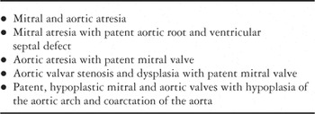
Table 1.

Phenotype of mitral and aortic atresia in the mid-trimester fetus. In the majority of fetuses, the left atrioventricular junction (yellow lines) has joined the muscular base of the atrial septum (*), walling the left atrium from the left ventricle. The presence of a ventricular septal defect (VSD) allows some blood flow to the left ventricle, resulting in a significant mitigation of the left ventricular hypoplasia.

Morphology of the prematurely closed interatrial septum. The top panel shows an example of mildly thickened flap valve, which is completely fused to the rims of the oval foramen, in a fetus with aortic valvar stenosis. The bottom panel shows thickening of the left atrial walls, the interatrial septum, and the pulmonary veins. The atretic mitral valve is overgrown by left atrial myocardium.

Left heart hypoplasia in the chick model. By the end of cardiac septation (incubation day 8 of 21), the four-chamber view shows that the left ventricle normally forms apex of the heart. In the heart subjected to left atrial ligation (right panel), the interatrial septum (ias) is deviated to the left, and the left ventricle is underdeveloped. As in the human situation, whole range of degrees of hypoplasia of the left heart structures is produced in this model, and the phenotype becomes progressively more severe. LA: left atrium; LV: left ventricle; RA: right atrium; RV: right ventricle. Left panel originally appeared in Reference #22.

Growth factor expression in left heart hypoplasia. In two-chamber views of the chick heart reacted with anti-fibroblast growth factor-2 antibody, the hypoplastic left ventricle shows a clear decrease in staining intensity. A similar situation can be observed in the fetal human heart immunostained for fibroblast growth factor-1. Pseudocolour display of fluorescent staining intensity is shown on the right. LV: left ventricle; RV: right ventricle. Top panels originally appeared in Reference #19.

The levo-atrial cardinal vein in a fetus with mitral and aortic atresia. Such veins allow pulmonary venous return to the right atrium in absence of interatrial communication (closed interatrial septum, seen on the bottom postnatal subcostal long-axis echocardiogram). In that patient, there were three decompressing veins draining to the right atrium, two of which are visible in this picture and indicated by arrows.

Aortic arch hypoplasia and coarctation in hypoplastic left heart syndrome. Various degrees of severity of the coarctation shelf are illustrated in the pathology specimens. In (a) a fetus with mitral and aortic atresia, the shelf is large, there is infolding of the aortic wall and the isthmus joins the side of the arterial duct. In (b), a fetus with aortic atresia and patent mitral valve, there is only slight infolding and the obstruction comprises mainly of a large intimal shelf continuous with the mouth of the arterial duct. In (c), a suprasternal long-axis view of a neonate, one can appreciate a diminutive ascending aorta. The dilated pulmonary trunk is seen in cross-section. Retrograde flow is seen on colour Doppler in the aortic arch (*) and ascending aorta. The coarctation is indicated by white arrow. The descending aorta is of normal size.

Staged surgical approach to left heart hypoplasia. The first stage, or the Norwood procedure, seeks to enlarge the oval foramen (1) to allow unrestricted interatrial blood flow, reconstructs the ascending aorta (2), resects completely the ductal tissues (3), and places a shunt to provide a controlled blood flow to the pulmonary circulation (4). Note that pulmonary trunk is disconnected from the right ventricle. Colours from blue to red indicate relative oxygen saturation. AAo: ascending aorta; AoA: aortic arch; ICV: inferior caval vein; LA: left atrium; LPu: left pulmonary artery; LV: left ventricle; Pu: pulmonary trunk; RA: right atrium; RPu: right pulmonary artery; RV: right ventricle; SCV: superior caval vein; DA: arterial duct.

Separation of systemic and pulmonary circulation completes the palliation. The second stage, or hemi-Fontan procedure, removes the shunt, and connects the superior caval vein from the right atrium to the right pulmonary artery. The last stage, completing the Fontan circulation, connects also the inferior caval vein to the pulmonary circulation, effectively separating systemic and pulmonary circulation. Colours from blue to red approximate increasing oxygen saturation. ICV: inferior caval vein; LPu: left pulmonary artery; RPu: right pulmonary artery; SCV: superior caval vein.

Prenatal interventions in the chick embryo. The left panel shows a silver microclip that was used to reduce the volume of the right atrium (RA). This resulted in distension of the left atrium (LA). In experimental left ventricular hypoplasia, such a procedure would restore the flow of blood and the loading of left heart structures, possibly reversing the already established hypoplasia in the remaining two-thirds of incubation period. The right panel shows targeted injection of green fluorescent protein-expressing adenovirus to the left ventricle (LV). Such an approach could be used to deliver growth factors that may restore decreased myocytic proliferation in the hypoplastic left ventricle. RV: right ventricle.

Bone marrow injection in the chick embryonic heart. The left panel shows donor cells of quail origin detected by quail-specific QH1 antibody in the chest wall and subepicardium (arrows). The right panel shows that these cells (green, confocal microscopy) continue to proliferate 48 hours after injection (2 hour pulse-labelling with 5-bromodeoxyuridine, BrdU, red). Our current goals are to optimise the conditions for differentiation of these cells into cardiomyocytes in an attempt to restore decreased myocardial mass in the experimental left heart hypoplasia. Scale bars 500 and 100 microns, respectively.
 You have
Access
- 49
- Cited by