Implications
Biotechnology in reproduction depends on the use of meiotically competent, fully grown oocytes which are capable of completing meiotic maturation. In the ovaries, there are, however, very limited amounts of them. Growing oocytes without meiotic competence represent a promising and abundant source. Mechanisms regulating the acquisition of oocyte meiotic competence are, however, still not clear. A gas with a short half-life, nitric oxide (NO), and its producer, NO-synthase (NOS) enzymes, can be involved in the growth and acquisition of meiotic competence. The role of NO/NOS in porcine oocyte meiotic competence was studied in this work, which could contribute to the development of reproductive biotechnology.
Introduction
At present, reproductive biotechnology is significantly developing in farm animal breeding, that is, in vitro fertilization, cloning by nuclear transfer, transgenic animal production, embryo transfer, etc. Biotechnology is advancing only slowly in pigs, and its effectiveness is still low. In view of the fact that pigs are used not only as farm animals but also as a significant biomedical model, it is necessary to increase the effectiveness of biotechnical methods used in this animal species.
A sufficiency of quality-matured oocytes is a key factor for biotechnological development (Ock et al., Reference Ock, Lee, Kim, Kumar, Balasubramanian, Choe and Rho2007). However, there are very few such oocytes in the ovaries of animals. A large number of growing oocytes represents a promising source for biotechnological progress.
Growing oocytes, however, are incapable of proceeding to meiotic maturation and completing it. This characteristic of oocytes is designated as meiotic competence. Its degree depends on the size of the oocyte. Pig oocytes up to 100 μm are completely meiotically incompetent and under in vitro conditions are incapable of breaking through the first meiotic block and exiting from the germinal vesicle stage (GV; Motlik and Kubelka, Reference Motlik and Kubelka1990). Those ranging from 100 to 110 μm in size have only partially developed meiotic competence for the time being. Under in vitro conditions, these oocytes undergo germinal vesicle breakdown (GVBD) and can only reach the metaphase I (MI) stage (Sorensen and Wassarman, Reference Sorensen and Wassarman1976). Pig oocytes are fully grown and meiotically competent if they reach a size of 120 μm. They are capable of completing meiotic maturation by reaching the metaphase II (MII) stage during cultivation in an in vitro environment (Motlik et al., Reference Motlik, Crozet and Fulka1984). In order to reach full meiotic competence, it is necessary for a growing oocyte to go through numerous precisely timed changes in the organization of the cell nucleus and cell cytoplasm. Many of them are not known in detail.
One of the regulatory substances is the free radical and diffusible gas, nitric oxide (NO), which is generated from oxygen by oxidation of l-arginine (Ignarro et al., Reference Ignarro, Buga, Wood, Byrns and Chaudhuri1987). This reaction is catalyzed by the NO-synthase enzyme (NOS), which occurs in cells in three main isoforms, grouped in two subclasses (Furchgott, Reference Furchgott1999; Ignarro et al., Reference Ignarro, Cirino, Casini and Napoli1999). There are inducible NOS (iNOS) and constitutive NOS including neuronal NOS (nNOS) and endothelial NOS (eNOS). nNOS and eNOS are calcium- and calmodulin-dependent isoforms, whereas iNOS is calcium- and calmodulin-independent and produces NO in larger amounts than constitutive isoforms (Moncada et al., Reference Moncada, Palmer and Higgs1991).
NOS isoforms are found in many cell types (Nathan and Xie, Reference Nathan and Xie1994), and these cells can express several NOS isoforms at the same time (Suschek et al., Reference Suschek, Rothe, Fehsel, Enczmann and Kolbbachofen1993). NOS isoforms also occur in the reproductive organs and oocytes of mammals (Tao et al., Reference Tao, Kodama, Kagabu, Fukuda, Murata, Shimizu, Hirano and Tanaka1997; Jablonka-Shariff et al., Reference Jablonka-Shariff, Ravi, Beltsos, Murphy and Olson1999; Tesfaye et al., Reference Tesfaye, Kadanga, Rings, Bauch, Jennen, Nganvongpanit, Holker, Tholen, Ponsuksili, Wimmers, Montag, Gilles, Kirfel, Herzog and Schellander2006). All three NOS isoforms likewise occur in pig oocytes during the growth period and meiotic maturation (Takesue et al., Reference Takesue, Hattori, Nishida, Kato and Fujihara2001 and Reference Takesue, Tabata, Sato and Hattori2003; Chmelíková et al., Reference Chmelíková, Sedmikova, Petr, Kott, Lanska, Tumova, Tichovska and Jeseta2009). The significance of NOS and NO for meiotic maturation was demonstrated in mammalian oocytes (Jablonka-Shariff and Olson, Reference Jablonka-Shariff and Olson1998; Schwarz et al., Reference Schwarz, Pires, De Bem, Adona and Leal2010).
The effect of NO on cells can vary depending on its concentration. In somatic cells, the high concentration of NO has a cytotoxic effect (Messmer et al., Reference Messmer, Lapetina and Brune1995) and low concentrations protect cells against oxidative stress (Kanner et al., Reference Kanner, Harel and Granit1991; Kuo and Abe, Reference Kuo and Abe1996). In mammalian oocytes, the high concentrations of NO inhibit meiotic maturation (Jablonka-Shariff and Olson, Reference Jablonka-Shariff and Olson1998 and Reference Jablonka-Shariff and Olson2000; Jablonka-Shariff et al., Reference Jablonka-Shariff, Ravi, Beltsos, Murphy and Olson1999), whereas low concentrations can stimulate oocyte maturation under in vitro conditions (Bu et al., Reference Bu, Xie, Tao, Wang and Xia2004; Tao et al., Reference Tao, Fu, Zhang, Xia, Yang and Xie2004 and Reference Tao, Xie, Hong, Chen, Jang and Xia2005).
The concentration of NO in the culture medium is important for the normal progression of meiotic maturation under in vitro conditions. NO may also play an important role in oocyte growth and the acquisition of meiotic competence.
The objective of this study was to determine the role of NO during the growth phase of pig oocytes and the relation of NO to meiotic competence of pig oocytes on the basis of monitoring the effect of (1) various NOS inhibitors with a different specificity to individual NOS isoforms, (2) NO donors and their combination even on oocytes in the growth phase and different levels of meiotic competence. Two inhibitors were used for inhibition, the non-specific competitive inhibitor l-NAME (n-nitro-l-arginine methyl ester), having the greatest affinity to eNOS, and further the iNOS-specific non-competitive inhibitor, aminoguanidine (AG), (Wolff and Lubeskie, Reference Wolff and Lubeskie1995). A stable analog of endogenic s-nitroso-n-acetylpenicillamine (SNAP) was used as the NO donor (Megson, Reference Megson2000; Yamamoto and Bing, Reference Yamamoto and Bing2000).
Material and methods
Collecting the ovaries
Pig ovaries were obtained at slaughterhouses from sows at an unknown stage of the estrous cycle. Within 1 h after slaughter, the ovaries were transported to the laboratory in a solution of 0.9% sodium chloride at a temperature of 39°C.
Collecting the oocytes and their culture
Growing oocytes were obtained from follicles smaller than 2 mm from thin strips from the cortex of the ovaries. The strips of tissue of the ovary were placed in 3.5 cm Petri dishes (Nunc, Roskilde, Denmark) with the modified culture medium M199 (Gibco BRL, Life Technologies, Paisley, Scotland) containing sodium bicarbonate (0.039 ml of a 7% solution per milliliter of the medium), calcium lactate (0.6 mg/ml), sodium pyruvate (0.25 mg/ml), gentamicin sulfate (0.025 mg/ml), HEPES (1.5 mg/ml), 13.5 IU eCG: 6.6 IU hCG/ml (P.G. 600, Intervet Boxmeer, Holland) and 10% fetal calf serum (GibcoBRL, Life Technologies, Karlsruhe, Germany). The oocytes were isolated from their follicles using a 25G needle. The isolated growing oocytes were separated according to the size of their internal diameter, that is, without the zona pellucida into three groups using an ocular micrometer on a microscope: 80 to 89, 90 to 99 and 100 to 110 μm. Oocytes with completed growth (120 μm) were acquired by aspiration of follicles 2 to 5 mm in size using a 20G needle.
Only oocytes with intact cytoplasm and compact cumulus or granulosa cells were selected for further experiments.
All oocytes were washed three times in the M199 culture medium before culture and were then cultured in Petri dishes containing 3 ml of the modified medium M199 at 39°C in a mixture of 5% CO2 in the air.
Evaluation of the stage of meiotic maturation
At the end of the culture, the oocytes were relieved of granulosa and cumulus cells by repeated pipetting through a narrow glass pipette. The denuded oocytes were mounted on slides, fixed with acetic acid and alcohol (1 : 3 v/v) for at least 24 h and then stained with 1% orcein. The oocytes were examined under a phase contrast microscope. The stages of meiotic maturation were determined according to the criteria published by Motlik and Fulka (Reference Motlik and Fulka1976). Oocytes with an intact nuclear membrane were assessed as being in the GV stage. The presence of clumps of chromatin indicates a stage of late diakinesis. The presence of a typical metaphase plate was assessed as the stage of MI. The presence of a metaphase plate and extruded polar body was assessed as the stage of MII. Activation was considered to have occurred if the oocytes were in the pronuclear stage and a polar body was visible.
Experimental design
Experiment 1
The effect of NOS inhibitors on the acquisition of meiotic competence of porcine oocytes was verified in this experiment.
Growing oocytes (80 to 89, 90 to 99 and 100 to 110 μm) and fully grown oocytes (120 μm) were cultured for 48 h in the modified M199 medium with the addition of the iNOS-specific inhibitor AG or of the NOS non-specific inhibitor l-NAME (Nω-nitro-l-arginine-methyl ester), which inhibits all NOS isoforms but has the highest specificity against eNOS. The concentrations of the inhibitors used were 2.5, 5, 7.5 and 10 mM. d-NAME (Nω-nitro-d-arginine-methyl ester) was used as a control of l-NAME toxicity. Since AG has no d-conformer, reversion was performed to exclude the possible toxic effect of the inhibitor. The oocytes were cultured for 48 h with the highest dose of the inhibitor and then for 24 h in a pure medium.
Another objective was to find out how the lack of NO caused by the NOS inhibitor influences the transition from MI to MII in fully grown oocytes. Fully grown oocytes (120 μm) were initially cultured for 24 h in the pure medium M199 and then for 24 h in a medium supplemented with 10 mM of AG. After 24-h culture in the pure medium, the control oocytes were transferred to a pure medium to eliminate the effects of mechanical damage. The oocytes were denuded of their surrounding cells after culture, fixed and the stage of meiotic maturation was evaluated.
The inhibitor was always dispersed in the medium 30 min before oocyte culture.
Experiment 2
The objective of the second experiment was to determine if NO added externally to a culture system could influence the meiotic competence of growing oocytes, and whether supplementation of culture medium with NO from an NO donor can reverse the inhibitory effect of the NOS inhibitor.
Growing oocytes (80 to 89, 90 to 99 and 100 to 110 μm) and fully grown oocytes (120 μm) were cultured for 48 h in the modified medium M199 with NO donor SNAP at a concentration of 0.03, 0.1, 0.3, 0.5, 1 and 2 mM or were cultured in M199 medium supplemented with the lowest effective dose of AG (5 mM) with the above-mentioned SNAP concentration.
The NO donor and the NOS inhibitor were dissolved in the medium 30 min before the culture of the oocytes. After cultivation, the oocytes were denuded of their surrounding cells, fixed and the stage of meiotic maturation was evaluated.
Statistical analysis
Each experimental group contained 120 oocytes, and all experiments were repeated four times. The data were analyzed by the least-squares analysis of variance using the program Statistica version 6.0. Differences between groups of oocytes were determined by Tukey's HSD test. P-values less than 0.05 were considered statistically significant. Statistically significant differences in stages of meiotic maturation of oocytes among different categories of oocytes are indicated by different alphabetical superscripts.
Results
Experiment 1
We studied how NOS inhibitors – AG and l-NAME – influence oocytes with different levels of meiotic competence.
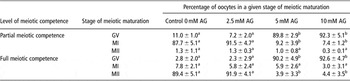
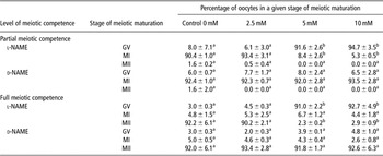
Both inhibitors at concentrations of 5, 7.5 and 10 mM disrupted meiotic maturation of growing oocytes with partial meiotic competence and an internal diameter of 100 to 110 μm, as well as in fully grown oocytes with fully developed meiotic competence (120 μm). Most of the oocytes were after 48-h culture arrested in the GV stage. A 2.5-mM concentration of the inhibitors did not influence the meiotic maturation of the oocytes with partially and fully developed meiotic competence. Oocytes with partial meiotic competence reached the MI stage, and those with fully developed meiotic competence reached the MII stage. The results are summarized in Tables 1 and 2. It was no surprise that l-NAME or AG at a concentration of 2.5, 5, 7.5 and 10 mM had any effect on the maturation of oocytes without meiotic competence with an internal diameter of 80 to 89 and 90 to 99 μm (data not shown).
Effect of AG on meiotic maturation of oocytes with partial (100 to 110 μm) and full meiotic competence (120 μm) cultured 48 h in vitro

AG = aminoguanidine; GV = germinal vesicle; MI = metaphase I; MII = metaphase II.
Data are presented as mean ± s.e.m. of four replicates. One hundred and twenty oocytes were analysed in each experimental group. There were four repetitions of the experiment.
Values with different superscripts within rows are significantly different at P < 0.05.
Effect of l-NAME and d-NAME on meiotic maturation of oocytes with partial (100 to 110 μm) and full meiotic competence (120 μm) cultured 48 h in vitro

GV = germinal vesicle; MI = metaphase I; MII = metaphase II.
Data are presented as mean ± s.e.m. of four replicates. One hundred and twenty oocytes were analysed in each experimental group. There were four repetitions of the experiment.
Values with different superscripts within rows are significantly different at P < 0.05.
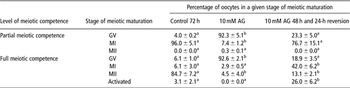
Although AG does not have an ineffective conformer, in contrast to l-NAME, it has been tested for toxicity to oocytes. After AG reversion, partially meiotically competent oocytes had a renewed ability to break down the first meiotic block. The reversion of AG in fully meiotically competent oocytes resulted in a large increase in the rate of parthenogenetically activated embryos, 26% v. 3% in the control group (Table 3).
24-h reversion of oocytes with partial meiotic competence (100 to 110 μm) and full meiotic competence (120 μm) after 48 h in vitro cultivation with 10 mM of AG

AG = aminoguanidine; GV = germinal vesicle; MI = metaphase I; MII = metaphase II.
Data are presented as mean ± s.e.m. of four replicates. One hundred and twenty oocytes were analysed in each experimental group. There were four repetitions of the experiment.
Values with different superscripts within rows are significantly different at P < 0.05.
Furthermore, we found that the inhibitor also affects the transition between the first and second meiotic metaphases, and after AG application it caused a block of meiotic maturation at the MI stage. Fully meiotically competent oocytes did not have the ability to reach the MII stage (Table 4).
Effect of AG on the transition of fully competent oocytes (120 μm) from the first to the second meiotic metaphase

AG = aminoguanidine; GV = germinal vesicle; MI = metaphase I; MII = metaphase II. Data are presented as mean ± s.e.m. of four replicates. One hundred and twenty oocytes were analysed in each experimental group. There were four repetitions of the experiment.
Values with different superscripts within rows are significantly different at P < 0.05.
Experiment 2
We studied whether NO from an external source added to a culture in vitro system by an NO donor influences the growth period and meiotic competence of porcine oocytes. We also studied whether the NO donor can reverse the inhibitory effect of the NOS inhibitor, AG.
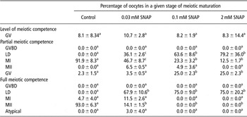
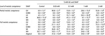
A reduction in the number of oocytes at the MI and II stages occurred after 48-h culture of partially and fully competent oocytes with the NO donor (Table 5). The oocytes did not continue in maturation after 24-h reversion in a pure medium. The donor concentration used disrupted the viability of the oocytes. One millimolar of SNAP influences porcine oocytes in the same manner as 2 mM. The concentration was therefore reduced to 0.1 mM; 0.03 mM of SNAP inhibited the oocytes much less. Fully grown oocytes matured up to 14% to the second meiotic stage after 48-h cultivation in the medium containing the NO donor in a concentration of 0.03 mM. NO externally applied to a culture medium had no effect on oocytes without meiotic competence.
Effect of NO donor SNAP on meiotic maturation of oocytes with partial (100 to 110 μm) and full meiotic competence (120 μm) cultured 48 h in vitro

NO = nitric oxide; SNAP = s-nitroso-n-acetylpenicillamine; GV = germinal vesicle; GVBD = germinal vesicle break down; LD = late diakinesis; MI = metaphase I; MII = metaphase II.
Data are presented as mean ± s.e.m. of four replicates. one hundred and twenty oocytes were analysed in each experimental group. There were four repetitions of the experiment.
Values with different superscripts within rows are significantly different at P < 0.05.
In the case of monitoring of the combined effect of the NOS inhibitor and NO donor, we found that in partially meiotic competent oocytes, as in fully meiotic competent oocytes, NO could actually reverse the effect of the NOS inhibitor. Oocytes with partial meiotic competence, just as oocytes with full meiotic competence, however, reached only the MI stage, and meiotically fully competent oocytes were not able to mature to the MII stage (Table 6).
Effect of a combination of NOS inhibitor and NO donor on oocytes with partial (100 to 110 μm) and full meiotic competence (120 μm) cultured 48 h in vitro

NOS = NO-synthase; NO = nitric oxide; SNAP = s-nitroso-n-acetylpenicillamine; GV = germinal vesicle; GVBD = germinal vesicle break down; LD = late diakinesis; MI = metaphase I; MII = metaphase II.
Data are presented as mean ± s.e.m. of four replicates. One hundred and twenty oocytes were analysed in each experimental group. There were four repetitions of the experiment.
Values with different superscripts within rows are significantly different at P < 0.05.
Discussion
In this study, we confirmed the role of NO during the process of acquisition of meiotic competence of pig oocytes using NOS inhibitors and NO donors.
Both of the inhibitors used, the selective competitive iNOS inhibitor AG and the non-selective NOS inhibitor l-NAME, blocked the maturation of both oocytes with partial meiotic competence and those with full meiotic competence at the end of the growth period. Their effect was reversible. After application of the inhibitors, GVBD breakdown was disrupted, which demonstrates the necessity of NO for the very beginning of meiotic maturation.
Regulation using the iNOS isoforms was shown to be critical for the meiotic maturation of pig oocytes in view of the fact that after its blocking, the maturation process is disrupted similar to that after application of non-selective inhibitors blocking all NOS simultaneously. It is probable that constitutive NOS isoforms are incapable of replacing this isoform by NO production. The reason for this may also be the partial effect of the iNOS-specific inhibitor on the iNOS and eNOS isoforms. One explanation may be the fact that NO production by iNOS is significantly higher; another reason may also be the differing regulation of iNOS and constitutive NOS, that is, calcium and calmoduline (Ignarro et al., Reference Ignarro, Cirino, Casini and Napoli1999).
The effects of both NOS inhibitors (AG, l-NAME) were quite strong, and it could seem that NOS and NO produced by NOS alone may be the sole factor controlling porcine oocyte maturation in vitro, and that only 10% of maturation is affected by some other factors. However, we cannot exclude an alternative explanation of the strong effect of NOS inhibitors. The higher meiotic reduction of porcine oocytes using higher concentrations of the inhibitors might be also associated with unspecific blocking, which can lead to the global transcription shutdown (Teodoro and O'Farrell, Reference Teodoro and O'Farrell2003). However, the global transcriptional shutdown cannot fully explain the observed effect of NOS inhibitors in fully grown oocytes which have transcriptionally inactive compact nucleoli (Crozet et al., Reference Crozet, Kanka, Motlik and Fulka1986).
The blockage of NOS in fully meiotically competent oocytes in the GV stage prevents the oocytes from reaching the meiotic MII stage, and this influence of NOS inhibitors is also clear when fully competent oocytes are blocked up to the meiotic MI stage. Inhibition of oocyte maturation by AG, however, was much more effective when it was added to the oocytes already in the GV stage. Pig oocytes probably do not need such a large quantity of NO in the transitional period between metaphases as oocytes in the GV stage, or they are much less perceptive to its effects, or they are regulated by NO originating from another NOS-inducible isoform during transition between metaphases. Blockage of meiotic maturation was also observed in mouse oocytes after NOS inhibition, where, however, meiotically competent oocytes regularly undergo GVBD and their meiotic maturation is blocked as early as between stages I and II of meiotic metaphase (Bu et al., Reference Bu, Xia, Tao, Lei and Zhou2003 and Reference Bu, Xie, Tao, Wang and Xia2004).
The changes observed in the meiotic maturation of oocytes after application of NOS inhibitors resembled those which were found during the culture of oocytes obtained from animals with a knockout gene for some NOS isoforms (Jablonka-Shariff and Olson, Reference Jablonka-Shariff and Olson1998). NOS inhibitors in the used dosage are not toxic for oocytes because after reversion, and also culture with the D non-effective conformer, they are capable of continuing maturation. However, full reversion was not observed in fully grown oocytes released from the block induced by AG. There was an increased portion of oocytes, which progressed only to the MI stage and did not complete maturation to the MII stage. This may indicate some detrimental effects of AG on oocytes. However, there was a significant portion of oocytes which underwent parthenogenetic activation after reversion, and this could indicate disturbed regulation either specific to NOS inhibition or due to the unspecific effect of the inhibitor. Different results of reversion, that is, block at the MI stage, maturation to the MII stage or parthenogenetic activation, could also reflect heterogenicity in the oocyte population, which was described in pig oocytes (Hunter, Reference Hunter2000).
The great increase in the share of parthenogenetically activated embryos after a 24-h reversion of fully grown oocytes in a pure medium was surprising. In view of the fact that AG belongs among competitive inhibitors, it is possible that the NOS enzyme, which lacks a substrate, accumulates during examination of the oocytes. After washing off the inhibitor, the production of NO begins in a greater degree, which is essential for the activation of oocytes (Petr et al., Reference Petr, Urbankova, Tomanek, Rozinek and Jilek2002). NO nitrosylates, for example, ryanodine receptors and their stimulation are capable of evoking parthenogenetic activation in fully grown matured oocytes (Xu et al., Reference Xu, Pommier, Arbov, Hutchings, Sotto and Foxcroft1998).
In addition to the inhibitors, we monitored the effect of the NO donor on oocytes with various degrees of meiotic competence. It was already known from experiments conducted earlier in our laboratory that if 2-mM SNAP is added to oocytes in MII, the result is parthenogenetic activation up to 71% during 10 h (Petr et al., Reference Petr, Rajmon, Lanska, Sedmikova and Jilek2005a). This concentration of the donor was therefore used as a starting point for the culture of oocytes with partial meiotic competence, also with full meiotic competence and a completed growth period. SNAP is a stable analog of endogenic s-nitrosothiols and after penetration to the cell, NO is released from it into the cell by endogenic enzymes and is therefore a source of NO in vivo (Megson, Reference Megson2000; Yamamoto and Bing, Reference Yamamoto and Bing2000). The dosage of the NO donor, which we used, 2.0 to 0.05 mM SNAP, disrupted the lifespan of the oocytes, which did not continue to mature after 24-h reversion in a pure medium. It is known that a high quantity of NO leads to toxic reactions of products in somatic cells (Messmer et al., Reference Messmer, Lapetina and Brune1995), which is probably also the case in pig oocytes. The fact that inhibition of NOS or addition of NO tends to have a similar effect on oocyte maturation might seem confusing. However, it could indicate that pig oocytes require quite balanced levels of NO for proper maturation progress. A lack or excess of NO due to inhibition of NOS or to the addition of an NO donor disturbs this balance, and the regulation of meiosis is impaired.
High concentrations of NO added to the in vitro system by the NO donor SNP inhibit meiotic maturation of mouse oocytes, whereas low concentrations of the NO donor stimulate maturation and prevent oocytes from aging and also stimulate mice embryo development (Jablonka-Shariff and Olson, Reference Jablonka-Shariff and Olson1998; Goud et al., Reference Goud, Goud, Diamond and Abu-Soud2005 and Reference Goud, Goud, Diamond, Gonik and Abu-Soud2008). We found that in pig oocytes, the NO donor at a low concentration is not able to affect the meiotic competence of oocytes; oocytes with partial meiotic competence were unable, in the presence of the NO donor, to break through the block of the MI stage. NO induces apoptosis and DNA damage in some types of cells. Goud et al. (Reference Goud, Goud, Diamond, Gonik and Abu-Soud2007) found that these changes of DNA in oocytes might be caused by reactive oxygen species (superoxide, hydrogen peroxide and hypochlorous acid), which are formed when high amounts of NO are present in cells. Our previous experiments showed that SNAP at 1-mM concentration activated the second stage of meiotic metaphase in 68% of pig oocytes (Petr et al., Reference Petr, Rajmon, Rozinek, Sedmikova, Jeseta, Chmelíková, Svestkova and Jilek2005b). This concentration of NO, however, has a toxic effect on partially and fully meiotically competent oocytes, suggesting that oocytes are more sensitive to NO at the beginning of oogenesis than at the end of their meiotic maturation. Another possible explanation is that oocytes in the early period of growth already contain a sufficient amount of NOS protein, which produces NO, and NO added from an external source is superfluous and harmful to oocytes.
Furthermore, we demonstrated that the inhibitory action of substances, which cause NO deficiency in oocytes, can be reversed in the pig oocyte by NO donors. The donor should be used in a very low concentration and cannot move the majority of oocytes from the MI stage to the MII stage.
The same amount of NO donor (0.1 mM) supplied to the culture medium was sufficient for the transition of blocked oocytes with partial and full meiotic competence to the stage of the MI stage.
NO binds in cells to the heme group of soluble guanylate cyclase (sGC) and thus increases the production of cyclic cGMP (Hofmann et al., Reference Hofmann, Ammendola and Schlossmann2000; Goud et al., Reference Goud, Goud, Diamond, Gonik and Abu-Soud2006). It is also probable that cGMP decreases cAMP levels and influences meiotic maturation by activation of cAMP-phosphodiesterase. However, there is very compelling evidence that NO can induce a number of biological effects outside of the cGMP signaling cascade, such as binding to a heme group of proteins different from cGMP or by nitrosylation of proteins such as Ras, which belongs to the MAPK pathway in oocytes, or to tyrosine nitrosylation (Rettori et al., Reference Rettori, Belova, Dees, Nyberg, Gimeno and Mccann1993). Oocytes with completed growth period and full meiotic competence and those with partial meiotic competence differ in some regulatory mechanisms, such as the content of cyclin B, cdc25C and Wee 1, or the ability to activate the InsP3/Ca2+ system (Hampl and Eppig, Reference Hampl and Eppig1995). Our experiments confirmed the involvement of NO in the regulation of the growth period and the process of meiotic competence acquisition. One question, however, remains: how NO acts in oocytes, and whether the mechanism of NO action is the same in both growing and fully grown oocytes.
Acknowledgments
We thank Mrs Lois Russell for her editorial assistance with this manuscript. The study was supported by Grants MSM 6046070901, MZE 0002701404 and GAČR 523/08/H064.