Case
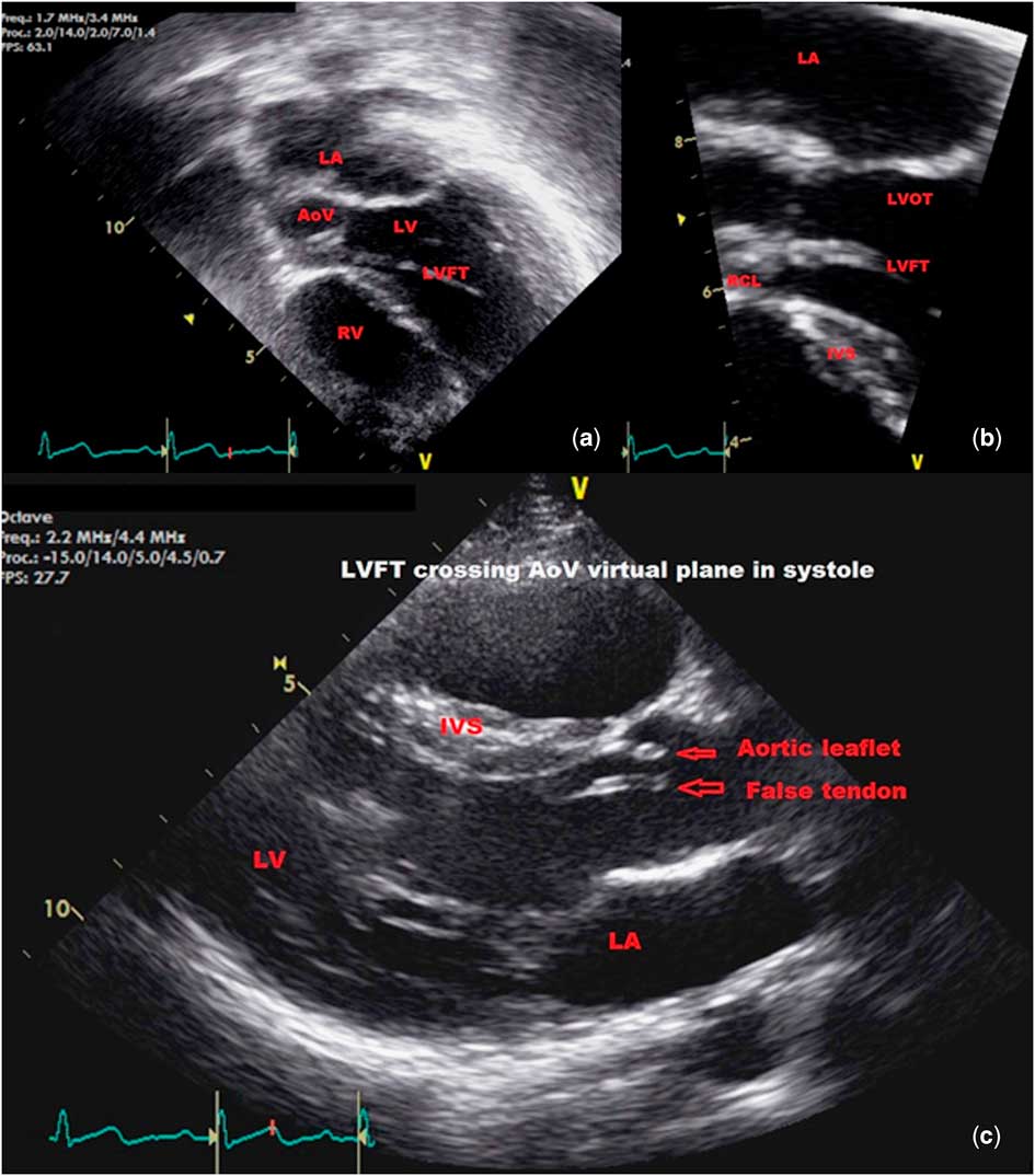
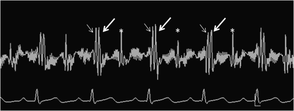
A previously healthy girl, aged 11 years, initially presented at 2 years of age for evaluation of a systolic ejection click heard on routine physical examination at the left upper sternal border. The electrocardiogram taken at that time showed normal sinus rhythm. An echocardiogram was performed, which ruled out the common causes for the click, such as an aortic valve with two leaflets, pulmonary valvar stenosis, or dilation of the pulmonary trunk. She remained asymptomatic, but the ejection click persisted, leading to repeated cardiac evaluation and echocardiography. The current echocardiogram confirmed normal cardiac anatomy and function, including normal aortic and pulmonary valves, along with normal features of the ascending aorta and pulmonary arteries. It also revealed a solitary linear structure, with the anticipated features of a false tendon, which took origin from the superolateral aspect of the apical segment of the left ventricular free wall, crossed the left ventricular cavity, and attached to the right coronary aortic valvar leaflet. The structure was seen to traverse the entrance to the aortic root during cardiac systole (Fig 1, Supplementary movie S1). The aortic valve, nonetheless, was shown to be functioning normally, without any evidence of stenosis or insufficiency. Phonocardiogram (Fig 2) shows early timing of the systolic click. We speculate that, as the tendon tauts in early systole, it may produce the ejection click heard on auscultation. It is unclear whether this will, in future, lead to aortic valve dysfunction. With this in mind, we have opted to follow-up the function of her aortic valve at 5-year intervals.
( a ) The apical four-chamber echocardiographic image shows a left ventricular false tendon (LVFT) crossing from the left ventricular (LV) free wall and attaching to the right coronary leaflet of the aortic valve (AoV); ( b ) the magnified echocardiographic view of the left ventricular outflow tract (LVOT) shows the attachment of the false tendon to the right coronary leaflet (RCL) of the AoV, ( c ) the parasternal long-axis echocardiographic view shows the false tendon (LVFT) crossing the virtual plane of the base of the aortic root (AoV) in systole. IVS=interventricular septum; LA=left atrium; RV=right ventricle.

Phonocardiogram recorded on iE 33 (Philips Medical Systems, Bothell, WA, USA): first heart sound (small arrow); ejection click (large arrow); and second heart sound (*).

Left ventricular false tendons are not uncommon in structurally normal hearts and have been implicated in the development of various clinical manifestations such as ejection systolic murmurs, monomorphic or polymorphic premature ventricular contractions, regional myocardial hypertrophy, and re-polarisation abnormalities, including ST elevation as seen on the electrocardiogram.Reference Kenchaiah, Benjamin, Evans, Aragam and Vasan 1 – Reference Loukas, Louis, Black, Pham, Fudalej and Sharkees 4 The false tendons are linear fibrous or fibromuscular structures that extend between different segments of the left ventricular free wall to the ventricular septum and papillary muscles, but are not usually described as attaching to the valvar leaflets.Reference Kenchaiah, Benjamin, Evans, Aragam and Vasan 1 They are made up of cardiomyocytes, with their supporting fibrous matrix, and usually include components of the ventricular conduction tissues. In terms of development, they are the remaining components of the trabecular layer of the developing ventricular myocardium.Reference Kervancioğlu, Ozbağ and Kervancioğlu 3 Usually found in the apical component of the left ventricle, they rarely extend into the sub-aortic area. Various anatomical classifications have been proposed,Reference Loukas, Louis, Black, Pham, Fudalej and Sharkees 4 with one suggesting that they can be simple, extending between two points, or complex, having three or more attachments.Reference Kenchaiah, Benjamin, Evans, Aragam and Vasan 1 To our knowledge, a tendon attaching to an aortic valvar leaflet, in association with an ejection click, has not been previously reported. Our described lesion has all the characteristics of a false tendon, but is attached to the aortic valvar leaflet rather than to the ventricular septum.
Our observation shows that a left ventricular false tendon may be considered as one of the lesions responsible for a systolic ejection click. As yet, we do not know whether it will have any impact on aortic valve function, but our finding does indicate the need for long-term counselling and follow-up of the patient.
Acknowledgement
None.
Financial Support
This research received no specific grant from any funding agency, commercial, or not-for-profit sectors.
Conflicts of Interest
None.
Supplementary material
To view supplementary material for this article, please visit http://dx.doi.org/10.1017/S1047951116000858