




No CrossRef data available.
The application of mass spectrometry to identify immunogenic components of excretory/secretory products from adult Dictyocaulus viviparus
Published online by Cambridge University Press: 12 May 2005
Abstract
Proteomics has come to the forefront in the post-genomic era. The ability to compare and identify proteins expressed in a particular cell type under specific physiological or pathological states requires a range of technologies, including separation of complex protein or peptide mixtures, densitometry-based or isotope-coded methods for comparison of multiple proteomes, and mass spectrometric methods for identification of individual low abundance proteins. Although an emergent technology, thus far, proteomics has provided new perspectives on many problems in biomedical science. In parasitology, proteomics has been used to answer specific biological questions relating to survival and development, and also to identify candidates for vaccines. Here, we describe an ongoing research programme in which proteomics is being used to identify potential vaccine candidates for the bovine lungworm, Dictyocaulus viviparus. This work is focusing on antibody responses to the adult parasite excretory/secretory (ES) products, with selection of candidate antigens based on differential screening with serum from immune versus non-immune animals to simplify the proteome and the ensuing analytical challenges. Thus far, we have identified seven candidate proteins using this strategy. Of these, one protein showed significant identity to a previously cloned gene from D. viviparus, whilst the other six proteins have shown no significant identities. Isolation of further peptide sequences is now warranted to facilitate cloning of the genes encoding these antigens.
Information
- Type
- Research Article
- Information
- Copyright
- © 2004 Cambridge University Press
INTRODUCTION
Dictyocaulus viviparus is an important parasitic nematode of cattle in temperate regions. The adult parasites inhabit the main stem bronchi and trachea. Together with the migrating pulmonary larvae, the adult worms cause an intense inflammatory reaction which results in bronchopneumonia (Jarrett, McIntyre & Urquhart, 1957). The most common clinical manifestations of this disease (known as parasitic bronchitis) are coughing, respiratory distress, weight loss and, in some cases, death (Jarrett et al. 1957). Immunity to D. viviparus is induced relatively easily and calves exposed to a limited number of experimental infections are rendered solidly immune (Jarrett et al. 1959). Based on this, an irradiated larval vaccine was developed (Jarrett et al. 1960), which involves the oral administration of irradiated third stage larvae (L3). After vaccination, irradiated larvae reach the lungs where they stimulate protective immunity without the parasites reaching patency or causing clinical disease. Immunity induced by the vaccine is not sterile: vaccinates are still susceptible to infection at a low level, even though they are protected against disease. These animals often have small numbers of parasites in their lungs and excrete low amounts of first stage larvae (L1) in their faeces, which maintains pasture contamination. In practice, such contamination acts to boost immunity in vaccinates and, without this, animals would become susceptible to parasitic bronchitis. The vaccine (Huskvac, Intervet) has been used with success in the field (Peacock, Menear & Poynter, 1970). Over the last fifteen years, the incidence of parasitic bronchitis has increased, particularly in adult cattle in which its effects are of major welfare and economic significance (David, 1993, 1996, 1997; Robinson, Jackson & Sarchet, 1993; Williams, 1996; Woolley, 1997). The rise in outbreaks reflects the growing tendency of farmers to stop vaccinating first season calves or to complement vaccination with anthelmintic treatments (Connan, 1993; Mawhinney, 1996; McKeand, 2000). Such treatments are highly effective in killing D. viviparus and can eradicate the parasite from a cattle population so that animals become susceptible to disease again.
A further disadvantage of the current vaccine is its instability. Because of this, it is not used abroad and its manufacture requires donor calves that are used each year for maintenance of the strain and production of the vaccine. With these factors in mind, it is clear that there is a need for an alternative D. viviparus vaccine that is more stable and, ideally, is capable of stimulating immunity without the need for boosting by re-exposure to natural infection. Currently, several studies, utilising genomic and proteomic technologies, have the identification of vaccine candidates for D. viviparus as their goal (Matthews et al. 2001; Kooyman et al. 2002; Lazari et al. 2003). Our approach has focused on immunoblotting to identify protective antigens, which were subsequently subjected to mass spectrometry analyses.
IDENTIFYING PROTECTIVE ANTIGENS OF D. VIVIPARUS
Protective immunity is associated with immune responses to antigens expressed in lung stage D. viviparus, i.e. fourth stage larvae (L4), fifth stage larvae (L5) and adult parasites (Jarrett & Sharpe, 1963; Michel, 1969). Thus far, the antigens that induce protection have not been identified, but molecules released by adult worms as excretory/secretory (ES) products appear to play an important role. In the guinea pig laboratory model, significant levels of protective immunity were obtained following immunisation with adult ES products when administered with Freund's complete adjuvant (McKeand et al. 1994, 1995). Antibody has a role in adult ES-induced immunity in guinea pigs, as protection can be transferred passively in serum from immunised animals to naïve recipients (McKeand et al. 1995). Similar observations had been made previously in calves exposed to experimental L3 infections, where serum was used to transfer immunity to naïve recipient calves (Jarrett et al. 1955). These studies strongly indicate that antibody plays an important role in immunity in dictyocaulosis and that analysis of such responses is pertinent to the identification of putative protective antigens. Hence, we have designed a strategy to identify vaccine candidates of the basis of their unique recognition by antibody in serum from immune cohorts of animals, compared to the recognition profiles observed in non-protected animals.
Adult ES products were separated by 2D-GE. Colloidal Coomassie staining indicated a complex mixture, consisting of at least 140 spots (Fig. 1). To identify the locations of candidate proteins, we performed immunoblots using sera from three strains of adult ES-immunised guinea pigs that showed different susceptibility to challenge following ES immunisation (McKeand et al. 1995). Of these strains, two (Strain 13 and Dunkin Hartley) were significantly protected compared to challenge control animals, whilst the third strain (Strain 2) remained susceptible to challenge (McKeand et al. 1994). Seven spots were selected on the basis of recognition by Dunkin Hartley and/or Strain 13 animals, but not by the susceptible Strain 2 animals. These spots have been subjected to mass spectrometry analysis as described below.

Fig. 1. Two-dimensional polyacrylamide gel of D. viviparus adult ES products harvested after 24 h culture (McKeand et al. 1994). For electrophoresis, 200 μg adult ES products were acetone precipitated and resuspended in 30 μl H2O and 170 μl rehydration buffer (3·4 mg DTT, 5·5 μl IPG buffer in 1 ml CUT buffer [7·7 M urea, 2·2 M thiourea, 4·4% w/v CHAPS]) and loaded onto a 11 cm, 1-dimension isoelectric focusing strip, pH 3–10 (Biorad) and focused overnight. The strips were then reduced and alkylated. For the second dimension, the proteins were separated on a 16 cm, 12·5% w/v polyacrylamide gel. The gel was stained with colloidal Coomassie blue. ‘MSP’ indicates the spot which gave significant identities to nematode major sperm proteins.
MASS SPECTROMETRY ANALYSES
Gel plugs coincident with the proteins of interest were excised and subjected to in-gel digestion with trypsin. The tryptic peptides were then mass-measured by MALDI-ToF mass spectrometry (Micromass M@LDI-ToF instrument, reflectron mode). Although the mass spectra were of high quality, database searching of the peptide mass fingerprints yielded no high-confidence hits for six of the seven proteins. This is perhaps unsurprising, as Dictyocaulus is poorly represented in existing databases such as SwissProt and TREMBL. Currently recorded are a total of 13 entries for D. viviparus, which correspond to a total of eight distinct proteins. For the seventh protein differentially identified by our guinea pig sera (marked ‘MSP’ on Fig. 1), we did obtain a significant hit. This protein showed significant similarity to the Caenorhabditis elegans major sperm protein (MSP, accession number AAA28115). Surprisingly, the predicted protein sequence of a previously cloned MSP from D. viviparus (Schneider 1993, accession number S64873) did not significantly match the spectra of its homologue in our adult ES products. On comparison of the predicted peptide sequence of the cloned D. viviparus MSP (Schnieder, 1993) with translated sequences of other nematode MSPs, it was clear that the alignment was imperfect. In particular, the C-terminal region of the cloned D. viviparus MSP protein sequence (Fig. 2, sequence S64873_F1) demonstrated virtually no similarity to the protein sequences from other organisms. On closer inspection of the cDNA sequence of the D. viviparus MSP clone, it is apparent that insertion of a single residue at position 181 corrects the reading frame, such that the resultant protein sequence (Fig. 2, sequence S64873corr_F1) becomes almost identical (89%) to the MSPs from other organisms. We subsequently confirmed this putative sequence error from our MALDI-ToF data. Once the revised C-terminal segment had been obtained by correction of the frame-shift, additional matching peptides located at the C-terminus were observed (Fig. 3). By preparing predicted tryptic maps for each of four putative DNA sequences encoding this region (Fig. 3, fragment T9), we were able to confirm that a G nucleotide would need to be inserted. The three other nucleotides predicted peptides for T9 that could not be located in our MALDI-ToF mass spectrum. Although the identification of this protein would not have required detailed analysis, this assignment indicates the difficulty of matching protein data, particularly to single pass cDNA inferred sequences, where frame-shifts in particular can drastically reduce the chance of complete coverage. Other interesting features were highlighted in analysis of the MSP protein. First, the N-terminal most tryptic peptide (Fig. 3, peptide T1) could not be matched without inferring removal of the initiator methionine and acetylation of the underlying alanine residue; a common post-translational modification. When these changes were factored in to mass mapping, a strong peak at approx 1567 Da provided confirmation of this change. We also note the necessity to search for masses corresponding to partial cleavage reactions, in which two tryptic peptides remained linked (a common observation) and particularly where there are pairs or cluster of arginine and lysine residues.

Fig. 2. Alignment of major sperm protein (MSP) sequences. Peptide mass fingerprinting of the spot marked ‘MSP’ in Fig. 1 yielded a tentative identification through peptides T2, T2–3, T4–5 and T5–6 to MSPs from other nematodes, including Onchocerca volvulus (MSP1_ONCVO and MSP2_ONCVO), Ascaris suum (MSP1_ASCSU and MSP2_ASCSU) and C. elegans (AAA28115). For the D. viviparus MSP (S64873) cloned previously (Schnieder, 1993), three sequences are presented here: the MSP sequence in reading frame 1 (S64873_F1), the corresponding sequence in reading frame 3 (S64873_F3) and the translation of the corrected full reading frame (S64873corr_F1). Superimposed on the alignment are the tryptic peptides (T1 to T18) that were matched (filled) or unmatched (open) to the corrected full reading frame of S64873. The circle indicates the approximate location where the frame-shift disrupted the open reading frame. Peptides marked with a range (e.g. T4–5) denote partial cleavages. Peptide T1 can only be matched if it is assumed to be des-Met and acetylated (Ac).

Fig. 3. MALDI-ToF spectrum of MSP. The MALDI-ToF spectrum obtained from in-gel tryptic digestion of the protein in the spot marked ‘MSP’ in Fig. 1 has been annotated to indicate the peptides that have been matched to the corrected primary sequence.
MALDI-ToF analysis is primarily used as an initial and rapid screen of the quality of the tryptic digests. Once the quality of our digests was confirmed, the tryptic peptides were subsequently resolved by microcapillary (75 μm) C18 reversed phase nanochromatography (200 nl/min, LC Packings Nanomate) and electrosprayed directly into the source of a QToF-Micro tandem mass spectrometer. The resultant product ion spectra were used for de novo sequencing. This was performed for all proteins except the putative MSP. For the remaining six proteins, the derived peptide sequence tags showed no significant matches to sequences from other nematodes, including C. elegans. From these preliminary analyses, these proteins have no obvious homologues in other nematodes. Indeed, in terms of the biology of the organism, these are potentially genes of great interest. However, we are currently repeating these experiments and subjecting the proteins to more extensive LC-MS/MS de novo sequencing. This will drive primer design for subsequent PCR and cloning experiments. Because these proteins are fragmented prior to being separated, it is not possible to ascertain the order of the different peptides. Thus, each sequenced peptide would have to be used to design a pair of degenerate primers to explore its order in relation to other peptides. To overcome this complication, we are working to derive peptide chemistry strategies that maximise the chances of obtaining the C- and N-terminal tryptic peptides to facilitate primer design for amplification of full-length transcripts. However, this strategy obviates a peptide-based approach, and requires prior separation of individual proteins, preferably in solution rather than in gel-based resolution. We have resolved native ES products according to charge and have obtained high-resolution separations. Peak-collected fractions have been analysed for immunoreactivity by western blot (Davidson, Beynon & Matthews, unpublished data), and fractions enriched in immunoreactive proteins have been digested and are currently being subjected to MALDI-ToF analysis.
CONCLUSIONS
We have identified seven antigens that are uniquely immunoreactive with sera from protected guinea pigs. One of these antigens is a putative MSP, whilst we await more analysis to ascribe putative functions and/or designations to the remaining six proteins. We have a conservative expectation of being able to design primers to obtain cDNA sequences for at least half of these, which would yield a total of four candidates for expression studies. These candidates will be prioritised on the basis of a composite figure of merit, taking into account the following: strength of immune response; broad disposition of immunoreactivity across all immune species, predicted function, size of expressed product, stage of expression and presence of putative glycosylation sites. These studies have shown that parasitic nematode-secreted molecules can be subjected to successful mass spectrometry analysis for the selection of candidate antigens for recombinant vaccine development.
ACKNOWLEDGEMENTS
We would like to thank the BBSRC (research project reference: 26/S17867) for funding this work and Dr Duncan Robertson for assistance with the mass spectrometry.
References
REFERENCES
CONNAN, R. M. (
1993).
Calfhood vaccination for Dictyocaulosis.
Veterinary Record
133,
554–555.CrossRefGoogle Scholar
DAVID, G. P. (
1996).
An epidemiological study of husk in adult cows in 32 UK herds (preliminary findings).
Cattle Practice
5,
295–297.Google Scholar
DAVID, G. P. (
1997).
Survey on lungworm in adult cattle.
Veterinary Record
141,
343–344.Google Scholar
JARRETT, W. F.,
JENNINGS, F. W.,
McINTYRE, W. I. M.,
MULLIGAN, W.,
THOMAS, B. A. C. &
URQUHART, G. M. (
1959).
Immunological studies on Dictyocaulus viviparus infection. The immunity resulting from experimental infection.
Immunology
2,
252–261.Google Scholar
JARRETT, W. F.,
JENNINGS, F. W.,
McINTYRE, W. I. M.,
MULLIGAN, W. &
URQUHART, G. M. (
1955).
Immunological studies on Dictyocaulus viviparus infection. Passive immunisation.
Veterinary Record
67,
291–296.Google Scholar
JARRETT, W. F.,
JENNINGS, F. W.,
McINTYRE, W. I. M.,
MULLIGAN, W. &
URQUHART, G. M. (
1960).
Immunological studies on Dictyocaulus viviparus infection. Immunity produced by administration of irradiated larvae.
Immunology
3,
145–151.Google Scholar
JARRETT, W. F.,
McINTYRE, W. I. M. &
URQUHART, G. M. (
1957).
The pathology of experimental parasitic bronchitis.
Journal of Pathology and Bacteriology
73,
183–193.CrossRefGoogle Scholar
JARRETT, W. F. &
SHARPE, N. C. C. (
1963).
Vaccination against parasitic disease: reactions in vaccinated and immune hosts in Dictyocaulus viviparus infection.
Journal of Parasitology
49,
177–189.CrossRefGoogle Scholar
KOOYMAN, F. N.,
YATSUDA, A. P.,
PLOEGER, H. W. &
EYSKER, M. (
2002).
Serum immunoglobulin E response in calves infected with the lungworm Dictyocaulus viviparus and its correlation with protection.
Parasite Immunology
24,
47–56.CrossRefGoogle Scholar
LAZARI, O.,
HUSSEIN, A. S.,
SELKIRK, M. E.,
DAVIDSON, A. J.,
THOMPSON, F. J. &
MATTHEWS, J. B. (
2003).
Cloning and expression of two secretory acetylcholinesterases from the bovine lungworm, Dictyocaulus viviparus.
Molecular and Biochemical Parasitology
132,
83–92.CrossRefGoogle Scholar
McKEAND, J. B. (
2000).
Vaccine development and diagnostics of Dictyocaulus viviparus.
Parasitology
120,
S17–S23.CrossRefGoogle Scholar
McKEAND, J. B.,
KNOX, D. P.,
DUNCAN, J. L. &
KENNEDY, M. W. (
1994).
Genetic control of the antibody repertoire against excretory/secretory products and acetylcholinesterases of Dictyocaulus viviparus.
Parasite Immunology
16,
251–260.CrossRefGoogle Scholar
McKEAND, J. B.,
KNOX, D. P.,
DUNCAN, J. L. &
KENNEDY, M. W. (
1995).
Immunisation of guinea pigs against Dictyocaulus viviparus using adult ES products enriched for acetylcholinesterases.
International Journal for Parasitology
25,
829–837.CrossRefGoogle Scholar
MATTHEWS, J. B.,
DAVIDSON, A. J.,
FREEMAN, K. L. &
FRENCH, N. P. (
2001).
Immunisation of cattle with recombinant acetylcholinesterase from Dictyocaulus viviparus and with adult worm ES products.
International Journal for Parasitology
31,
307–317.CrossRefGoogle Scholar
MICHEL, J. F. (
1969).
The epidemiology and control of some nematode infections in grazing animals.
Advances in Parasitology
7,
211–282.CrossRefGoogle Scholar
PEACOCK, P.,
MENEAR, H. C. &
POYNTER, D. (
1970).
The prevention and treatment of husk.
Veterinary Record
86,
148–160.Google Scholar
ROBINSON, T. C.,
JACKSON, E. R. &
SARCHET, R. W. (
1993).
Husk in heifers.
Veterinary Record
132,
143.CrossRefGoogle Scholar
SCHNIEDER, T. (
1993).
The diagnostic antigen encoded by gene fragment Dv3-14: a major sperm protein of Dictyocaulus viviparus.
International Journal for Parasitology
23,
383–389.CrossRefGoogle Scholar
WOOLLEY, H. (
1997).
The economic impact of husk in dairy cattle.
Cattle Practice
5,
315–317.Google Scholar

Fig. 1. Two-dimensional polyacrylamide gel of D. viviparus adult ES products harvested after 24 h culture (McKeand et al. 1994). For electrophoresis, 200 μg adult ES products were acetone precipitated and resuspended in 30 μl H2O and 170 μl rehydration buffer (3·4 mg DTT, 5·5 μl IPG buffer in 1 ml CUT buffer [7·7 M urea, 2·2 M thiourea, 4·4% w/v CHAPS]) and loaded onto a 11 cm, 1-dimension isoelectric focusing strip, pH 3–10 (Biorad) and focused overnight. The strips were then reduced and alkylated. For the second dimension, the proteins were separated on a 16 cm, 12·5% w/v polyacrylamide gel. The gel was stained with colloidal Coomassie blue. ‘MSP’ indicates the spot which gave significant identities to nematode major sperm proteins.
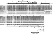
Fig. 2. Alignment of major sperm protein (MSP) sequences. Peptide mass fingerprinting of the spot marked ‘MSP’ in Fig. 1 yielded a tentative identification through peptides T2, T2–3, T4–5 and T5–6 to MSPs from other nematodes, including Onchocerca volvulus (MSP1_ONCVO and MSP2_ONCVO), Ascaris suum (MSP1_ASCSU and MSP2_ASCSU) and C. elegans (AAA28115). For the D. viviparus MSP (S64873) cloned previously (Schnieder, 1993), three sequences are presented here: the MSP sequence in reading frame 1 (S64873_F1), the corresponding sequence in reading frame 3 (S64873_F3) and the translation of the corrected full reading frame (S64873corr_F1). Superimposed on the alignment are the tryptic peptides (T1 to T18) that were matched (filled) or unmatched (open) to the corrected full reading frame of S64873. The circle indicates the approximate location where the frame-shift disrupted the open reading frame. Peptides marked with a range (e.g. T4–5) denote partial cleavages. Peptide T1 can only be matched if it is assumed to be des-Met and acetylated (Ac).
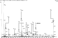
Fig. 3. MALDI-ToF spectrum of MSP. The MALDI-ToF spectrum obtained from in-gel tryptic digestion of the protein in the spot marked ‘MSP’ in Fig. 1 has been annotated to indicate the peptides that have been matched to the corrected primary sequence.
 You have
Access
- 5
- Cited by