Introduction
In 1906, Doctor Gustav Killian described for the first time an antrochoanal polyp, a benign tumour originating from the maxillary sinus mucosa. It usually grows through the maxillary ostium into the nasal cavity, without bone erosion or expansion, and extends posteriorly towards the choana.Reference Bakshi and Vaithy1 The antral portion is usually cystic, and it frequently originates from the posterior, inferior, lateral or medial wall of the maxillary sinus.Reference Frosini, Picarella and De Campora2
The antrochoanal polyp is almost always unilateral. It is more common in children and young adults, representing around 10 per cent of all nasal polyps in adults, and 33 per cent of all paediatric nasal polyps.Reference Yuca, Bayram, Kiroglu, Etlik, Cankaya and Sakin3 Histologically, an antrochoanal polyp has a vascular and oedematous stroma, covered with pseudostratified ciliated epithelium, and containing inflammatory cells.Reference Yaman, Yilmaz, Karali, Guclu and Ozturk4
Surgery is the only possible treatment for antrochoanal polyp. Nowadays, functional endoscopic sinus surgery (FESS) is considered the ‘gold standard’ technique.Reference Bakshi and Vaithy1,Reference Yuca, Bayram, Kiroglu, Etlik, Cankaya and Sakin3,Reference El-Sharkawy5 However, other external approaches, such as the Caldwell–Luc procedureReference Orvidas, Beatty and Weaver6 or the mini Caldwell–Luc procedure, might be used alone or with FESS.Reference Kizil, Aydil, Ceylan, Uslu, Baştürk and İleri7,Reference Al-Mazrou, Bukhari and Al-Fayez8
The chance of antrochoanal polyp recurrence in children remains high, although surgical techniques are now more effective and safer. At present, there is no accepted indication for revision surgery or ‘wait and see’ policy.Reference Comoglu, Celik, Enver, Sen, Polat and Deger9 Recently, Galluzzi et al., in their systematic review, observed that no data were available regarding the failure of revision surgery performed for antrochoanal polyp recurrence, and no study had compared endoscopic with combined techniques for the treatment of recurrence.Reference Galluzzi, Pignataro, Maddalone and Garavello10
This study aimed to compare FESS with a combined approach (FESS plus a Caldwell–Luc procedure) for the treatment of paediatric antrochoanal polyp, in terms of antrochoanal polyp recurrence and safety.
Materials and methods
This study represents a retrospective case series.
Data collection
A database was created using the surgical records of the paediatric patients (aged less than 18 years old) who were diagnosed and treated for antrochoanal polyp from January 2010 to January 2018.
The following data were recorded for each patient: age, sex, side of the lesion, exposure to farm or domestic animals, related diseases, passive smoking, number of surgical procedures performed, operative technique, recurrence, the length of time between surgery and recurrence, complications, follow-up duration, and outcomes.
Data management and statistics
Data were collected on forms, and were computerised and analysed using Stata Statistical Software: Release 13 (StataCorp, College Station, Texas, USA). Continuous variables are presented as the mean (± standard deviation) and categorical variables are presented as counts. The Fisher's exact test was performed to compare recurrences in relation to surgical techniques and pre-surgical features. A p-value of less than 0.05 was considered statistically significant.
Ethics
This study has been carried out in accordance with the World Medical Association Declaration of Helsinki on medical research protocols and ethics. The need for informed consent was waived by the medical ethics committee of our institution, given the retrospective nature of the study.
Results
A total of 27 paediatric patients were included in the study. All patients underwent surgical resection of the antrochoanal polyp, and the diagnosis was confirmed histologically. The average age of patients at the time of diagnosis was 10.4 ± 2.49 years.
All patients underwent a pre-operative radiological assessment, involving axial and coronal computed tomography (CT) scans of the nose and paranasal sinuses (Figure 1).
(a) Coronal computed tomography (CT) scan showing an antrochoanal polyp arising from the right maxillary antrum through the widened natural ostium of the sinus. (b) Coronal CT scan of the polyp filling the right choana. (c) Axial CT scan of the polyp extending into the right nasal cavity. (d) Sagittal CT scan showing the polyp extending to the nasopharynx.

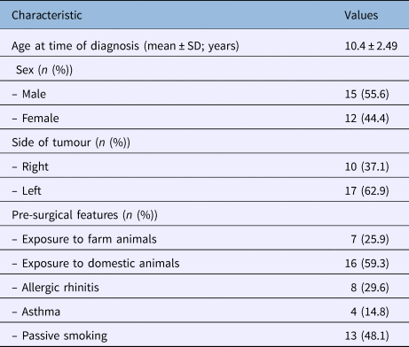
Male patients accounted for 55.6 per cent of all cases (Table 1). The location of the lesion was on the left side in 62.9 per cent of the cases. More than half of the patients had a history of exposure to domestic or farm animals, while 48.1 per cent of the patients were passive smokers. Allergic rhinitis and asthma were the most common morbidities identified.
Baseline characteristics of patients

SD = standard deviation
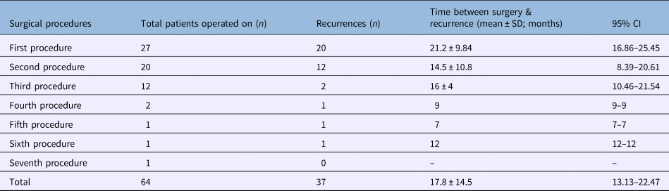
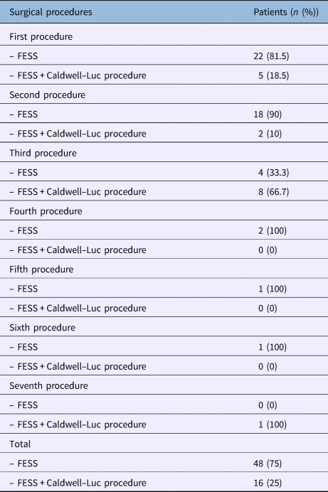
A total of 64 surgical procedures were performed, all executed by the same surgical team in a paediatric tertiary care hospital (Table 2). The average time between surgery and recurrence was 17.8 ± 14.5 months (95 per cent confidence interval (CI) = 13.13–22.47). The recurrence rate was recorded for each patient, with up to seven surgical procedures in one patient before there was control of the disease (Table 2). The overall follow-up time after the final surgical procedure was 26.9 ± 11.4 months. There were no complications intra-operatively or during follow up in any of the patients. There was a tendency to perform FESS alone in the primary surgical procedure, and a tendency to carry out the combined approach (FESS plus Caldwell–Luc procedure) as more recurrences occurred (Table 3).
Recurrences after primary and revision surgical procedures

SD = standard deviation; CI = confidence interval
Number of patients who underwent each surgical technique

FESS = functional endoscopic sinus surgery
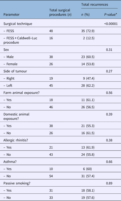
The recurrence rate after FESS alone was 72.9 per cent, compared with 12.5 per cent after FESS plus the Caldwell–Luc procedure (p < 0.00001). No significant differences were found in recurrence rates in terms of age, side of the lesion, exposure to animals, allergic rhinitis, asthma or passive smoking (Table 4).
Recurrences by surgery technique and pre-surgical features

*Recurrences rates were compared using Fisher's exact test; a p-value of less than 0.05 was considered statistically significant. FESS = functional endoscopic sinus surgery
Discussion
Antrochoanal polyps are solitary soft tissue masses, generally originating from the maxillary sinus and extending towards the choana by passing through the sinus ostium. Typically, the polyp passes through the ostium into the middle meatus, without causing bone deformation or destruction. It fills the middle meatus between the middle turbinate and the lateral nasal wall, and grows posteriorly to reach the choana.Reference Kizil, Aydil, Ceylan, Uslu, Baştürk and İleri7 Previous studies have shown that antrochoanal polyp is more common in males than in females (with a male-to-female ratio of 1.7:1), and there is a slight predominance for the compromise of the left maxillary sinus.Reference Lee, Yoon, Lee and Lim11
Most of the antrochoanal polyps are broad-based; however, up to 92 per cent of polyps originate from the posterior wall of the maxillary sinus.Reference Lee and Huang12 Frosini et al., in their study of 200 cases, suggested that antrochoanal polyp occurred from the combination of an antral cyst and an obstructed maxillary ostium secondary to an anatomical abnormality, such as a deviated nasal septum or a turbinate alteration.Reference Frosini, Picarella and De Campora2 Although the exact aetiology of antrochoanal polyp is undefined, allergic rhinitis, chronic sinusitis and lower respiratory tract disorders stand suspicious.Reference Maldonado, Martínez, Alobid and Mullol13,Reference Kelles, Toplu, Yildirim and Okur14 In this study, half of the patients were passive smokers or had a history of exposure to animals. One-third of the patients had allergic rhinitis, while one-sixth had asthma.
Clinically, antrochoanal polyp usually presents with unilateral nasal obstruction; however, other symptoms might be present, including epistaxis, purulent rhinorrhoea, post-nasal drip, snoring, dysphonia and dysphagia.Reference Maldonado, Martínez, Alobid and Mullol13,Reference Aydin, Keskin, Ustundag, Iseri and Ozkarakas15 Antrochoanal polyp may mimic a wide range of pathologies, although certain clinical and radiological features should raise the index of suspicion.Reference Choudhury, Hariri, Saleh and Sandison16
Nasal endoscopy and CT are excellent tools for diagnosing antrochoanal polyp.Reference Yaman, Yilmaz, Karali, Guclu and Ozturk4,Reference Lee, Yoon, Lee and Lim11 On nasal endoscopy, antrochoanal polyp typically appears as a smooth polypoid mass originating in the middle meatus, extending into the choana and nasopharynx, and eventually to the oral cavity.Reference Frosini, Picarella and De Campora2 The CT scanning can frequently help, and usually shows: a soft tissue mass filling the maxillary sinus, opacification of the middle meatus, and extension into the choana. A CT scan is, in most cases, sufficient to identify the base of the polyp; however, the presence of another sinonasal abnormality may hinder proper identification of the site. On magnetic resonance imaging, antrochoanal polyp shows hypointensity on a T1-weighted image and hyperintensity on a T2-weighted image.Reference Yaman, Yilmaz, Karali, Guclu and Ozturk4,Reference Lee, Yoon, Lee and Lim11
Antrochoanal polyp differential diagnoses include ipsilateral nasal tumours such as angiofibroma, nasal glioma, encephalocele, mucocele, retention cyst, inverted papilloma or haemangioma.Reference Lee, Yoon, Lee and Lim11,Reference Lee and Huang12
Surgery is the only feasible treatment for antrochoanal polyp. Functional endoscopic sinus surgery is one of the main available procedures. However, sometimes the base of the polyp cannot be completely removed with this technique. This increases recurrence rates,Reference Kelles, Toplu, Yildirim and Okur14 as recurrence of antrochoanal polyp is most probably due to regrowth of some missed remaining polypoid lesion within the maxillary sinus.Reference Atighechi, Baradaranfar, Karimi and Jafari17 In a recent systematic review, the average recurrence rate for antrochoanal polyp surgery in children was 15 per cent (95 per cent CI = 11.0–21.0).Reference Galluzzi, Pignataro, Maddalone and Garavello10 Additionally, Chaiyasate et al. found a higher recurrence rate in children than in adults, with a recurrence time of 1.2 ± 0.6 years.Reference Chaiyasate, Roongrotwattanasiri, Patumanond and Fooanant18 The authors also recommended a minimum follow up of two years after surgery in order to detect 95 per cent of recurrences.Reference Chaiyasate, Roongrotwattanasiri, Patumanond and Fooanant18 In the present study, the average time between procedures and recurrence was 17.8 ± 14.5 months, with an overall follow-up time of 26.9 ± 11.4 months after the final surgery.
Recurrent antrochoanal polyp is not always innocent, and may cause an evident facial asymmetry due to maxillary expansion. Therefore, the identification of techniques that reduce the recurrence rate is of great importance.Reference Nour19 Combined approaches like transcanine sinoscopy, the Caldwell–Luc procedure and the mini Caldwell–Luc procedure are proposed to enable the correct identification and resection of the polyp's antral base.Reference Franche, Granzotto and de Borba20
In the present study, the recurrence rate was 5.8 times lower in those who underwent FESS plus the Caldwell–Luc procedure (p < 0.00001), probably secondary to better exposure and radical excision from the base of the polyp. These findings are similar to those of Kelles et al., who found that patients treated with FESS plus the Caldwell–Luc procedure had a lower rate of recurrence; although, unlike our study, more than 70 per cent of the cases in their series were adult patients.Reference Kelles, Toplu, Yildirim and Okur14 They also found higher rates of synechia and bleeding associated with the mucosal injury in the septum and the inferior concha, due to excessive manipulation of the endoscope and surgical instruments.Reference Kelles, Toplu, Yildirim and Okur14 Additionally, introducing the endoscopes through the window of a combined approach allows better exposure of the maxillary sinus walls, and the remnant polyp is better resected with the help of forceps or a curette, with less damage to the sinus mucosa. A mini-Caldwell approach also makes it possible to pass an endoscope easily through the anterior wall of the maxillary sinus for better visualisation of its medial wall and floor.Reference Atighechi, Baradaranfar, Karimi and Jafari17
Ozer et al. observed recurrence in patients after FESS and no recurrence after combined surgical procedures.Reference Ozer, Ozer and Cagici21 However, Atighechi et al. performed FESS with a mini-Caldwell approach in their patients, and reported minimal recurrences and a low complication rate.Reference Atighechi, Baradaranfar, Karimi and Jafari17 A combined approach could ensure the complete removal of the polyp through the two openings according to the position of insertion (posterior, anterior, lateral, medial or inferior wall of the maxillary sinus), and the treatment of the obstructed osteomeatal complex. A wide antrostomy allows good ventilation to the sinus and avoids ostial stenosis.Reference Eladl and Elmorsy22 Furthermore, the use of angled endoscopes (45 and 70 degrees) and angled instrumentations can guarantee a complete resection of the polyp by identifying its base in the maxillary sinus.Reference Eladl and Elmorsy22
• Antrochoanal polyp is a benign tumour originating from maxillary sinus mucosa, growing through the maxillary ostium into the nasal cavity, towards the choana
• It is more common in children and young adults, representing 10 per cent of all adult and 33 per cent of all paediatric nasal polyps
• Surgery is the only viable treatment; functional endoscopic sinus surgery (FESS) is considered the ‘gold standard’ for primary surgery
• Recurrence is likely due to regrowth of some missed remaining polypoid lesion within the maxillary sinus
• If the whole polyp cannot be excised endoscopically, a combined approach (FESS plus Caldwell–Luc) is recommended, to minimise recurrences
• Patient follow up over two years may detect 95 per cent of recurrences
Disadvantages for the combined approach include the small distance between the developing teeth buds and the infraorbital bundles, which should be accurately studied before opening the anterior wall.Reference Eladl and Elmorsy22 Previous studies have demonstrated that maxillary sinus volume in paediatric patients is not affected after the canine fossa puncture approach in paediatric patients with antrochoanal polyp.Reference Lee, Baek, Kim, Byun, Lee and Hong23 Orvidas et al. found facial paraesthesias in 10 per cent of their patients treated with the Caldwell–Luc approach.Reference Orvidas, Beatty and Weaver6 In the present study, no complications were reported during surgery or follow up in any of the patients undergoing a combined approach.
Conclusion
The recurrence rate after surgery performed for the treatment of antrochoanal polyp is high. Nowadays, FESS is the gold standard for treatment. However, if the whole polyp cannot be excised from its base endoscopically, authors suggest a combined approach, even in primary surgery, to minimise the chance of recurrence. An exclusive external open approach is not recommended, given the importance of restoration of maxillary sinus ventilation (achieved by opening the ostium), to guarantee good results and low recurrence rates.
Competing interests
None declared