Case description
A 3-year-old boy presented with an outside echocardiogram reading of a “left atrial aneurysm”. According to his caregiver, the patient was symptomatic with exertional dypsnoea, decreasing activity levels over the past few months, and occasional nausea and vomiting. All of these were changes from baseline, 1 year prior, and had been noticeably worse over the preceding few months. Although it was stated that his heart “beat very fast sometimes and he has to stop and rest”, the electrocardiogram revealed a normal sinus rhythm, and no formal arrhythmia was ever documented. A computed tomography scan and transthoracic echocardiogram revealed a giant left atrial appendage aneurysm, and the patient was put forward for surgery.
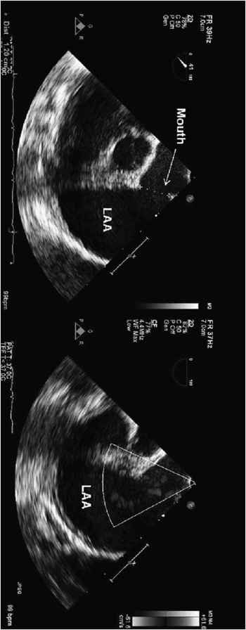
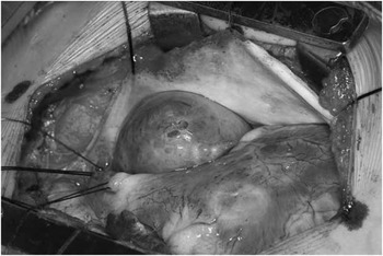
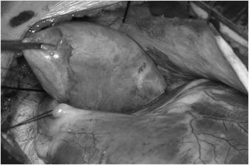
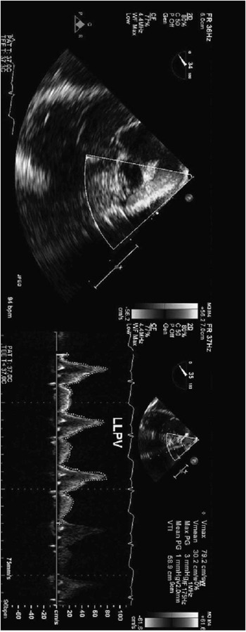
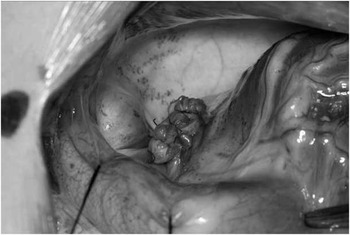
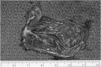
After induction, a transoesophageal echocardiography was performed, which confirmed the diagnosis and revealed no clot burden within the left atrial appendage aneurysm (Fig 1). A cardiopulmonary bypass standby was used with the lines brought up and secured in the field. Through a median sternotomy, the left atrial appendage aneurysm was identified and exposed with stay sutures on the left lateral aspect of the pulmonary artery and the left side of the pericardium (Figs 2 and 3). A 4-0 polypropylene suture was placed as a purse string around the base of the left atrial appendage aneurysm, and this was snared with a tourniquet. With the snare firmly down, transoesophageal echocardiography was repeated and ensured no impingement on the pulmonary venous drainage (Fig 4) as well as a normal left ventricular function. The aneurysm was sharply resected and an additional 4-0 polypropylene was used to doubly oversew the base of the aneurysm (Fig 5). The resected specimen was opened and examined on the back table (Fig 6). The patient was discharged home on post-operative day 4 and remains asymptomatic at follow-up.
A two-dimensional transoesophageal echocardiogram showing an LAAA (∼3 × 4 cm) with a mouth measuring 1.2 cm ( a ). There were no echo densities suggestive of a thrombus within the appendage. A Doppler colour flow revealed a low-velocity flow into and out of the appendage ( b ). LAAA = left atrial appendage aneurysm.

Intraoperative visualisation of the LAAA in situ once stay sutures are placed in the leftward pericardium and pulmonary artery. LAAA = left atrial appendage aneurysm.

Intraoperative visualisation of the LAAA elevated out of the pericardium with forceps. LAAA = left atrial appendage aneurysm.

Intraoperative transoesophageal echocardiographic assessment of the left-sided pulmonary veins after the off-pump snaring of the base of the LAAA revealed no obstruction by colour flow ( a ) or spectral Doppler interrogation (mean gradient 1 mmHg, b ). LAAA = left atrial appendage aneurysm.

Intraoperative appearance of the snared and doubly oversewn base of the LAAA after resection of the body of the aneurysm. LAAA = left atrial appendage aneurysm.

The resected body of the LAAA. LAAA = left atrial appendage aneurysm.

Discussion
A PubMed search for “left atrial appendage aneurysm” reveals only 40 citations, beginning with a case report in Circulation in 1975. 1 , Reference Krueger, Ferlic and Mooring 2 A recently reported exhaustive MEDLINE search could confirm only 78 patients since 1922.Reference Chowdhury, Diplomate and Seth 3 The term “congenital” left atrial appendage aneurysm is colloquially used to differentiate primary cases – thought to be caused by congenital dysplasia of the pectinate musculature – from those caused by a variety of secondary causes such as a severe long-standing mitral regurgitation. The vast majority of these PubMed citations describe one case managed with the use of a cardiopulmonary bypass. There have been two citations of left lateral thoracotomy utilising off-pump techniques.Reference Zhao, Ge and Yan 4 , Reference Baburaj, Remeshwara and Vellachamy 5 Chowdhury and associates summarise the literature as follows, “Use of the extracorporeal circulation via median sternotomy is the widely accepted approach…it provides a safe excision in a motionless field and precludes embolization. Despite the report of some investigators on the use of left lateral thoracotomy without cardiopulmonary bypass and by Burke and associates on stapling of the aneurysm, we consider these techniques as risk procedures”.Reference Chowdhury, Diplomate and Seth 3
We agree that the use of thoracotomy introduces undue risk secondary to the limited support options if the aneurysm is torn or ruptured or if there is difficulty exposing or managing the base of the resection. However, many if not most left atrial appendage aneurysms can probably be managed off-pump with a cardiopulmonary bypass standby, as in this case. The keys to this approach are the initial transoesophageal echocardiographic documentation of the lack of a clot burden (Fig 1) and transoesophageal echocardiographic confirmation of no pulmonary vein impingement by the purse string suture at the base of the left atrial appendage aneurysm (Fig 2). A recent report on neonatal repair stresses the possible tension on the circumflex coronary artery as another source of morbidity for this repair. By snaring the purse string and observing pulmonary venous flow and left ventricular function on the echocardiogram, the absence of these complications can be verified, and the resection can proceed with reinforcement of the base. The cardiopulmonary bypass lines are on the table and ready in case of bleeding or instability/arrhythmia secondary to the required exposure. The latter can likely be minimised by well-placed stay sutures.
Financial Support
This research received no specific grant from any funding agency, commercial or not-for-profit sectors.
Conflicts of Interest
None.