INTRODUCTION
Ornamental fish have become important in global trade, reaching about one billion dollars of wholesale and three billion dollars of retail annually (Olivotto et al., Reference Olivotto, Zenobi, Rollo, Migliarini, Avella and Carnevali2005). Marine species corresponded to 10% of this amount, with a great potential to increase in coming years (Lem, Reference Lem, Cato, Corbin and Wilhelm2001). From more than 1400 marine ornamental fish species traded (Wabnitiz et al., Reference Wabnitiz, Taylo, Green and Razak2003) only 72 species are raised in captivity (Moe, Reference Moe, Cato and Brown2003), characterizing the activity as extractive (Tlusty, Reference Tlusty2002). Due to the highly selective nature of this trade with the large amount of animals collected, the potential for over-exploitation can be high (Friedlander, Reference Friedlander2001; Rubec et al., Reference Rubec, Cruz, Pratt, Oellers, McCullough and Lallo2001). Furthermore, scientific literature on rearing marine ornamental fish is scarce, and several advances already obtained are industry protected.
The Gobiidae family is part of the international marine ornamental fish market, being responsible for 5 to 7% of sales (Wabnitiz et al., Reference Wabnitiz, Taylo, Green and Razak2003). This family is considered the largest among marine fish, having an important role in temperate and tropical reef fish communities (Nelson, Reference Nelson1994). According to Colin (Reference Colin1975), some features presented by Gobiidae fish facilitate their rearing in captivity, such as, natural spawning ability, large egg size, and small adult size. Additionally, fish of the genus Elacatinus are monogamist, another quality that facilitates broodstock maintenance in captivity (Harding et al., Reference Harding, Almany, Houck and Hixon2003; Whiteman & Côté, Reference Whiteman and Côté2003). They are also iteroparous, presenting a long breeding season with repeated spawning events by individual fish (Moyle & Cech, Reference Moyle and Cech1988).
The barber goby Elacatinus figaro (Sazima et al., Reference Sazima, Moura and Rosa1996) is a coastal species, endemic to the Brazilian coast, distributed as far north as the State of Paraíba (07°S–34°W) to their southernmost limit, the State of Santa Catarina (27°S–48°W) (Sazima et al., Reference Sazima, Moura and Rosa1996). Members of this species are small, with an elongated body, and present nice colours, in narrow yellow and black stripes. They are benthonic and have the habit of cleaning interactions, mainly feeding on ectoparasites of fish (Sazima et al., Reference Sazima, Moura and Rosa1996, Reference Sazima, Sazima, Francini-Filho and Moura2000) and zooplankton. According to Gasparini et al. (Reference Gasparini, Floeter, Ferreira and Sazima2005), E. figaro was one of the most important ornamental marine fish exported by Brazil. However, little is known of its reproduction in captivity, besides it is currently listed as an endangered species and can no longer be legally collected from the wild (IBAMA, 2004).
It is expected that once the technology for production of juvenile barber goby is mastered, fishing pressure could be reduced through increasing availability of fish reared in captivity. Effective stock enhancement programmes can also be attempted, once successful production of fish is achieved.
The study of reproduction and early life stages of a new aquaculture species is important to help establish optimal protocols for rearing it in captivity. Production of larvae of marine ornamental fish follows techniques developed for table fish and includes the use of rotifers and Artemia along with green water (Ostrowski & Laidley, Reference Ostrowski and Laidley2001; Borges et al., Reference Borges, Faria, Gil, Gonçalves and Almada2003; Olivotto et al., Reference Olivotto, Zenobi, Rollo, Migliarini, Avella and Carnevali2005, Reference Olivotto, Rollo, Sulpizio, Avella, Tosti and Carnevalli2006; Wittenrich et al., Reference Wittenrich, Turingan and Creswell2007).
In this study, the reproductive biology, embryonic development, first feeding and larviculture of barber goby E. figaro until settlement of juveniles was examined in laboratory conditions.
MATERIALS AND METHODS
Reproduction
Adult barber goby from the coast of Bahia (Brazil) were obtained from a local pet shop and taken to the Laboratory of Marine Fish Culture of the Federal University of Rio Grande. Total length of males was 3.7 ± 0.1 cm and their weight was 528 ± 50 mg, while females measured 3.7 ± 0.4 cm and weighed 580 ± 14 mg.
Broodstock aquaria (84 l, 60 × 35 × 40 cm) with no substrate provided were connected to a recirculating aquaculture system composed of: protein skimmer, biological filter type dry–wet, UV filter/15W, live rocks and heater with thermostat (Aquarium Systems, Italy). Natural seawater, previously filtered (5 µm) was maintained at 26°C, pH 8.2, salinity 34‰, ammonia and nitrite concentrations never exceeded 0.02 mg.l−1 and 2 mg.l−1, respectively. Photoperiod was set at 12L:12D, and light intensity provided by fluorescent lamps (20W) was equal to 950 lx at the water surface. Adult barber goby were fed with a commercial diet for marine fish (INVE, NRD, 500–800 µm, 59% crude protein and 16% lipids) twice every day.
In order to establish the experimental pairs, three barber gobies were placed randomly in four aquaria containing a spawning shelter composed of a 10 cm long PVC pipe (32 mm internal diameter) filled with a plastic film. A few days later, the pairs were formed, being confirmed by the exclusion of one fish from close contact with the other two fish in the aquarium. This fish was then removed from the aquarium.
Four pairs of barber goby were observed daily during 40 days. Four spawns were observed for each pair; these spawning events were filmed in order to measure their duration. Once spawning was completed, the plastic film with eggs adhered was removed for counting and biometrics using a stereomicroscope equipped with a micrometric eye piece (Wild, M5A), and then they were returned to the broodstock aquarium. When hatching started, the plastic film containing adhered eggs was moved into a beaker of 1 l with gentle aeration for approximately 1 hour, when all larvae had already hatched. The newly hatched larvae were counted, and hatching percentage was estimated as the ratio between the number of hatched larvae and the number of eggs.
Embryonic development
Two clutches of eggs, from two different pairs, were followed for embryonic development. They were photographed with a digital camera (Canon, Power Shot A85, 4MP) attached to a microscope (Nikon E200) or to a stereomicroscope (Wild, M5A). The photographs were taken at 2 hour intervals during the first 48 hours after fertilization (HAF), at 6 hour intervals between 48 and 120 HAF, and thereafter, every 12 hours until hatching, at 40 and 100×magnification. Newly hatched larvae were photographed under the stereomicroscope.
First feeding and larviculture
Water for larviculture was taken from the broodstock recirculating system, it was held under the same environmental conditions, but under continuous photoperiod at 200 lx.
Two groups of 100 larvae from three different spawns were used to determine the time for first feeding and the time for death by starvation. Just after hatching, larvae were stocked in six cylinder tanks (15 l) painted black on the walls and white on the bottom, water was gently aerated and no substrate was provided. Larvae were either deprived of food (three tanks) or offered rotifers Brachionus plicatilis (10 rotifer ml−1) along with the microalgae Nannochloropsis oculata (500,000 cells ml−1) until all unfed larvae died of starvation. Every morning, 3 to 5 larvae were sampled from the fed tanks and examined under the stereomicroscope for the presence of rotifers. It was difficult to accurately register the number of dead larvae due to the lack of contrast between the dead bodies and the white tank bottom. In order to overcome this problem, time to 100% death by starvation was estimated by the presence of swimming larvae.
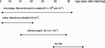
Growth and survival of the fed groups were followed until all larvae settled to the bottom of the tanks. Larvae were offered exclusively rotifers (10 rotifer ml−1) in green water until 14 days after hatching (DAH). Thereafter, larvae were fed on a diet composed of rotifer and Artemia nauplii (0.1 Artemia ml−1), but 21 DAH rotifer was discontinued and only Artemia nauplii (1 Artemia ml−1) was offered (Figure 1). Every morning the water was siphoned out through a screen of 800 µm, allowing for the removal of residual prey. New preys were replaced daily to keep the desired food density. After individuals settled to the bottom, they were offered a dry diet (INVE, NRD, 400–600 µm, 59% crude protein and 16% lipids) along with Artemia, and it was readily accepted.
Feeding protocol of barber goby Elacatinus figaro during larviculture.

Growth of larvae was followed for 28 days, at this moment larvae begin to settle to the bottom of the tank. Ten larvae were sampled from each tank every week for growth measurements. They were anaesthetized with benzocaine 30 ppm, measured under a stereomicroscope equipped with an eyepiece micrometer, and their standard length was measured. Fish were not returned to their tanks, therefore, the number of larvae sampled during the experiment was subtracted from the initial number of larvae stocked to calculate the survival rate. The survival rate was estimated counting the number of individuals present in each tank at the end of the growth trial in relation to the initial number of larvae.
RESULTS
Reproduction
The first egg clutches were laid between 24 and 31 days after barber goby pairs were formed. Fish began to spawn within 2 to 3 hours after light was turned on and spawning lasted about 1.5 hours. The interval between spawns of each pair ranged between 8 to 10 days. Eggs were deposited in a monolayer on the ceiling of the PVC pipe. Each clutch of eggs was considered to contain all eggs of a given spawn, and as such it resembled the actual fecundity. Fecundity averaged 648 ± 183 eggs, ranging from 430 to 1020 eggs per spawn and hatching rate ranged from 62 to 80% with an average of 69 ± 6%.
There is no clear sexual dimorphism in barber goby. Females have a swollen abdomen before spawning and their genital papillae are evident when eggs are about to be released. However, a behavioural dimorphism could be observed, since males present constant parental care, guarding the nest and ventilating the eggs until hatching, while females do not tend the eggs. There was no parental care for the newly hatched larvae.
Embryonic development
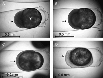
Barber goby eggs were elliptical in shape, measuring 1.81 ± 0.1 mm total length and 0.61 ± 0.03 mm total width. They were adhered to the nest by filaments present at the proximal end. These eggs are telolecithal, denoting that yolk is concentrated at the distal end, close to the five protuberances that emerge 2 HAF in the chorion surface. At this moment the number of blastomeres was large and could not be counted, there were also a large number of small oil globules (Figure 2A). Blastula was already observed 4 HAF and mid-gastrula stage was achieved 12 HAF, when the rim of migrating germ cells covered one-third of the yolk (Figure 2B), late gastrula was reached 18 HAF (Figure 2C). Blastopore was closed between 20 and 22 HAF. By 24 HAF the early neurula stage was observed, the head fold was facing the proximal end and optical placodes were beginning to develop (Figure 2D).
Early embryonic development of barber goby Elacatinus figaro. (A) Four hours after fertilization (HAF), blastula was formed, and the egg presents 5 protuberances in the distal part of the egg, yolk sac (Y) already formed; (B) 12 HAF, gastrula stage; (C) 18 HAF, blastopore is closed; (D) 24 HAF starts up the embryo formation and eyes formation (*).

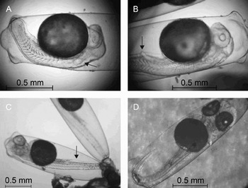
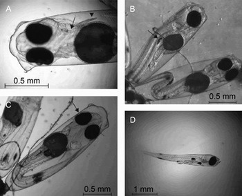
Thirty-two HAF the tail bud was formed and slightly protruded from the yolk. At 36 HAF the tail bud was protruded from the yolk, myomeres were present, and auditory placodes were observed (Figure 3A). Most of the embryos reversed their position in the egg 42 HAF, and development proceeded with their heads facing the distal end of the adhesive side. The anal pore became visible 46 HAF (Figure 3B). By 48 HAF the heart beating could be observed and the tail was already larger than the yolk, it was long enough to bend inside the egg at 66 HAF. By 72 HAF yolk sac volume was clearly reduced, body length was increased, and melanophores were present in the caudal area (Figure 3C). Retina was slightly pigmented 86 HAF, but 96 HAF pigmentation was more evident, caudal fin was also developing (Figure 3D). Poorly developed pectoral fins and gill arches were observed 130 HAF (Figure 4A). By 144 HAF embryos presented well pigmented eyes and the gas bladder was already formed (Figure 4B). From this moment on, manipulation of the eggs could result in the early emergence of larvae. Eyes were well pigmented 156 HAF, and there were melanophores along the whole body extension. Tail was long enough to reach gill arches (Figure 4C). Intense embryo movement was observed 168 HAF, chorion was then ruptured at the distal end, and larvae immediately hatched (Figure 4D).
Mid-embryonic development of barber goby Elacatinus figaro. (A) 36 HAF, hearing placodes formation; (B) 46 HAF, anal pore visible; (C) 72 HAF, formation of the tail fins and pigmentation; (D) 96 HAF, eyes and tail fins well pigmented.

Late embryonic development and newly hatched larvae of barber goby Elacatinus figaro. (A) 130 HAF branchial arches and pectoral fins are visible; (B) 144 HAF eyes are already well pigmented and gas bladder presents formed; (C) 156 HAF the eyes and body are well pigmented, and the tail reaches branchial arches; (D) after hatching the larvae are very active, presenting the mouth open and functional anus.

Pectoral fins poorly developed and gill arches were observed 130 HAF (Figure 4A). At 144 HAF, embryos presented well pigmented eyes and the gas bladder was already formed (Figure 4B). From this moment, manipulation of the eggs could result in the early emergence of larvae. Eyes were well pigmented 156 HAF, and there were melanophores along the whole body extension. The tail was long enough to reach gill arches (Figure 4C). Intense embryo movement was observed 168 HAF, chorion was then ruptured at the distal end, and larvae immediately hatched (Figure 4D).
First feeding and larviculture
Newly hatched larvae were colourless with total length of 3.15 ± 0.07 mm. Both mouth and anus were already open and the gas bladder was inflated soon after hatching, mouth gape measured 0.35 ± 0.02 mm in high and 0.23 ± 0.02 mm in width. Newly hatched larvae showed a very active behaviour, positive phototaxis, and their eyes were well pigmented. They had a continuous finfold (dorsal, pelvic and caudal) and one pair of pectoral fins (Figure 4D).
Larvae had already consumed most of their yolk sac reserves and were already feeding on rotifers 1 DAH. Yolk sac could no longer be observed under stereomicroscope 2 DAH and larvae deprived of food began to die between 2.5 and 3 DAH. Severe mortality was observed by the end of the third day, and all larvae were dead 4 DAH. Survival of larvae fed on rotifers was not estimated because we did not want to harm them by individual counting and compromise the larviculture, but a large number of larvae died within the first week after hatching.
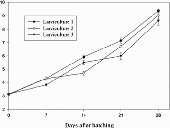
Total length of larvae increased almost 3 fold, from 3.15 ± 0.07 mm at hatching to 8.50 ± 0.18 mm four weeks after hatching, just before they begin to settle to the bottom (Figure 5). Survival during larviculture ranged between 2 and 20% with an average of 6.7 ± 7.1%.
Growth (mean ± standard error) during 28 days of larviculture of barber goby Elacatinus figaro.

Larvae began to settle to the bottom of the tanks between 28 and 35 DAH. The characteristic adult colours were evident two days after fish settled to the bottom, showing a black body with a yellow longitudinal stripe well-defined.
DISCUSSION
Most fish sold for the marine aquarium trade is taken from the wild. The development of techniques to produce viable eggs and raise larvae of endangered species in captivity can help to alleviate their risk of extinction, because the pressure to collect wild fish will be diminished, allowing the recovery of natural stocks (Ostrowski & Laidley, Reference Ostrowski and Laidley2001; Sarkar et al., Reference Sarkar, Deepak, Negi, Singh and Kapoor2006).
Our results indicate that spawning, reproductive frequency and parental care of E. figaro are similar to other small gobiids such as Elacatinus evelynae, Elacatinus Oceanops Jordan and Priolepis nocturna (Olivotto et al., Reference Olivotto, Zenobi, Rollo, Migliarini, Avella and Carnevali2005; Wittenrich, Reference Wittenrich2007; Wittenrich et al., Reference Wittenrich, Turingan and Creswell2007).
Fecundity of E. figaro was higher than previously observed for other species of Elacatinus. Fecundity of E. oceanops, Elacatinus puncticulatus, Elacatinus multifasciatus and E. evelynae typically range from 200 to 450 eggs per spawn (Valenti, Reference Valenti1972; Olivotto et al., Reference Olivotto, Zenobi, Rollo, Migliarini, Avella and Carnevali2005; Wittenrich, Reference Wittenrich2007). However, it is important to notice that fecundity may vary with female size and pre-spawning conditions (Colin, Reference Colin1975), and despite that broodstock used in this experiment was acclimated to the laboratory, their history is not known.
Egg morphology can be used as a specific characteristic. The protuberances found at the distal end of the eggs of E. figaro can be used to distinguish this species from E. evelynae. Both species are quite similar and have similar egg size, but according to Colin (Reference Colin1975) and Olivotto et al. (Reference Olivotto, Zenobi, Rollo, Migliarini, Avella and Carnevali2005), eggs of E. evelynae do not have such protuberances.
Embryos of E. figaro undergo a reorientation inside the chorion. The head is originally oriented towards the proximal end of the egg, but a couple of days before hatching the embryo changes its position, and the head starts to face the distal end. Time for reorientation of E. figaro embryos was smaller than reported for E. oceanops (50 HAF) by Valenti (Reference Valenti1972). The same author observed that embryos did not hatch without this reorientation. On the other hand, there is no relationship between reorientation and hatching for P. nocturna (Wittenrich et al., Reference Wittenrich, Turingan and Creswell2007).
In the present study, hatching occurred at 5 DAF when eggs were handled without care. Natural hatching happened only 7 DAF. This fact showed that stress was directly associated to the hatching period of E. figaro. According to Olivotto et al. (Reference Olivotto, Zenobi, Rollo, Migliarini, Avella and Carnevali2005), E. evelynae eggs also hatch at 7 DAF.
The hatching rate presented here was lower than that found to other Gobiidae. The hatching rate of goby Oxyeleotris marmoratus ranged from 80 to 90% (Tan & Lam, Reference Tan and Lam1973; Tavarutmaneegul & Lin, Reference Tavarutmaneegul and Lin1988), and regarding P. nocturna, it ranged from 92 to 99.8% (Wittenrich et al., Reference Wittenrich, Turingan and Creswell2007). In particular, the low hatching rate of barber goby can be, among other factors, related to poor nutrition of the broodstock. Fish were fed on a commercial diet for marine fish larvae, which was not specifically developed for barber goby. Moreover, Brooks et al. (Reference Brooks, Tyler and Sumpter1997) reported that broodstock fed on natural food laid better quality eggs, which was reflected in higher hatching rate and survival of larvae. That is because accumulation of nutrients, such as essential amino acids, fatty acids, and vitamins in eggs are dependent on the diet provided to the female during the period of gonadal development (Bell et al., Reference Bell, Famdale, Bruce, Navas and Carrillo1997; Lavens et al., Reference Lavens, Lebegue, Jaunet, Brunel, Dhert and Sorgeloos1999).
Positive phototaxis observed for E. figaro was also described for larvae of other Gobiidae (Senoo et al., Reference Senoo, Kaneko, Cheah and Ang1994; Wittenrich, Reference Wittenrich2007) and this characteristic can be used as a tool to collect larvae, when the eggs are incubated in the presence of the broodstock.
Death of larvae by starvation, due to inappropriate supply of food, is a common feature in marine fish larvae during first feeding, when larvae rely on their yolk reserves until adequate prey is available (Holt, Reference Holt, Cato and Brown2003; Sheng et al., Reference Sheng, Lin, Chen, Shen and Lu2007). In this study, time to death by starvation and the period of high mortality during first feeding were coincident, suggesting that first feeding is also a critical period for barber goby.
Different from P. nocturna, which could not prey on rotifers at first feeding and died of starvation (Wittenrich et al., Reference Wittenrich, Turingan and Creswell2007), barber goby can be reared on rotifers at first feeding. This difference between species is likely to be due to the larger mouth gape of E. figaro.
Even though barber goby larvae survived beyond 5 DAH when fed only on rotifers, alternative preys, such as copepods and enriched rotifers should also be evaluated for rearing barber goby larvae, since these zooplankton preys are known to improve growth and survival of other marine ornamental fish (Olivotto et al., Reference Olivotto, Rollo, Sulpizio, Avella, Tosti and Carnevalli2006).
The results of the present investigation demonstrate the viability to produce naturally spawned eggs of the barber goby E. figaro in captivity. Natural spawns are observed every 8–10 days and larvae emerge 7 days after fertilization at 26°C. Rotifers are effectively used at first feeding and larvae can start preying on Artemia two weeks after hatching. Larvae completed metamorphosis and settled to the bottom 28 to 35 DAH, when total length was equal to 8.5 mm in average. The protocol proposed here seems to be appropriate for mass production of juvenile barber goby and the aim to reduce illegal fishing pressure on wild stocks can be addressed through laboratory culture of fish for the ornamental industry. It can also be helpful for evaluation of the feasibility of stock enhancement programmes for this species.
ACKNOWLEDGEMENTS
The authors would like to acknowledge the support of Sarlo Better and Produtos Tornado. R.V. Rodrigues is a graduate student of Aquaculture at FURG and is supported by ANP, K.C. Miranda-Filho is a research fellow of PRODOC/CAPES and L.A. Sampaio is a research fellow of CNPq (process 301673/2006-3).