At present, several authors have presented evidence suggesting the efficacy of real-time three-dimensional echocardiography over standard cross-sectional interrogation for better orientation of complex intra-cardiac anatomy, including defects permitting interatrial shunting.Reference Van den Bosch, Ten Harkel and McGhie1 For defects within the oval fossa, it is essential to know the diameter of the defect, and its relation to the rims of the oval fossa, before undertaking device closure. Sinus venosus superior defects can be visualised well by two-dimensional transthoracic or transoesophageal echocardiography; however, correct identification of the right upper pulmonary vein and its spatial relationship with the atrial communication and left atrium for future surgical correction, while maintaining a widely patent right upper pulmonary-to-left atrium pathway, may not be clear. Similarly, it is often difficult to differentiate between the sinus venosus inferior defect and an absent posterior rim in oval fossa defects based on conventional two-dimensional echocardiography. Similar to the previous type, understanding the precise spatial relationship of right pulmonary vein(s) and the atrial communication is crucial for a good long-term outcome of surgical repair. On several occasions, we have experienced incorrect reporting of postero-inferior defects as sinus venosus inferior defects. The coronary sinus defect can be missed on routine echocardiography, particularly if the defect is small or very large, or indeed if the roof of the coronary sinus is missing. The correct identification of the size and position of defects within the roof of the coronary sinus using two-dimensional echocardiography remains challenging.
Transoesophageal real-time three-dimensional echocardiography might well help in improving the quality of imaging and in presenting cardiac anatomy in an orientation similar to the surgeon's view. Furthermore, it has been shown that real-time three-dimensional echocardiography has potential for better selection of patients for catheter intervention, and may potentially replace the use of balloons for sizing the device chosen for closure.Reference Zhu, Cao, Rhodes and Hijazi2 Initial experience has also shown that the technique can show the dynamic behaviour of the rims of the oval fossa, helping both standardReference Suematsu, Takamoto and Kaneko3, Reference Handke, Heinrichs and Moser4 and robotic surgical closure.Reference Suematsu, Kiaii, Bainbridge, del Nido and Novick5 In this report, we describe our own experience using three-dimensional post-processing to create detailed imaging of different lesions permitting interatrial shunting, showing the size of the defects, and their relationship to adjacent cardiac structures. We aimed to examine four patients with four different interatrial communications, assessing the extent to which currently existing transthoracic real-time three-dimensional echocardiography can determine intra-atrial structures, and how the images should be correctly interpreted.
Patients
Patient ST
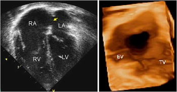
An asymptomatic 9-year-old girl presented for the first time following recurrent respiratory infections. Her chest X-ray revealed an increased cardiothoracic ratio, and the electrocardiogram showed sinus rhythm with right axial deviation and right ventricular hypertrophy. The transthoracic cross-sectional echocardiography revealed a large defect in the floor of the oval fossa (Fig 1, left panel), with the rims of the fossa itself being mildly deficient. There was a significant left-to-right shunt, with a ratio of systemic and pulmonary flows of 4 to 1 revealed by the continuity equation, moderate right ventricular hypertrophy, a flattened septum with paradoxical motion, and moderate tricuspid regurgitation due to poor coaptation of the valvar leaflets. An attempt was made to close the defect in the catheterisation laboratory, with the intra-procedural transoesophageal echocardiography showing the defect to have a diameter of 18 millimetres, and stop-flow balloon sizing suggesting a diameter of 22 millimetres. On the basis of this information, several attempts were made to deploy a 20-millimetre Amplatzer device, although without success, and the device was subsequently removed. The patient was then referred for real-time three-dimensional echocardiography (Fig 1, right panel) before anticipated surgical closure. Acquisition of the full-volume data set took 2 minutes, and the time required for post-processing was 5 minutes. The findings confirmed the presence of a large defect in the floor of the oval fossa, which had a deficient inferior rim, with the hole being in close contact with the Eustachian valve. The communication was successfully closed at surgery.
An atrial septal defect within the oval fossa demonstrated in a cross-sectional four-chamber view (left panel), and as seen from right atrial aspect in a ‘face to face’ view using three-dimensional imaging (right panel). Notice the large defect (arrow) with a small inferior rim and close attachment of Eustachian valve (EV; RA = right atrium; RV = right ventricle; LV = left ventricle; LA = left atrium; TV = tricuspid valve).

Patient SD
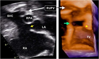
This 2-year-old asymptomatic female child presented following the discovery of a murmur upon admission to hospital with fever and a respiratory infection. The examination revealed a fixed split second heart sound and an ejection murmur. The transthoracic cross-sectional echocardiography showed a superior sinus venosus defect (Fig 2, left panel), with overriding of the orifice of the superior caval vein, and the right upper pulmonary vein connecting to superior caval vein while retaining its communication with the roof of the left atrium. The ratio of pulmonary to systemic flow, as revealed by the continuity equation, was 2.4 to 1, and there was moderate-to-severe overload of the right heart. There was no significant tricuspid valvar regurgitation. Real-time three-dimensional echocardiography clearly showed the location, size, and relations of the defect (Fig 2, right panel); the defect appears outside the confines of the oval fossa evolving the roof of atrial wall and junction of superior caval vein. Acquisition of the full-volume data set took 3 minutes, whereas the post-processing time was 16 minutes. The patient underwent a Warden procedure, with enlargement of the venoatrial communication and reconnection of the right upper pulmonary vein exclusively to the left atrium.
A superior sinus venosus defect with partially anomalous pulmonary venous drainage documented in the cross-sectional bicaval view (left panel), and as seen from right atrial aspect in a ‘face to face’ view (right panel). Notice the moderate-sized defect (yellow arrow) at the level of the superior caval vein (SVC) and its junction with the right atrium, with the right upper pulmonary vein (RUPV) connected with both the SVC and the roof of the left atrium (SVC, white arrows). Three-dimensional imaging clearly shows that atrial communication is outside the confines of the oval fossa evolving roof of atrial wall (asterisk) and the junction of SVC (green arrow; RPA = right pulmonary artery; RA = right atrium; TV = tricuspid valve; LA = left atrium).

Patient HO
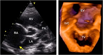
This 9-month-old male infant was referred as an outpatient for evaluation of a murmur with recurrent coughs and colds. On examination, he had normal pulses, a prominent right ventricular impulse, a pulmonary flow murmur, and a widely split second heart sound. The transthoracic cross-sectional echocardiography revealed a large defect located postero-inferiorly within the oval fossa, adjacent to the orifice of the inferior caval vein surrounded by hyperplastic Eustachian valve (Fig 3, left panel). The continuity equation showed the ratio of pulmonary to systemic flow to be 4 to 1. The right heart was markedly dilated, with mild right ventricular hypertrophy and mild tricuspid valvar regurgitation. Acquisition of a real-time three-dimensional echocardiographic full-volume data set took 2 minutes, and post-processing required 8 minutes, with the reconstruction showing a large defect close to the inferior caval vein (Fig 3, right panel). It was possible to create a view of the septal surface as would be obtained by the surgeon, showing how the hyperplastic Eustachian valve partially obscured access to the defect. The patient then underwent successful surgical closure of the defect using a pericardial patch, with the post-operative course proving uneventful.
A defect within the infero-posterior part of the oval fossa visualised in the parasternal short-axis view on cross-sectional echocardiography (left panel), and as seen from the right atrial aspect in a ‘face-to-face’ view (right panel). Notice the large defect (yellow arrow) close to the junction of the inferior caval vein (IVC) partly hidden behind the hyperplastic Eustachian valve (EV), separating the defect from the right atrial vestibulum as clearly seen on three-dimensional echocardiography (PA = pulmonary trunk; LA = left atrium; RA = right atrium; RV = right ventricle; TV = tricuspid valve).

Patient NJ
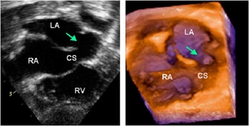
This 3-year-old patient was transferred for elective surgery from overseas. He was a twin, born at 34 weeks weighing 1.5 kilogram, and had been diagnosed postnatally to have an atrial septal defect. Later, a more detailed cross-sectional echocardiography performed locally showed a large defect in the roof of the coronary sinus (Fig 4, left panel), with a transoesophageal study showing contrast entering the left atrium from the systemic venous channel. On admission, he was well and active, but prone to colds and chest infections. On examination, there was no evidence of cyanosis or heart failure at rest; the shape of the chest was normal, and an ejection murmur graded at 3 from 6 was heard along the left sternal border. The previous diagnosis was confirmed on detailed echocardiography, including three-dimensional imaging. The acquisition of the full-volume data set took 3 minutes, and post-processing required 12 minutes, with the reconstructions showing a circular defect between the cavity of a persistent left superior caval vein and the left atrium (Fig 4, right panel). There was significant left-to-right shunting across the defect, with the ratio of pulmonary to systemic flows estimated as 3.4:1. The child underwent elective surgical closure, with the hole being closed so that the left superior caval vein drained exclusively to the right atrium. The transoesophageal echocardiography confirmed a good repair, but atrioventricular sequential pacing was initially required because of intermittent complete atrioventricular block. Implantation of a pacemaker was contemplated; however, 24-hour electrocardiographic monitoring carried out on the 19th post-operative day showed sinus rhythm, and thus implantation of a pacemaker was deemed to be unwarranted. All other aspects of recovery were unremarkable.
Defect within the roof of coronary sinus (CS) visualised in subcostal cross-sectional four-chamber view (left panel), and as seen by subcostal three-dimensional echocardiogram from the right inferior aspect (right panel). The coronary sinus is shown ‘face to face’ with a circular-shaped defect seen in the middle of the roof (arrow; RA = right atrium; RV = right ventricle; LA = left atrium).

Discussion
Echocardiography is now usually the ideal tool for diagnosis of cardiac lesions in younger patients. Standard diagnostic measures for those with suspected interatrial shunting now include transthoracic and transoesophageal cross-sectional imaging. Multiple views are necessary to obtain the complete spatial information required by the operator to mentally reconstruct the three-dimensional format. In the patients described in this report, such reconstruction produced the correct diagnosis, and there were no false-positive or -negative findings in either of our patients. However, as we have shown, subcostal real-time three-dimensional echocardiography provided this reconstructed information directly, while simultaneously providing unique views of the location and shape of interatrial communications, thus optimising planning for the most appropriate intervention. Since 2006, three-dimensional echocardiography has been the routine clinical tool at Great Ormond Street Hospital; between 2006 and 2009, we have examined a total of 292 patients. Of these patients, atrial communications were analysed from the subcostal approachin 45 (13%), eight (17.7%) patients had sinus venosus superior defect, two (4.4%) patients had a defect within the roof of coronary sinus, four (8.9%) patients had postero-inferior oval fossa defect, and the remaining 31 (68.9%) patients had central oval fossa defect. Very good or satisfactory results were achieved in all patients with sinus venosus defect, coronary sinus defect, and postero-inferior atrial communications. Unsatisfactory results were achieved in eight of the patients with central oval fossa using three-dimensional echocardiographies, partly because of patient age versus probe frequency mismatch, and partly because we were unable reconstruct full volume data sets.
Subcostal views are beneficial for using low-frequency probes in small children, as they avoid near-field artefacts from ribs; furthermore, the presence of soft tissue (liver) from the subcostal approach enables better penetration from longer distance with more near-field volume, which is very important for three-dimensional reconstruction in small children. Post-processing reconstruction was carried on at the end of the session by optimising image quality, that is, volume gain adjustment and zooming in all patients, and cropping. All images were cropped in a manner so as to view atrial structures preferentially from the right atrial aspect; image colourisation then enabled to adjust optimal volume for obtaining the best possible depth perception. The post-processed clip was then viewed from multiple sites to understand the spatial relationship of all atrial structures, coronary sinus, as well as systemic and pulmonary venous connections. Frame-by-frame rotational and translational motion was finally used for detailed visualisation of septal margins, coronary sinus orifice and its walls – in patient 4 – as well as systemic and pulmonary venous connections.
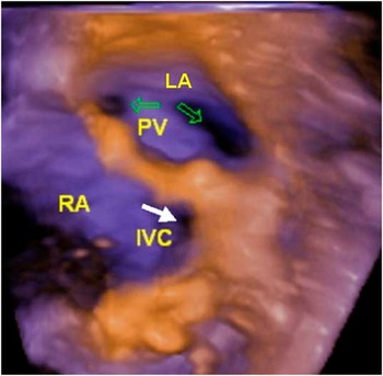
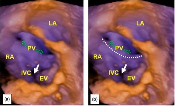
For the defects confined within the oval fossa, the three-dimensional reconstructions showed the view of the floor of the fossa as would be visualised by the surgeon, and also supported suggestions that this technique can be an effective alternative for guiding deployment of closure devices introduced through catheters.Reference Van den Bosch, Ten Harkel and McGhie1, Reference Roman, Nii, Golding, Benson and Smallhorn6, Reference Chen, Hsiung, Hsieh, Li and Chou7 In a patient with the superior sinus venosus defect, the reconstruction clearly showed that the right upper pulmonary vein was connected to the superior caval vein while retaining its attachment to the roof of the left atrium, thus producing the overriding of the caval venous orifice relative to Waterstone's or Sondergaard's groove,Reference Beerman and Zuberbuhler8 which was hence converted to a tube rather than a groove. Knowledge of the location and size of the venoatrial communication then helped the surgeon to repair the defect while avoiding obstruction to either the superior caval vein or the anomalously connected right upper pulmonary vein. Defects opening postero-inferiorly within the oval fossa are rare, and need to be distinguished from inferior sinus venosus defects, particularly when associated with a hyperplastic Eustachian valve. When looking at the defect shown in our third patient only from the right atrium, one might gain the impression that the hole is an inferior sinus venosus defect. The value of the three-dimensional approach, however, is that it permits us to examine the relationships of the caval and pulmonary veins to the defect. If the defect was truly an inferior sinus venosus defect, then the right inferior pulmonary vein would be connected to the inferior caval vein, while retaining its connection to the left atrium. However, as our additional illustrations show, the pulmonary veins are exclusively connected to the left atrium (Fig 5), showing that the defect is within the oval fossa but has excavated its posterior rim towards the orifice of the inferior caval vein. The view we obtained showing the right atrial aspect (Fig 6a and b), however, was invaluable to the surgeon, permitting him to close the defect so that the inferior caval vein retained its connection to the right atrium. In situations such as this, with persistence of a large Eustachian valve, it is very easy to mistake the Eustachian valve for the septum, and to patch the inferior caval vein into the left atrium diverting the inferior caval venous return into the left atrium,Reference Morishita, Yamashita, Yamada, Arikawa and Taira9 or totally obstructing the orifice of the inferior caval vein.Reference Becker, Buss, Sebening, Meisner and Döhlemann10 In a patient with coronary sinus defect, the correct diagnosis had been made by cross-sectional imaging, but the subsequent three-dimensional reconstruction showed the entirety of the communication and its relation with the surrounding structures.Reference Acar, Arran and Paranon11
‘Face-to-face’ view from the anterior aspect across the atrial septum, facing the posterior left and right atrial walls. The right and left pulmonary veins (PV) can be viewed within the left atrium (LA) and inferior caval vein (IVC) within the right atrium (RA).

A similar view to the previous image with the plane rotated towards right, facing posterior left (LA) and the right (RA) atrial wall predominantly from the RA aspect. The right and left pulmonary veins (PV) are visible through a large atrial communication behind the hyperplastic Eustachian vein (EV). Note the complete absence (a) of the postero-inferior rim between PV and inferior caval vein (IVC). The hypothetic margins of the posterior rim are highlighted in the right panel for better understanding of the spatial relationships (b).

Other groups have already described how real-time three-dimensional echocardiography creates a “virtual sense of depth”, showing the dynamic qualities of cardiac lesions.Reference Chen, Hsiung, Nanda, Hsieh and Chou12, Reference van den Bosch, Ten Harkel and McGhie13 Cross-sectional imaging is limited in determining the true maximum diameter of a defect at various points during systole and diastole from only one plane of imaging. Real-time three-dimensional echocardiography has the potential to limit error by imaging the entire depth, width, and height of the septum throughout the cardiac cycle.Reference Morgan, Casey, Craig and Sands14 Balloon-sizing aims to convert asymmetric defects into a circle of maximum diameter. The three-dimensional imaging data obtained thus far suggest that the maximum reconstructed diameter correlates well with balloon sizing,Reference Zhu, Cao, Rhodes and Hijazi2 and thus may preclude the need for this procedure, which is still practised by most units and is known to be not entirely risk free.Reference Sivasankaran, Harikrishnan, Narayanan and Jaganmohan15 The accuracy of sizing on real-time three-dimensional echocardiography also marries with comparisons of three-dimensional echocardiography made relative to transoesophageal echocardiography, suggesting that the former method alone will prove to be adequate perioperatively, without the need for costly and comparatively risky transoesophageal procedures.
Conclusion
Despite the fact that standard cross-sectional two-dimensional echocardiography performed by experienced echocardiographer can establish correct diagnosis in all types of atrial communications, real-time three-dimensional reconstruction may provide additional value to the surgeon and interventionist for better understanding of spatial intracardiac morphology.