Introduction
Distant metastases of differentiated thyroid carcinoma are known to present at diagnosis but can also occur decades after initial treatment. The site and nature of metastases is known to have a significant impact on the morbidity, mortality and final outcome of these patients. Complete cure is rare.Reference Sampson, Brierley, Le, Rotstein and Tsang1 The majority of metastases from differentiated thyroid carcinoma occur in the lymph nodes, lungs and bones. Uncommon sites include the brain, liver, kidneys, spleen, choroid plexus and a few other rare sites.
There are very few reports in the literature on renal metastases from differentiated thyroid carcinoma; while some occur at presentation,Reference Graham and Roe2, Reference Borde, Basu, Kand, Arya and Shet3 others present decades after detection of the primary.Reference Moudouni, En-Nia, Rioux-Leclerq, Manunta, Guille and Lobel4–Reference Davis and Corson6 In this study, we analysed the characteristics of renal metastases from differentiated thyroid carcinoma in four patients managed at our centre. In view of its rare occurrence and limited description in the literature, we believed that a detailed study and analysis of follow up would provide greater insights into the management of this disease entity.
Materials and methods
This was a retrospective audit of four patients with renal metastases from differentiated thyroid carcinoma who had received radioiodine therapy at our centre, with follow-up information available in all cases.
Results
Patient characteristics
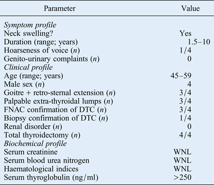
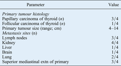
Table I shows detailed information on the patients. The characteristics of the primary thyroid tumour and the metastatic lesions are described below.
Patients' clinical profile

FNAC = fine needle aspiration cytology; DTC = differentiated thyroid carcinoma; WNL = within normal limits
Primary tumour
Differentiated thyroid carcinoma was demonstrated in all patients on histopathological analysis (papillary carcinoma of the thyroid in three patients and follicular carcinoma of the thyroid in one) (Table II). Tumour–node–metastasis staging was undertaken in all patients. Histology records revealed a tumour size of 4 cm or more in all patients (range, 4–14 cm).
Patients' histological and metastatic profile
Extn = extension
Associated metastatic burden
Three patients had lymph node metastases. Two patients had isolated metastases to both kidneys but no other distant metastases; both underwent ultrasound-guided histological confirmation of their renal lesions. The remaining two patients had extensive, multi-organ metastasis. Computed tomography (CT) scanning of the neck and thorax revealed extension of the primary disease into the superior mediastinum in three patients, with bilateral pulmonary metastasis in two of these three. One patient had metastasis to the brain together with macro-nodular metastasis to the lungs and both kidneys. One patient had macro-nodular metastasis to the lungs, liver and both kidneys. In these latter two patients, it was not possible to obtain adequate samples for histological confirmation. Hence, the diagnosis was primarily confirmed by 131I whole body scintigraphy. Both patients were over the age of 45 years.
Renal metastasis imaging
Abdominal ultrasonography
This was performed in all four patients. All the lesions detected on ultrasonography were well circumscribed, iso-echoic, exophytic in appearance and uncalcified. Occasional lesions demonstrated an internal vascularity. Eleven lesions were demonstrated in the four patients, with no evidence of other renal pathology. All 11 lesions were located at either pole of the kidneys. In all patients, both kidneys were involved. No sub-centimetre lesions were noted on ultrasonography in any of the patients.
Abdominal computed tomography
This was performed in one patient, and demonstrated three well defined, heterogeneous, exophytic lesions involving both kidneys. Multiple small, sub-centimetre lesions were noted bilaterally.
Radionuclide imaging
The patients underwent 99mTc diethylene triamine penta-acetic acid renography and 99mTc (III) dimercaptosuccinic acid renal scanning for evaluation of renal function. Three patients had normal findings in all three phases of the furosemide-enhanced 99mTc diethylene triamine penta-acetic acid renogram, and their glomerular filtration rate was well-maintained (range, 70–90 ml/min). One patient demonstrated bilaterally symmetrical, reduced cortical extraction with compromised glomerular filtration rate (50 ml/min) and maintained drainage following furosemide injection. In this patient, the 99mTc (III) dimercaptosuccinic acid scan demonstrated 16 photon-deficient, well circumscribed, cortical lesions involving both kidneys, with a primary polar distribution pattern. Except for these latter lesions, normal functioning cortical mass was observed in all four patients.
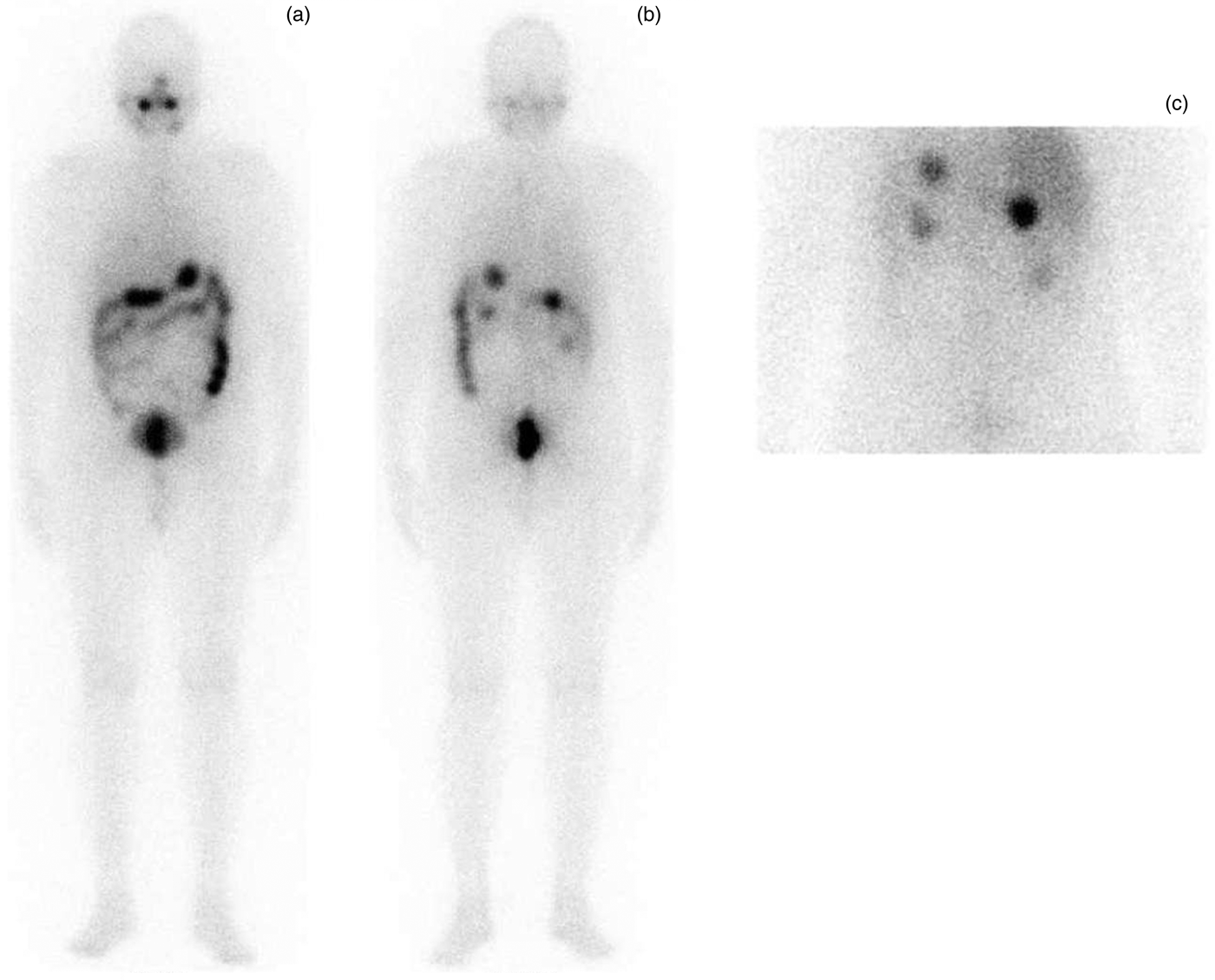
131I scanning was performed in all patients (Figures 1 to 4) both prior to and after high dose radioiodine therapy (the latter at the point of discharge from hospital). A total of 19 well circumscribed lesions, concentrating varying degrees of radioiodine, were noted in the four patients, involving both kidneys, on the post-therapy scan.
(a) Anterior and (b) posterior whole-body 131I scan images, together with (c) posterior delayed spot images of the abdomen, demonstrating radioiodine-concentrating metastases in both kidneys.
Concordant, metastatic differentiated thyroid carcinoma lesions seen in both kidneys on: (a) fluoro-deoxyglucose positron emission tomography; (b) 131I post-therapy scanning; and (c) coronal computed tomography scanning.
131I-concentrating differentiated thyroid carcinoma metastases seen in both kidneys on (a) anterior and (b) posterior spot images of the abdomen, which correspond to photon-deficient, space-occupying lesions noted on (c) anterior and (d) posterior 99mTc(III) dimercaptosuccinic acid renal scans.
(a) Whole body 131I scan and (b) whole body 18F-fluoro-deoxyglucose (FDG) positron emission tomography computed tomography scan (posterior view), demonstrating concordant radioiodine- and FDG-concentrating differentiated thyroid carcinoma metastases in both kidneys and both lungs, together with a few skeletal lesions.
Fluoro-deoxyglucose positron emission tomography
In one patient, there was evidence of ‘flip-flop’ between 131I study and fluoro-deoxyglucose (FDG) positron emission tomography. The remaining three patients demonstrated concordant lesions showing both radioiodine and FDG uptake (Figure 2).
Follow up
The patients received a cumulative average therapy dose of 21.83 GBq (range, 9.176–37.666 GBq) over 3 to 4 divided doses of 131I. Three patients demonstrated a stable disease status, following radioiodine therapy, for a minimum of four years after the diagnosis of renal metastasis. One patient expired due to a poorly differentiated lung carcinoma that developed subsequently over a period of two years.
Discussion
Distant metastasis from differentiated thyroid carcinoma may be diagnosed at initial presentation, and complete cure is rarely achieved.Reference Sampson, Brierley, Le, Rotstein and Tsang1 In our group of patients with renal metastasis, none demonstrated genito-urinary symptoms at presentation; the diagnosis of metastasis was established on initial pre-treatment investigation alone. All patients were men over the age of 45 years with extensive disease at the primary site (with retro-sternal extension of a large goitre in 3 of the 4 patients). Lymph node metastasis was recorded in three patients, pulmonary metastasis in two, brain metastasis in one and liver metastasis in one. Thus, our observations indicate that renal metastasis can present alone or together with metastasis to other organ systems. Also, in patients with isolated renal metastasis, the effect of the prognostic indicators age, sex and primary disease extent appears to match that seen for other metastatic lesions.
In patients with differentiated thyroid carcinoma who have undergone total thyroidectomy, measurement of serum thyroglobulin and ablation of any residual thyroid remnants is of paramount importance; in this context, thyroglobulin concentration has significant specificity and additional value as a prognostic survival marker.Reference Phan, Jager, van der Wal, Sluiter, Plukker and Dierckx7, Reference Eustatia-Rutten, Smit, Romijn, van der Kleij-Corssmit, Pereira and Stokkel8 In the presented series, all four patients had significantly raised serum thyroglobulin levels (i.e. >250 ng/ml), including those with isolated renal metastasis. This indicates the thyroglobulin-secreting nature of these lesions. In both patients with isolated renal metastasis, significantly elevated serum thyroglobulin levels mandated aggressive, careful and comprehensive investigation in order to determine the cause of the elevation (especially in the absence of differentiated thyroid carcinoma metastases in commonly encountered sites). In the other two patients with multi-organ metastasis of differentiated thyroid carcinoma, renal metastasis was detected during initial investigation and confirmed on post-therapy scanning.Reference Kumar, Nadig, Patra, Srivastava, Verma and Bal9–Reference Spies, Wojtowicz, Spies, Shah and Zimmer11
Imaging plays an important role in the management of thyroid carcinoma. Anatomical and functional imaging assist both treatment decision-making and follow-up management, especially in patients with renal metastasis from differentiated thyroid carcinoma who belong to a high risk group and are asymptomatic at presentation. Ultrasonography is very useful to detect macroscopic lesions and guide fine needle aspiration cytology (FNAC) where appropriate; however, it may not detect all lesions. In the current series, macroscopic renal metastases from differentiated thyroid carcinoma were bilateral, well circumscribed and exophytic and showed a polar distribution. Bilateral occurrence of lesions is a hallmark of renal metastasis from thyroid carcinoma.Reference Davis and Corson6 In the present series, ultrasound-guided FNAC proved to be a safe and feasible diagnostic procedure for patients with this condition. Computed tomography, performed in two of our four patients, demonstrated multiple, bilateral, sub-centimetre lesions in both kidneys; hence, this modality could be useful for imaging small, indolent lesions (which will harbour germinal centres capable of developing into recurrent or new renal metastatic lesions, although these would also be expected to respond favourably to radioiodine, due to their small size).
• Renal metastasis from differentiated thyroid carcinoma almost always demonstrates bilateral, multifocal involvement
• Ultrasound-guided fine needle aspiration cytology plus 131I scintigraphy are optimal for confirmation of diagnosis
• Radioiodine therapy can achieve durable, symptom-free palliation
• Genito-urinary symptoms and reduced renal function are unusual
• Patients usually present with a relatively large primary with extensive locoregional disease; nearly 50 per cent have distant, multi-organ involvement
When imaging renal metastases of differentiated thyroid carcinoma, 131I scintigraphy plays a pivotal role in establishing and confirming the diagnosis of renal metastasis, and in demonstrating the expression of sodium iodide symporter within the metastatic lesions, a feature which makes them amenable to high-dose 131I therapy (in contrast to lesions demonstrating low expression).Reference Smallridge, Castro, Morris, Young, Reynolds and Merino12 In our total series (four patients), 19 lesions were seen to demonstrate 131I concentration, in comparison with 11 lesions seen on ultrasonography and 16 seen on 99mTc(III) dimercaptosuccinic acid scan. This observation confirms the important role of 131I scintigraphy in imaging renal metastases from differentiated thyroid carcinoma, in addition to its utility in predicting the efficacy of radioiodine therapy.
Conclusion
The majority of differentiated thyroid carcinoma patients with renal metastasis can be managed effectively with radioiodine, achieving a stable disease status. In the presented series, ultrasound-guided FNAC and 131I scintigraphy had significant reliability in detecting renal metastasis from differentiated thyroid carcinoma.