INTRODUCTION
Schistosomiasis ranks second, behind malaria, among human parasitic diseases in terms of public health and socio-economic importance in tropical and subtropical areas (Greenwald, Reference Greenwald2005). The egg-induced fibrosis can lead to portal hypertension, which causes much of the morbidity and mortality associated with this disease (Magalhaes et al. Reference Magalhaes, Miranda, Miranda, Araujo, Jesus, Silva, Santana, Pearce, Carvalho and Jesus2004; Wynn et al. Reference Wynn, Hesse, Sandler, Kaviratne, Hoffmann, Chiaramonte, Reiman, Cheever, Sypek and Mentink-Kane2004). However, the therapy for reversing liver fibrosis is not yet well established. Recently, research for new drugs has refocused on natural products (Sun et al. Reference Sun, Wei, Wu, Gui and Wang2007). Traditional Chinese Medicine has been practiced widely in China and other Asian countries for thousands of years and is a potential source of pharmaceutical remedies (Sun et al. Reference Sun, Wei, Wu, Gui and Wang2007). PAE, a monoterpene glucoside, is one of the main bioactive components of total glucosides of paeony extracted from the root of Paeonia lactiflora (Takagi and Harada, Reference Takagi and Harada1969; Kimura et al. Reference Kimura, Kimura, Takahashi, Muroi, Yoshizaki, Kanaoka and Kitagawa1984, Reference Kimura, Kimura and Nojima1985; Chu et al. Reference Chu, Luo, Li, Gao, Yu, Wei, Wu and Shen2007). Many studies (Chan et al. Reference Chan, Liu, Jiang, Zhou, Wong, Xu and Liu2006; Chu et al. Reference Chu, Luo, Li, Gao, Yu, Wei, Wu and Shen2007; Hsu et al. Reference Hsu, Chiu, Cheng, Wu, Lin and Huang2007; Huang et al. Reference Huang, Chang, Lee, Kim, Kang and Kim2008; Lee et al. Reference Lee, Shin, Bae, Han, Kim, Kang and Kim2008; Liu et al. Reference Liu, Wei and Song2006; Wu et al. Reference Wu, Wei, Song, Zhang, Chen and Hu2007; Zhang et al. Reference Zhang, Li, Leung, Liu, Xu and Bian2008 b) suggested that PAE contribute to the main bioactivity of Moutan cortex such as anti-inflammatory, anti-allergic, and immunoregulatory effects. Our previous studies have confirmed that PAE can effectively attenuate hepatic fibrosis induced by CCL4 in animals (Wang et al. Reference Wang, Wei, Wang, Wu, Yan, Yue, Zhang and Xu2005; Gui et al. Reference Gui, Wei, Wang, Wu, Sun, Chen and Wu2006; Sun et al. Reference Sun, Wei, Wu, Gui and Wang2007). These studies have resulted in considerable interest in PAE as a therapeutic agent in chronic liver disease caused by schistosomiasis.
HSCs are presently regarded as one of the key cell types involved in the progression of liver fibrosis. The activation of HSCs to a proliferative, myofibroblastic phenotype plays a key role in hepatic fibrogenesis, since these cells are the principal cellular source of the excess collagen synthesis during hepatic fibrosis. Hepatic fibrosis in Schistosomiasis japonica is initiated by egg deposition and followed by inflammation, which stimulates production of Th2 cytokines. The latter, in turn, activates ‘resting’ HSC, which secretes collagens (mainly collagen I and III)-rich matrix into the extracellular space. In the murine model of schistosomiasis, several Th2-associated cytokines, mainly IL-13, are associated with an increased risk of fibrosis (Kaplan et al. Reference Kaplan, Whitfield, Boros and Grusby1998; Chiaramonte et al. Reference Chiaramonte, Schopf, Neben, Cheever, Donaldson and Wynn1999 b; de Jesus et al. Reference De Jesus, Magalhaes, Miranda, Miranda, Araujo, De Jesus, Silva, Santana, Pearce and Carvalho2004; Magalhaes et al. Reference Magalhaes, Miranda, Miranda, Araujo, Jesus, Silva, Santana, Pearce, Carvalho and Jesus2004; Alves Oliveira et al. Reference Alves Oliveira, Moreno, Gazzinelli, Martins-Filho, Silveira, Gazzinelli, Malaquias, Loverde, Leite and Correa-Oliveira2006; Talaat et al. Reference Talaat, El-Bassiouny, Osman, Yossif, Charmy and Al-Sherbiny2007; Wilson et al. Reference Wilson, Mentink-Kane, Pesce, Ramalingam, Thompson and Wynn2007). Furthermore, human S. mansoni and S. japonicum fibrosis is linked with high levels of IL-13 and thus IL-13 is a major regulator of liver fibrosis in S. mansoni and S. japonicum-infected people and perhaps other Th2-mediated inflammatory responses (de Jesus et al. Reference De Jesus, Magalhaes, Miranda, Miranda, Araujo, De Jesus, Silva, Santana, Pearce and Carvalho2004; Magalhaes et al. Reference Magalhaes, Miranda, Miranda, Araujo, Jesus, Silva, Santana, Pearce, Carvalho and Jesus2004; Alves Oliveira et al. Reference Alves Oliveira, Moreno, Gazzinelli, Martins-Filho, Silveira, Gazzinelli, Malaquias, Loverde, Leite and Correa-Oliveira2006; Bartley et al. Reference Bartley, Ramm, Jones, Ruddell, Li and Mcmanus2006; Coutinho et al. Reference Coutinho, Acosta, Wu, Mcgarvey, Su, Langdon, Jiz, Jarilla, Olveda, Friedman and Kurtis2007). IL-13 mediates its effects via a complex receptor system IL-4Rα/IL-13Rα1 and IL-13Rα2 (Hershey, Reference Hershey2003). IL-13 activates both IL-4Rα chain and IL-13Rα1 chain to stimulate JAK1, JAK2 and TYK2, leading to selective activation of STAT6 (Kelly-Welch et al. Reference Kelly-Welch, Hanson and Keegan2005; Wong and Leong, Reference Wong and Leong2004). IL-13Rα2 acts as a decoy receptor and is a critical down-regulatory factor of IL-13–mediated tissue fibrosis induced by S. mansoni (Chiaramonte et al. Reference Chiaramonte, Mentink-Kane, Jacobson, Cheever, Whitters, Goad, Wong, Collins, Donaldson, Grusby and Wynn2003).
The aim of our present work was to evaluate the anti-fibrotic activity of PAE. It was designed to investigate the effects of PAE administration on S. japonicum egg-induced liver fibrosis in mice in vivo. In addition, the effects of PAE on proliferation and collagen synthesis in primary cultures of mouse HSCs were evaluated in vitro.
MATERIALS AND METHODS
Animals and treatment
Female BALB/c mice, 6–8 weeks old, were obtained from the Laboratory Animal Center of University of Science and Technology of China (Anhui, China). These mice were infected percutaneously with approximately 25 S. japonicum cercariae. The cercariae were obtained from laboratory-raised and infected Oncomelania hupensis (Jiangsu Institute of Parasitic Diseases, China). All animals for each experiment were infected on the same day with the same preparation of cercariae. These infected animals were divided into 3 groups randomly, group 1 were uninfected mice, group 2 were infected/untreated mice, and group 3 were infected/PAE-treated mice (Normal=Uninfected, Model=Untreated, Group 3=Treated). In order to decrease the deaths of infected mice, each group, except for the normal group, was orally given praziquantel (PZQ, 500 mg/kg/day) on the 42nd day (2 days of successive administration) after infection. In the PAE (Nanjing Zelang Medical Technology Co. Ltd, China; purity, 98%; melting point, 196°C; Voucher number: ZL080410) treated group, the PAE (30 mg/kg/day) was administrated orally on the 12th day (30 days of successive administration) after infection; the mice in the model group were synchronously given the same volume of solvent only. On the 102nd day following infection, the livers were obtained from mice under ether anaesthesia. Liver tissue was preserved for histological analysis in 4% paraformaldehyde, the remaining tissue was homogenized for analyses of IL-13 and hydroxyproline.
Liver histopathological examination
Measurements of granuloma and fibrosis
Liver tissue was fixed in 4% (v/v) paraformaldehyde in PBS and embedded in paraffin, and then processed for histology. From each liver tissue, serial sections (5 μm) were stained with haematoxylin and eosin (H&E) for light microscopic evaluation to examine the size of the granulomas. The size of each granuloma in the section was counted and the average granuloma size was calculated for each section. Masson trichrome staining for collagen in each liver tissue section was evaluated in order to score hepatic fibrosis. The severity of fibrosis was individually determined using the semi-quantitative grading system according to histological assessment of collagen staining described by Chen (Reference Chen, Cai, Chen, Chen and Liu2002) . The scores of fibrosis in liver specimens were graded from − to +++ and correspondingly numbered from 0 to 3: Grade 0 (20=1), normal; Grade I (21=2): collagen around granulomas and penetrating into them; Grade II (22=4): liver fibrosis increases in the portal tracts, little collagen appears among interlobular tissue; Grade III (23=8): liver fibrosis forms interlobular tissue. The entire liver section was reviewed at a magnification of 100×. Each of the 5 random microscopic fields per section were detected and a score ranging from 0 to 3 was assigned. The results of fibrosis were determined as the mean of 5 different fields on each section, respectively. All assessments were performed in blind fashion by 3 independent investigators.
Immunohistochemistry (IHC)
Col I protein was stained with goat anti-mouse polyclonal antibody (Santa Cruz Biotechnology, Inc., CA, USA), then the IHC test was performed using a Histostain-Plus kit (Zhongshan Biotechnology Co., Beijing, China) according to the manufacturer's instructions. The α-SMA proteins were stained with anti-α-SMA monoclonal antibody (Sigma-Aldrich, Inc., St Louis, MO, USA), the IHC test was conducted following the instructions of the Powervision Two-Step detection system (Zhongshan Biotechnology Co., Beijing, China). By use of the point counting stereological method, quantitative histological analyses were made in a blinded manner under a light microscope and with a computer image analysis system (Nikon Corporation, Japan). Briefly, 5 photographs (200×magnification) were taken from each non-overlapping liver section per mouse. Then a unified image correction factor was set up, and the mean optical density value (MOD) was chosen. The results of MOD were determined as the mean of 5 different fields on each section, respectively. All assessments were performed in blind fashion by 3 independent investigators.
Detection of hepatic collagen
The fresh liver was homogenized in 1 ml of PBS per 0·1 g tissue, and the supernatant of this homogenate was used for analysis of hydroxyproline content. A colourmetric assay was used to determine hepatic hydroxyproline content according to the manufacturer's instructions (Nanjing Jiancheng Bioengineering Institute, Nanjing, China). Results were expressed as μg/g wet tissue.
Detection of IL-13
Levels of IL-13 in liver homogenates were determined using commercial enzyme-linked immunosorbent assay (ELISA Kits, R&D system, Inc., Minneapolis, USA).
Isolation and cultivation of HSCs
HSCs were isolated from normal mice livers by sequential in situ perfusion with collagenase (Sigma-Aldrich, Inc., St Louis, MO, USA) and pronase (Merck, Chelles, France) as previously described (Chu et al. Reference Chu, Luo, Li, Gao, Yu, Wei, Wu and Shen2007) and were separated from the resulting cell suspension by density-gradient centrifugation through a single layer of 18% Nycodenz (Sigma-Aldrich, Inc., St Louis, MO, USA). After centrifugation (1200 g for 20 min at 4°C), purified HSCs were collected from the top of the Nycodenz cushion and resuspended in Dulbecco's Modified Eagle's Medium (DMEM; Gibco, Grand Island, NY, USA) containing 20% fetal calf serum (FCS, Gibco, Grand Island, NY, USA), penicillin (100 U/ml), streptomycin (100 mg/ml). Then the isolated HSCs were seeded at a density of 1·0×105 cells/ml in 96-well culture plates. After the cells became subconfluent (at 70–80% confluence), the cells were cultured with DMEM without FCS for 24 h (serum starvation) before the start of all experiments.
Measurement of HSC proliferation and Col I concentration
Effect of PAE on the proliferation of HSCs with rIL-13
HSCs in primary passage were plated at a density of 1·0×106 cells/ml in 96-well culture plates. Subsequently, The confluent cells were incubated with 50 ng/ml rIL-13 (Sigma-Aldrich, Inc., St Louis, MO, USA) and various concentrations of PAE (0, 30, 60, 120 mg/l) or colchicine (1 μmol/l, Sigma-Aldrich, Inc., St Louis, MO, USA) in DMEM medium (DMEM containing 0·5% FCS) for 48 h. Colchicine is an inhibitor of cell proliferation and was used as the negative control in the experiment. HSC proliferation was detected by MTT (3-4,5-dimethylthiazol-2-yl)-2,5-diphenyltetrazolium bromide) colourimetric assay. The MTT cell proliferation test measures the reduction of a tetrazolium component (MTT) into an insoluble formazan product by the mitochondria of viable cells. The cells are read using a plate reader at a wavelength of 570 nm. The amount of colour produced is directly proportional to the number of viable cells. The absorbance was read on the reader at a wavelength of 570 nm. Three cultures in each group were examined.
Effect of PAE on ECM production in response to rIL-13
To evaluate ECM production, we measured the concentration of Col I in the HSC culture supernatant. The cells (1×10⁵) were seeded into 96-well plates and incubated with various concentrations of PAE (0, 15, 30, 60, 120 mg/l) or AG490 (50 μmol/l, as a positive control, Sigma-Aldrich, Inc., St Louis, MO, USA) for 24 h in DMEM medium with 0·5% FCS. AG490 (Sigma-Aldrich, Inc., St Louis, MO, USA), the Janus-activated kinase (JAK)-selective inhibitor (Wong and Leong, Reference Wong and Leong2004), tyrphostin AG490, was used to inhibit phosphorylation of JAK and the signal transducer and activator of transcription 6 (STAT6), and subsequently reduce the ECM produced by HSCs. Then the cells were incubated with rIL-13 (at a final concentration of 50 ng/ml) for 2 h. At the end of the experiment, cell culture media were collected and centrifuged for 20 min at 450 g, at 4°C. The Col I concentration in supernatants was then determined by ELISA using a Col I ELISA kit (R&D system, Inc., Minneapolis, USA) according to the instructions of the manufacturer.
RNA isolation and PCR
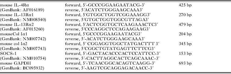
The purpose of PCR experiments is to examine the transcriptional response of HSCs to the treatment with PAE. Total RNA of all the HSCs was isolated using Trizol reagent (Invitrogen, Carlsbad, CA, USA) following the manufacturer's instruction. The quantity of total RNA was assessed by spectrophotometry with an A 260/A 280 ratio of more than 2·1 and the quality of RNA was determined by Sepharose gel electrophoresis. Then, DNase I digestion and first-strand cDNA synthesis (adjusting all total RNA to the same concentration) were performed using Reverse Transcription System (A3500, Promega Corporation, USA) according to the manufacturer's protocol. Next, the cDNAs were amplified with various specific primers (Table 1). All primers were designed by Primer Premier 5.0 software and PCR products were visualized on agarose gels. Mouse glyceral dehyde-3-phosphatedehydrogenase (GAPDH) was used as an internal control. The amplification was performed in thermal cycling with 30 cycles using a procedure of denaturation at 94°C for 45 s, annealing at 50°C for 45 s, and extension at 72°C for 45 s followed by a final extension step for an additional 10 min. PCR products were quantified relative to GAPDH internal controls.
All primers for RT-PCR

Western blotting analysis
The treated HSCs were lysed and the cell proteins were extracted using the method described previously (Chu et al. Reference Chu, Luo, Li, Gao, Yu, Wei, Wu and Shen2007). Total HSC protein concentrations in the supernatants were then measured by the Enhanced BCA (bicinchoninic acid) Protein Assay Kit (Nanjing Jiancheng Bioengineering Institute, China). The HSC proteins from each sample were analysed by SDS-polyacrylamide gel electrophoresis and transferred onto a nitrocellulose membrane (Millipore, Billerica, MA, USA) by semi-dry transfer. Blots were probed overnight at 4°C with the following primary antibodies: anti-Col I, anti-SOCS-1, anti-STAT6, anti-p-STAT6 and anti-β-actin (Santa Cruz Biotechnology, Inc., California, USA), followed by incubation with the appropriate horse-radish peroxidase-conjugated secondary antibody at various dilutions for 2 h. Detection was achieved by enhanced chemiluminescence using SuperSignal West Femto Maximum Sensitivity Substrate (Thermo Fisher Scientific Inc., Rockford, USA) and exposed to film. Filters were quantified by using the JD-801 gel image analysis system (Jiangsu JEDA Science-Technology Development Co. Ltd, China).
Statistical analysis
Data were expressed as means±S.D. Statistical significance of the difference between groups was determined by one-way analysis of variance (using SPSS 11.5 software package). P<0·05 indicated a statistically significant difference.
RESULTS
Effect of PAE on liver histopathology
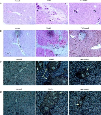
Histological examination showed a significant change in the profile of collagen fibre deposition in the liver sections of mice pre-treated with PAE compared to model mice. Liver sections from the normal mice showed no fibrosis. As shown in Fig. 1A and B, Masson trichrome and H&E staining revealed enormous schistosome granulomas surrounded by an inflammatory cell accumulation and fibril aggregation in the portal area of the livers. The cord-like fibril aggregation extended from the portal area to the hepatic sinus and dissected the hepatic lobule in the model group. In contrast, there were only scattered egg nodules of S. japonicum in the livers of the mice in the PAE-treatment group, with fewer and smaller granulomas and less significant fibrosis; moreover, the normal structure of hepatic lobules was maintained, with fibrogenesis confined to the portal area. As shown in Table 2, in the PAE-treatment group the mean area of granuloma and degree of fibrosis were reduced by nearly 53·4% and 38·5% compared with the model group.
Hepatic pathology in normal, model, and PAE pre-treatment mice infected with Schistosoma japonicum. (A) Haematoxylin and eosin-stained sections of livers from infected model mice show enormous schistosome granulomas surrounded by inflammatory cell accumulation. Only fewer and smaller egg granulomas were found in the livers of the mice in PAE pre-treatment group, ×100. The arrow identifies the egg. (B) Masson trichrome staining of sections of livers demonstrates that the cord-like fibril aggregation extended from the portal area to the hepatic sinus and dissected the hepatic lobule (blue staining) in model mice. There is less significant fibrosis in PAE-treated mice compared with wild-type model mice. Original magnifications, ×100. The arrow identifies deposition of collagen. (C) IHC staining of sections of liver shows the dramatically decreased content of Col I in the PAE group compared with the model group. The band of collagen fibrils extends from the portal area to the hepatic sinus and dissected liver lobules in model mice. Col I in the PAE group is confined to the portal area. Original magnifications, ×200. Col I proteins were stained brown in sections and the arrow identifies positive staining. (D) IHC staining of sections of liver demonstrates positive expression of α-SMA not only in the portal area, but also in the hepatic sinus in the model mice. In the PAE pre-treatment group, positive expression of α-SMA was clearly inhibited and limited only to the portal area. Original magnifications, ×200. α-SMA proteins were stained brown in sections and the arrow identifies positive staining.

Effect of PAE on hepatic granuloma, fibrosis, hydroxyproline, Col I, α-SMA, and IL-13 ( , n=24)

* P<0·05; ** P<0·01, compared with the corresponding model group.
Effect of PAE on the expression of α-SMA and Col I in liver
IHC staining demonstrated that the content of α-SMA and Col I in the model group was dramatically increased compared with the normal mice. The content of α-SMA and Col I was found to extend from the portal area to the hepatic sinus, dissecting liver lobules, in the model mice. However, α-SMA and Col I were limited only to the portal area in the PAE-treatment group and the degree of α-SMA and Col I expression was markedly decreased by 30·3% and 46·6% compared with the model group (Fig. 1C and D, Table 2).
Effect of PAE on hydroxyproline in liver homogenates
Analysis of hepatic hydroxyproline content was carried out as an index of liver fibrosis. Elevated hydroxyproline levels were measured in model animals with respect to normal mice (Table 2). Following treatment with PAE, hydroxyproline content in liver tissue was reduced by nearly 54·5% compared with model mice.
Effect of PAE on the IL-13 level in liver homogenates
In model mice, the level of IL-13 was dramatically increased in the hepatic homogenate whereas it was decreased by 43·0% in the PAE-treated mice (Table 2).
Effect of PAE on HSC proliferation stimulated by IL-13
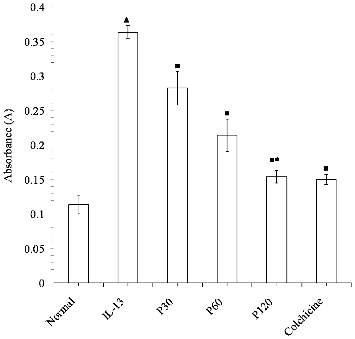
Our study showed that (i) co-culture of HSCs with IL-13 induced a significant proliferative response as illustrated in Fig. 2; (ii) pre-incubation with increasing concentrations of PAE significantly reduced the proliferative response in a dose-dependent manner; (iii) no statistical significance was noted in reduction of the proliferative response between colchicines (1 μmol/l) and PAE (P>0·05) when the concentration of PAE was increased to 120 mg/l.
Effect of PAE on proliferation of HSCs stimulated by rIL-13. HSCs were treated with PAE (0, 30, 60, 120 mg/l) or colchicine (1 μmol/l) for 48 h before being treated with IL-13 for 24 h, and then HSC proliferation was detected by the MTT colourimetric assay. Co-culture of HSCs with rIL-13 induced a significant proliferation of HSCs, and pre-incubation with increasing concentrations of PAE significantly reduced the proliferative response in a dose-dependent manner, especially when the concentration of PAE was increased to 120 mg/l, there is no statistical significance in the reduction of proliferation between colchicines (1 μmol/l) and PAE. The negative control contained PAE at 0 mg/l; P<0·01 compared with the normal group; P<0·05 compared with the IL-13-treated alone group; P>0·05 compared with the colchicine group; Each bar represents the mean and standard deviation of triplicate determinations and data presented are representative of 3 independent experiments.

PAE inhibits IL-13-induced collagen production in HSCs
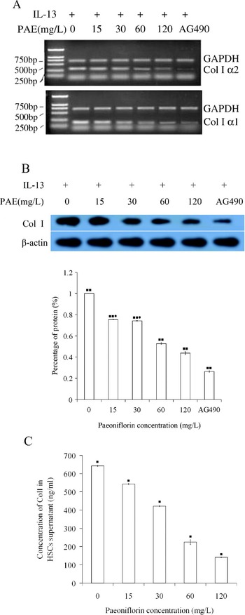
To assess the effect of PAE on IL-13-induced Col I production, RT-PCR and Western blotting analysis were performed for Col I in HSCs, respectively. Pre-treatment with PAE led to a dose-dependent suppression of Col I mRNA and protein, which was confirmed as shown in Fig. 4A and B, and this effect was significantly inhibited by the specific JAK kinase inhibitor, AG490. Furthermore, when HSCs were cultured with various concentrations of PAE, Col I secretion by IL-13-stimulated HSCs was inhibited significantly (Fig. 3C). In summary, Col I produced by IL-13-stimulated HSCs was suppressed by PAE at the gene and protein level in a concentration-dependent manner.
Effect of PAE on rIL-13-induced Col I production in HSCs. Primary HSCs (1·0×105) were seeded in 24-well plates in growth medium containing 15% FCS for 24 h, then serum-starved for an additional 24 h. Subsequently, HSCs were pre-incubated with PAE (0, 15, 30, 60, 120 mg/l), or AG490 (at 50 μmol/l) for 24 h before being stimulated with rIL-13 (50 ng/ml) for 2 h. (A) RT-PCR analysis of Col I α1 and α2 gene expression in HSCs. Levels of Col I α1 and α2 mRNA from HSCs induced by rIL-13 were down-regulated by PAE in a concentration-dependent manner. (B) Western blotting analysis of Col I expression in HSCs. Levels of Col I protein from HSCs induced by rIL-13 were down-regulated by PAE in a concentration-dependent manner. (C) ELISA analysis of Col I protein secretion in HSCs culture media. Levels of Col I protein in HSC superatant induced by IL-13 were down-regulated by PAE in a concentration-dependent manner. ▪▪ There was statistical significance among these groups (P<0·01); ▪ there was statistical significance among these groups (P<0·05). • There was no statistical significance among the two groups (P>0·05). Results are the mean±S.D. from 3 independent experiments. Each bar represents the mean and standard deviation of triplicate determinations and data presented are representative of 3 independent experiments.

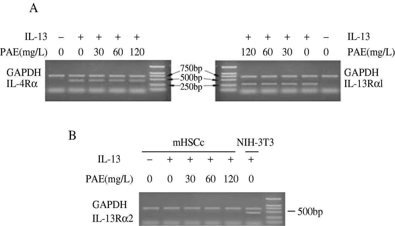
Composition of the receptor on mouse hepatic stellate cells that were treated with different PAE concentrations for 24 h before rIL-13 incubation for RT-PCR. Amplification of IL-4Rα, IL-13Rα1, and IL-13Rα2 mRNA was performed on total RNA from HSCs. (A) PAE did not alter IL-4Rα and IL-13Rα1 levels induced by rIL-13. (B) Neither rIL-13 nor PAE treatment induced the transcript of IL-13Rα2. Experiments were set up in triplicate (data not shown). Amplified PCR products for IL-4Rα and IL-13Rα1 are shown in their expected positions/sizes.

Analysis of IL-4/IL-13 receptor components on HSCs
To examine whether PAE exerts its inhibitory effects by down-regulating the expression of IL-13 receptor, we ran RT-PCR on the transcripts of IL-4Rα/IL-13Rα1 and IL-13Rα2. In this study, all experiments were performed with HSCs. By using the RT-PCR method, the transcripts of IL-4Rα and IL-13Rα1 on HSCs were induced by treatment with IL-13 whereas IL-13Rα2, a critical down-regulatory factor of IL-13–mediated tissue fibrosis induced by S. mansoni and S. japonicum. (Chiaramonte et al. Reference Chiaramonte, Cheever, Malley, Donaldson and Wynn2001; McKenzie and Fallon, Reference Mckenzie and Fallon2003; Wynn et al. Reference Wynn, Hesse, Sandler, Kaviratne, Hoffmann, Chiaramonte, Reiman, Cheever, Sypek and Mentink-Kane2004), was not detectable (Fig. 4). Furthermore, the expression of IL-4Rα and IL-13Rα1 mRNA did not change in HSCs treated with increasing concentrations of PAE (data not shown). The results suggested that IL-13 could stimulate the expressions of IL-4Rα and IL-13Rα1 mRNA, but not IL-13Rα2. So, we presumed that IL-13 induces the expression of Col I mainly through the IL-4 Rα/IL-13Rα1 signal transduction pathway and PAE inhibits the production of collagens by interfering with the IL-13 signalling pathway. The following experiments were set up to test this hypothesis.
PAE inhibits IL-13-induced STAT6 phosphorylation
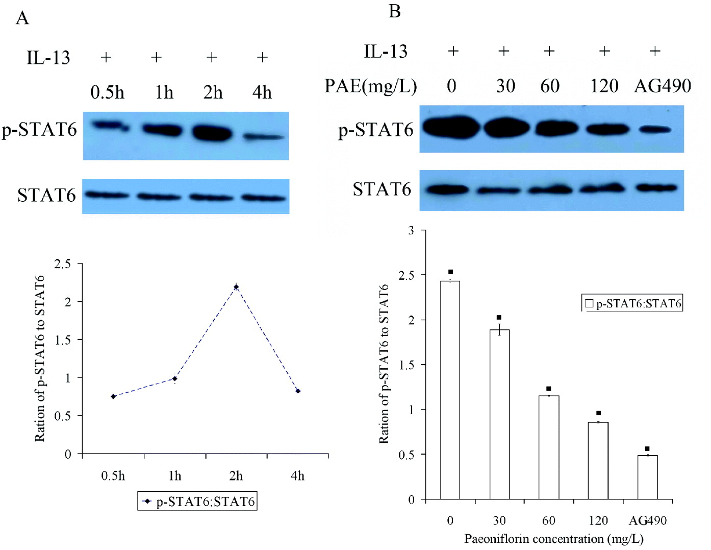
To determine whether PAE can inhibit activation of STAT-6 induced by IL-13 in HSCs, we examined the expression of STAT-6 and p-STAT6 by use of Western blotting. As shown in Fig. 5A and B, IL-13-induced STAT6 phosphorylation, and this effect did not peak until 2 h but was significantly attenuated at 4 h after addition of IL-13, and IL-13 had no effect on STAT6 protein expression in HSCs. PAE, however, effectively reduced phosphorylation of STAT6, but had no effect on STAT6 protein in HSCs. Furthermore, levels of STAT6 phosphorylation were markedly down-regulated by PAE in a concentration-dependent manner, and AG490, a inhibitor of JAKs, was shown to inhibit phosphorylation of STAT6, but had no effect on STAT6 protein.
Effect of PAE on rIL-13-induced STAT6 activation in HSCs. Primary HSCs (1·0×105) were seeded in 24-well plates in growth medium containing 15% FCS for 24 h, then serum-starved for an additional 24 h. Subsequently, HSCs were either left alone as a control or pre-incubated with PAE or AG490 for 24 h before being stimulated with rIL-13 for 2 h. (A) Time-course of STAT6 phosphorylation stimulated by rIL-13. rIL-13 caused transient STAT6 phosphorylation, and STAT6 phosphorylation did not peak until 2 h but significantly attenuated at 4 h after addition of rIL-13. rIL-13 had no effect on STAT6 protein expression in HSCs. (B) Effects of PAE and AG490 on rIL-13-induced STAT6 phosphorylation. PAE effectively reduced the phosphorylation of STAT6, but had no effect on STAT6 protein in HSCs. Levels of STAT6 phosphorylation were down-regulated by PAE in a concentration-dependent manner, and AG490, an inhibitor of JAKs, was shown to inhibit phosphorylation of STAT6, but had no effect on STAT6 protein. For Fig. 5A and B, cell extracts were subjected to Western blotting using phospho-specific antibodies. The band intensities of phosphorylation of STAT6 were normalized to that of the corresponding non-phosphorylated. ▪ There was statistical significance among these groups (P<0·05). Results are the mean±S.D. from 3 independent experiments. Each bar represents the mean and standard deviation of triplicate determinations and data presented are representative of 3 independent experiments.

PAE elevated the expression of SOCS-1
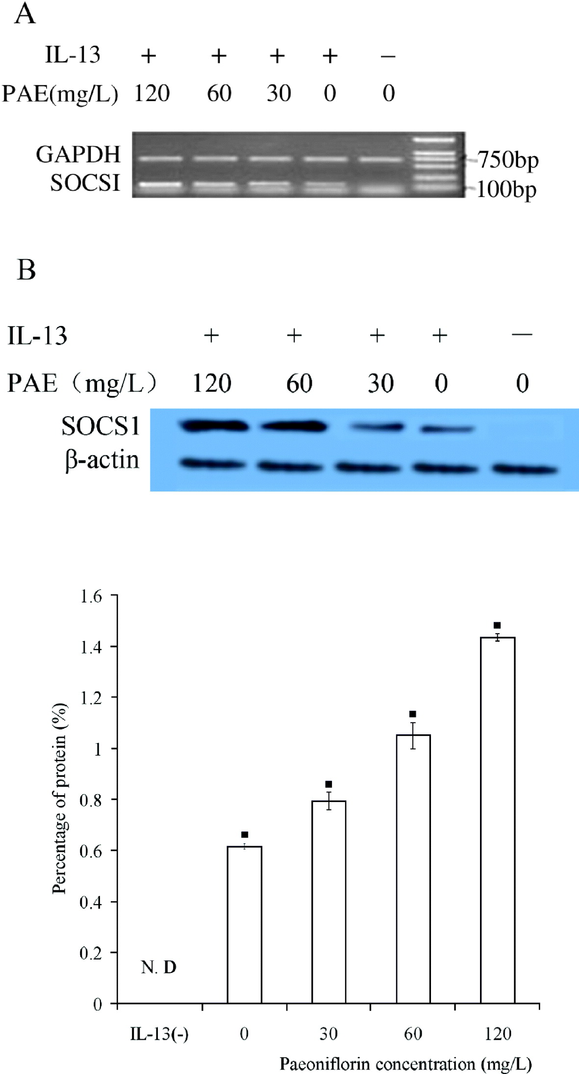
To determine whether IL-13 causes expression of SOCS-1 (a suppressor of cytokine signalling-1) in HSCs, SOCS-1 mRNA and protein were semi-quantified by RT-PCR and Western blotting analysis in our study. Our results demonstrated that SOCS-1 mRNA levels peaked after 1 h in comparison with the control. After a long incubation of the cells, SOCS-1 mRNA and protein expression were still at elevated levels (data not shown). Moreover, we detected whether SOCS-1 could be affected by PAE at various concentrations in all groups. As shown in Fig. 6A and 6B, levels of SOCS-1 mRNA and SOCS-1 protein were up-regulated following PAE treatment relative to IL-13 treatment alone. Additionally, levels of SOCS-1 mRNA protein were up-regulated by PAE in a concentration-dependent manner.
Effect of PAE on rIL-13-induced gene and protein levels of SOCS-1 in HSCs. Primary HSCs (1·0×105) were seeded in 24-well plates in growth medium containing 15% FCS for 24 h, then serum-starved for an additional 24 h. Subsequently, HSCs were either left alone as a control or pre-incubated with PAE (0, 30, 60, 120 mg/l) for 24 h before being stimulated with rIL-13 (50 ng/ml) for 1 h when various concentrations of PAE (0, 30, 60 and 120 mg/l) were added to medium containing rIL-13. Levels of SOCS-1 mRNA and protein were markedly up-regulated by PAE in a concentration-dependent manner. (A) RT-PCR analysis of SOCS-1 gene expression in HSCs. (B) Western blotting of SOCS-1 protein expression in HSCs. ▪ there was statistical significance among these groups (P<0·05). N.D.: not detected. Results are the mean±S.D. from 3 independent experiments. Each bar represents the mean and standard deviation of triplicate determinations and data presented are representative of 3 independent experiments.

DISCUSSION
Natural drugs have made a significant contribution to the treatment of liver fibrosis. Use of herbal drugs in the treatment of liver diseases (Sun, Reference Sun, Wei, Wu, Gui and Wang2007) has a long tradition, especially in Eastern medicine. PAE is an active monomer extracted from paeony, one of the well-known traditional herbs in China (Takagi and Harada, Reference Takagi and Harada1969). PAE has immunoregulatory (Takayama et al. Reference Takayama, Arima, Kanaji, Toda, Tanaka, Shoji, Mckenzie, Nagai, Hotokebuchi and Izuhara2006; Chu et al. Reference Chu, Luo, Li, Gao, Yu, Wei, Wu and Shen2007; Hung et al. Reference Hung, Yang, Tsai, Huang and Huang2008) and anti-inflammatory effects (Zhang et al. Reference Zhang, Wei, Wang, Wang, Chen, Chen, Wu and Hu2008 a); Huang et al. Reference Huang, Chang, Lee, Kim, Kang and Kim2008; Smith, Reference Smith2008). Preparations of many traditional Chinese herbs used in anti-hepatic fibrosis contain paeony root (Li et al. Reference Li, Luo, Li, Cheng, Huang, Liu and Waalkes2003; Chu et al. Reference Chu, Luo, Li, Gao, Yu, Wei, Wu and Shen2007). The present study demonstrated that PAE had therapeutic effects on liver fibrosis caused by S. japonicum in mice. We studied the effect of PAE on the formation of hepatic granuloma and fibrosis, and the changes of fibrotic markers in liver tissues including hydroxyproline, α-SMA and Col I. The results indicated that PAE, administrated in a safe dosage with minimal side effects (Chu et al. Reference Chu, Li, Yang, Wu, Li, Ding, Luo and Shen2008), not only diminishes the dimension and degree of hepatic granuloma and fibrosis, but also decreases the level of hydroxyproline, α-SMA and Col I in liver. Furthermore, our study also revealed that no impact of PAE on the worm and egg burden was seen in the infected mice with or without PAE treatment, suggesting that the lower hepatic hydroxyproline and collagen deposition in PAE-treated mice was not associated with parasite killing.
Interleukin 13 is an immunoregulatory cytokine predominantly secreted by activated Th2 cells (Wynn, Reference Wynn2003). It has similar functions to IL-4 and both of them share a common receptor. However, unlike IL-4, IL-13 appears to be necessary in the effector phase of inflammation and fibrogenesis (McKenzie et al. Reference Mckenzie, Culpepper, De Waal Malefyt, Briere, Punnonen, Aversa, Sato, Dang, Cocks, Menon, De Vries, Banchereau and Zurawski1993, Reference Mckenzie, Bancroft, Grencis and Mckenzie1998; Minty et al. Reference Minty, Chalon, Derocq, Dumont, Guillemot, Kaghad, Labit, Leplatois, Liauzun, Miloux, Minty, Casellas, Loison, Lupker, Shire, Ferrara and Caput1993; Granel et al. Reference Granel, Chevillard and Dessein2007). IL-13 ablation experiments and studies with IL-13–deficient mice as well as IL-13 antagonist in wild mice demonstrated that IL-13 and IL-13-/IL-4Rα, and the Stat6-dependent pathway represented a central mechanism regulating fibrosis in murine schistosomiasis (Chiaramonte et al. Reference Chiaramonte, Donaldson, Cheever and Wynn1999 a, Reference Chiaramonte, Cheever, Malley, Donaldson and Wynn2001; Magalhaes et al. Reference Magalhaes, Miranda, Miranda, Araujo, Jesus, Silva, Santana, Pearce, Carvalho and Jesus2004; Alves Oliveira et al. Reference Alves Oliveira, Moreno, Gazzinelli, Martins-Filho, Silveira, Gazzinelli, Malaquias, Loverde, Leite and Correa-Oliveira2006; Ta, Reference Ta2008).
In our study, we found that the expression of IL-13 in liver homogenate was elevated significantly more in model mice than in normal mice and that PAE could markedly lower the level of IL-13. Therefore, one of the mechanisms of the suppressive effects of PAE on collagen synthesis may be a direct decrease of IL-13 production in liver, presumably related to the suppression of Th2 cells. Our hypothesis is that the anti-fibrotic activity of PAE might be associated with regulating the IL-13 signal pathway and simultaneously suppressing the proliferation of HSC.
HSCs are the primary cell-type in the liver responsible for excessive collagen synthesis during hepatic fibrosis (Bartley et al. Reference Bartley, Ramm, Jones, Ruddell, Li and Mcmanus2006; Tsukada et al. Reference Tsukada, Parsons and Rippe2006). Following liver injury, the HSC undergoes a complex transformation or activation process in which the cell changes from a quiescent, vitamin A-storing cell to that of an activated, myofibroblast-like cell with remarkable proliferative, contractile, pro-inflammatory, and fibrogenic properties, which is characterized by de novo expression of α-smooth muscle actin (α-SMA). Metabolically, an increase in DNA synthesis and cellular proliferation occurs following HSC activation (Bartley et al. Reference Bartley, Ramm, Jones, Ruddell, Li and Mcmanus2006; Chang et al. Reference Chang, Ramalho, Ramalho, Martinelli and Zucoloto2006; Tsukada et al. Reference Tsukada, Parsons and Rippe2006). HSCs play a crucial role in liver fibrosis, as they are responsible for excessive deposition of extracellular matrix proteins, of which Col I predominates. Activated HSCs have now been clearly identified as the primary cellular source of ECM components, thus HSCs have been identified as a ‘signal target’ of hepatic fibrosis (Gressner, Reference Gressner1998). Because IL-13 was the primary fibrogenic mediator in schistosomiasis (Chiaramonte et al. Reference Chiaramonte, Cheever, Malley, Donaldson and Wynn2001, Reference Chiaramonte, Schopf, Neben, Cheever, Donaldson and Wynn1999 b) and IL-13 was shown to directly stimulate Col I production by fibroblasts and LI-90 HSCs cell lines in vitro (Chiaramonte et al. Reference Chiaramonte, Schopf, Neben, Cheever, Donaldson and Wynn1999 b; Jinnin et al. Reference Jinnin, Ihn, Yamane and Tamaki2004; Sugimoto et al. Reference Sugimoto, Enjoji, Nakamuta, Ohta, Kohjima, Fukushima, Kuniyoshi, Arimura, Morizono, Kotoh and Nawata2005). Some studies reported that PAE significantly inhibited HSC proliferation and collagen synthesis stimulated by TGF-β in vitro (Chu et al. Reference Chu, Luo, Li, Gao, Yu, Wei, Wu and Shen2007; Sun, Reference Sun, Wei, Wu, Gui and Wang2007). So we here intended to explore whether PAE could directly interfere with collagen production induced by IL-13 from isolated murine primary HSCs.
The present study demonstrated that PAE could inhibit the HSC proliferation induced by IL-13, suggesting that PAE may decrease collagen production by suppressing HSC activation. Furthermore, the HSCs used in this study expressed one type of receptor complex for IL-13, IL-4Rα/IL-13Rα1 heterodimer, and did not express IL-13Rα2 under the experimental conditions. Moreover, the expression of IL-4Rα/IL-13Rα1 was not affected by PAE, indicating that it is a constitutive expression and is not up-regulated. The IL-4Rα/IL-13Rα1 complex responded to IL-13 in HSCs and was probably linked to intracellular signalling pathways in our study. Additionally, we found that treatment of HSCs with IL-13 could induce STAT-6 phosphorylation, but had no effect on expression of STAT6. Pre-treatment with PAE decreased the amount of total p-STAT6 observed in response to IL-13, without altering the total level of STAT6 protein in the cells. The decoy receptor, IL-13Rα2, acts as a negative feedback inhibitor of IL-13 by reducing its interaction with the signalling type-2 IL-4 receptor. In the progression of schistosomiasis-induced liver fibrosis, IL-13Rα2 played a critical role in the inhibition of IL-13–mediated tissue fibrosis. Moreover, the decoy IL-13Rα2 can also regulate the magnitude of Th2-type cytokine production in vivo (Chiaramonte et al. Reference Chiaramonte, Mentink-Kane, Jacobson, Cheever, Whitters, Goad, Wong, Collins, Donaldson, Grusby and Wynn2003; Wynn et al. Reference Wynn, Hesse, Sandler, Kaviratne, Hoffmann, Chiaramonte, Reiman, Cheever, Sypek and Mentink-Kane2004). Our results showed that neither IL-13 nor PAE treatment induced the expression of the decoy receptor IL-13Rα2 and, thus, this receptor does not seem to contribute to the inhibitory effects of PAE. However, the reason why PAE can reduce the amount of p-STAT6 in the absence of the decoy receptor IL-13Rα2 remains unknown.
Proteins of the suppressors of cytokine signalling (SOCSs) family have important functions as negative regulators of cytokine signalling (Yoshimura and Kubo, Reference Yoshimura and Kubo2007). Another study revealed that higher amounts of Col I and lower levels of SOCS-1 mRNA were produced by fibroblasts from lungs of patients with IPF compared to healthy lungs (Nakashima et al. Reference Nakashima, Yokoyama, Onari, Shoda, Haruta, Hattori, Naka and Kohno2008). Furthermore, the deficiency of SOCS-1 in murine fibroblasts resulted in increased collagen production, whereas over expression of SOCS-1 suppressed collagen production in vitro (Hiroyasu Shoda et al. Reference Hiroyasu Shoda, Ryouhei Nishino, Taku Nakashima, Nobuhisa Ishikawa, Noboru Hattori and Tetsuji Naka2007). Previous investigators (Hebenstreit et al. Reference Hebenstreit, Luft, Schmiedlechner, Regl, Frischauf, Aberger, Duschl and Horejs-Hoeck2003) reported that SOCS-1 inhibits pulmonary inflammation and fibrosis, and SOCS-1 expression can be induced in human epithelial lung cell line A549 by IL-4 and IL-13. Other reports (Yoshida et al. Reference Yoshida, Ogata, Kamio, Joo, Shiraishi, Tokunaga, Sata, Nagai and Yoshimura2004; Zhi-Xin Zhao et al. Reference Zhi-Xin Zhao, Xiao-Mou Peng, Yu-Tian Chong and Zhi-Liang Gao2008) demonstrated that expression of SOCS-1, a suppressor of liver fibrosis, had obvious correlations to the hepatic fibrosis stage. The investigation of SOCS-1 molecule has given important insights into the regulatory mechanisms of cytokine signalling. These regulatory systems offer a fascinating explanation for our results, and the interaction between these cytokines and the SOCS family is warranted.
To date, however, no published studies have examined the down-regulation of SOCS-1 expression by IL-13 in HSCs. We here report for the first time, that IL-13 could induce expression of SOCS-1 both at mRNA and protein levels in HSCs. Meanwhile, we found that PAE pre-treatment induces higher levels of SOCS-1 expression in HSCs in a concentration-dependent manner, and the elevated expression of this protein coincides with decreased STAT6 phosphorylation. SOCS-1 can bind to the Jak kinase domain and inhibit the kinase activity (Dickensheets et al. Reference Dickensheets, Venkataraman, Schindler and Donnelly1999; Yoshimura, Reference Yoshimura and Kubo2007) and SOCS-1 promoter contains 3 functional STAT6 binding sites (Hebenstreit et al. Reference Hebenstreit, Luft, Schmiedlechner, Regl, Frischauf, Aberger, Duschl and Horejs-Hoeck2003). It suggested that PAE's attenuating impact on the level of p-STAT6 protein might be partly through the elevation of SOCS-1 gene expression. However, the reason for PAE's elevation effect on the expression of SOCS-1 gene in the presence of IL-13 in HSCs remains to be clarified. This also suggested that SOCS-1 may be a potential therapeutic targeting molecule against fibrogenesis. Additionally, PAE obviously suppresses the proliferation of HSCs caused by IL-13 in our study. This is another reason for the decreased expression of Col I in HSCs.
We could conclude from our work that the level of p-STAT6 inversely coincides with SOCS-1 and that PAE inhibits IL-13-induced activation of STAT6 and Col I expression at least in part by increasing expression of SOCS-1. So we hypothesize that increased expression of SOCS-1 may attenuate STAT6 phosphorylation in our experimental model, which in turn leads to decreased proliferation and collagen production in response to IL-13 in HSCs. PAE has marked anti-fibrotic effects in the liver of mice with chronic Schistosomiasis japonica. Investigation of the potential of PAE therapy for management of various stages in the progress of fibrosis-associated chronic schistosomiasis is currently in progress.
ACKNOWLEDGEMENTS
The present work conforms to Chinese legal requirements and accepted international ethical standards, including those relating to animal conservation and welfare, and to the journal's policy on these matters. We thank Dr Deyong Chu at the Department of Parasitology, Anhui Medical University (Anhui, China) for his technical assistance.
FINANCIAL SUPPORT
The project was funded by the Natural Science Foundation of China (Nos 30571631 and 30872209), Doctoral Program of Higher Education of China (No. 200803660003), and Natural Science Foundation of Anhui (No. 090413089).