Introduction
In general, most culture media for in vitro production (IVP) of porcine embryos contain serum or bovine serum albumin (BSA), especially in the case of in vitro maturation (IVM) adding porcine follicular fluid (pFF). Serum or BSA provides oocytes with growth factors, hormones, amino acids, vitamins, trace elements and antioxidants (Chang et al., Reference Chang, Jones, Ellefson and Ryan1976; Rui-Hua et al., Reference Rui-Hua, Yong-Hai, Li-Hong, Xiao-Ning, Hong and Wei-Hua2002). These substances stimulate oocytes and embryos during IVM and in vitro culture (IVC); however, the compositions of pFF and serum are complicated and the precise effects of these substances on oocytes or embryos become unclear. The addition of pFF, serum or BSA to a culture medium may lead to variability in the efficacy of outcomes among batches, which makes reliable experimental repeatability impossible. In particular, utilization of these substances is unsuitable for studies on the basic molecular mechanisms such as oocyte and embryo metabolism, the significant effects of growth factors on oocyte cytoplasmic maturation and embryonic development.
Recently, several researchers have been reported on the establishment of a defined medium for both IVM and IVC (Holm et al., Reference Holm, Booth, Schmidt, Greve and Callesen1999; Iwasaki et al., Reference Iwasaki, Kimura and Totsukawa1999; Yoshioka et al., Reference Yoshioka, Suzuki, Tanaka, Anas and Iwamura2002; Kishida et al., Reference Kishida, Lee and Fukui2004; Yang et al., Reference Yang, Han, Niwa and Iannaccone2004). On bovine IVP, Oyamada & Fukai (Reference Oyamada and Fukui2004b) reported blastocyst formation using defined IVM and IVC media throughout the process of IVP. However, there have been limited reports on porcine defined IVP. Until recently, for porcine IVP, Kishida et al. (Reference Kishida, Lee and Fukui2004) used a defined IVM medium and produced blastocysts following intracytoplasmic sperm injection (ICSI) and Yoshioka et al. (Reference Yoshioka, Suzuki, Tanaka, Anas and Iwamura2002) reported that porcine zygotes matured and fertilized in vivo can develop into blastocysts in a chemically defined medium and resulted in the birth of piglets. Moreover, Iwasaki et al. (Reference Iwasaki, Kimura and Totsukawa1999) and Yoshioka et al. (Reference Yoshioka, Suzuki, Itoh, Kikuchi, Iwamura and Rodiguez-Martinez2003) reported that porcine blastocysts could be produced by in vitro fertilization (IVF) and IVC in chemically defined media and obtained piglets after transfer. But, the developmental rates of porcine oocytes matured and cultured in defined media are low compared to those cultured in media containing serum or BSA and, to our best knowledge, blastocysts from porcine oocytes both matured and cultured in vitro in defined media following ICSI has not been reported.
Numerous studies have been reported on cytoplasmic glutathione (GSH) in various domestic animals such as cattle, sheep, pigs and goats (Abeydeera et al., Reference Abeydeera, Wang, Prather and Day1998a; Takahashi et al., Reference Takahashi, Nagai, Hamano, Kuwayama, Okamura and Okano1993; de Matos et al., Reference de Matos, Gasparrini, Pasqualini and Thompson2002; Gonzales et al., Reference Gonzales, Bejar, Mertens and Paramio2003). These reports indicate that oocytes containing a high GSH concentration promote subsequent embryonic development after IVF. And, it is generally known that the in vivo matured oocytes yield higher developmental rates (Nagashima et al., Reference Nagashima, Grupen, Ashman and Nottle1996) and higher concentrations of GSH than those of in vitro matured oocytes (Brad et al., Reference Brad, Bormann, Swain, Durkin, Johnson, Clifford and Krisher2003; Gonzales et al., Reference Gonzales, Bejar, Mertens and Paramio2003). Glutathione synthesis in cytoplasm depends on the availability of cysteine in a culture medium. Cysteine is one of the constituent amino acids of GSH. Cysteine is unstable and easily oxidized to cystine in the medium. However, when thiol compounds such as β-mercaptoehtanol and cysteamine (Cys) are added to the medium, oocytes can use the cysteine because thiol compounds reduce cystine to cysteine.
It was reported that epidermal growth factor (EGF) added to IVM media for various domestic animals such as cattle, sheep, pigs and cats (Abeydeera et al., Reference Abeydeera, Wang, Cantley, Pieke, Prather and Day1998b, Reference Abeydeera, Wang, Cantley, Rieke, Murphy, Prather and Day2000; Grazul-Bilska et al., Reference Grazul-Bilska, Choi, Bilski, Weigel, Kirsch, Kraft, Reynolds and Redmer2003; Sirisathien et al., Reference Sirisathien, Fonseca and Brackett2003; Oyamada et al., Reference Oyamada, Iwayama and Fukui2004a; Merlo et al., Reference Merlo, Iacono, Zambelli, Prati and Belluzzi2005) improved the rates of nuclear maturation and blastocyst formation. Moreover, Abeydeera et al. (Reference Abeydeera, Wang, Prather and Day2000) reported that the 10 ng/ml EGF addition to protein-free tissue culture medium (TCM)199 supplemented with 0.1% polyvinyl alcohol (PVA) for IVM medium resulted in a significantly increased intracellular GSH concentration. Although the additional effect of EGF in IVM media has been reported by many researchers (Abeydeera et al., Reference Abeydeera, Wang, Cantley, Pieke, Prather and Day1998b, Reference Abeydeera, Wang, Cantley, Rieke, Murphy, Prather and Day2000; Miyoshi et al., Reference Miyoshi, Rzucidlo, Pratt and Stice2002; Grazul-Bilska et al., Reference Grazul-Bilska, Choi, Bilski, Weigel, Kirsch, Kraft, Reynolds and Redmer2003; Sturmey et al., Reference Sturmey and Leese2003; Kishida et al., Reference Kishida, Lee and Fukui2004; Oyamada et al., Reference Oyamada, Iwayama and Fukui2004a; Pujol et al., Reference Pujol, Bejar and Paramio2004; Merlo et al., Reference Merlo, Iacono, Zambelli, Prati and Belluzzi2005), there have been few studies on the beneficial effect of EGF on preimplantation embryo development (Paria & Dey, Reference Paria and Dey1990; O'Neill, Reference O'Neill1997; Wei et al., Reference Wei, Park, Day and Prather2001).
In our previous study (Kobayashi et al., Reference Kobayashi, Lee and Fukui2006), we found that the addition of Cys and cystine to a defined IVM medium (modified TCM199) enhanced the blastocyst formation rate after ICSI. However, in that study, cystine was also added to the control medium. Therefore, it was unclear if additional cystine was necessary for the IVM culture medium, because TCM199 has already contained small amounts of cysteine and cystine.
In the present study, we have investigated: (1) the efficacy of additional cystine and/or Cys to a defined IVM medium on intracellular GSH concentration and subsequent embryonic development following ICSI; and (2) the additional effect of EGF in a defined IVC medium, a modified porcine zygote medium (mPZM).
Materials and Methods
Oocyte collection
Porcine ovaries were obtained at a local slaughterhouse and transported to the laboratory in sterile 0.9% NaCl containing penicillin G (Meiji Seika) and streptomycin (Meiji Seika) at about 37 °C within 3 h of slaughter. Cumulus–oocyte complexes (COCs) were aspirated from follicles (2–6 mm diameter) using an 18-gauge needle fixed to a 10 ml disposable syringe. Subsequently, only COCs with a compact cumulus mass with more than three layers were selected for IVM culture. The COCs were washed three times in HEPES-buffered Tyrode's medium (Hagen et al., Reference Hagen, Prather, Sims and First1991) with 0.05% (w/v) polyvinyl alcohol (PVA; Sigma; TLH–PVA).
In vitro maturation
The base medium for IVM was a modified TCM199 (Sigma) supplemented with 3.05 mM glucose (Wako Pure Chemical Industries), 0.91 mM sodium pyruvate (Wako Pure Chemical Industries), 10 ng/ml epidermal growth factor (EGF; Sigma), 0.05% (w/v) PVA (Sigma) and 75 mg/l kanamycin (Sigma). In Experiments 1 and 2, this medium was further supplemented with 100 µM Cys (Sigma: M-6500) or with 0.6 mM cystine (Wako Pure Chemical Industries) according to the experimental design.
Selected COCs were washed in an IVM medium and 23 to 38 COCs in a group were transferred into each well of a Nunc 4-well multidish (Nunc) containing 500 µl of IVM medium and cultured at 39 °C in a humidified atmosphere of 5% CO2 in air. During the first 21 to 23 h, the COCs were cultured in the IVM medium supplemented with 10 IU/ml PMSG (Teikoku-Zouki) and 10 IU/ml hCG (Teikoku-Zouki). Then, COCs were washed three times in the IVM medium without hormones and cultured for an additional 21 to 23 h in the same medium.
Oocyte and sperm preparation
After IVM culture, COCs were denuded of their cumulus cells by repeated pipetting in 0.1% (w/v) hyaluronidase (Sigma) in the TLH–PVA. The oocytes were washed three times in TLH–PVA and remained in the same medium until ICSI in Experiments 1 and 2. In Experiment 3, after washing in the TLH–PVA, oocytes maintained in the IVM medium at 39 °C in a humidified atmosphere of 5% CO2 in air until ICSI.
Semen from an individual boar was used for ICSI through all the experiments. Frozen pelleted semen was thawed at 39 °C and sperm suspension was placed under 1.2 ml of Dulbecco's phosphate-buffered saline (PBS; Gibco-BRL) containing 0.1% PVA (PBS–PVA) in a microcentrifuge tube and the mixture was incubated for 30 min at 39 °C in 5% CO2 in air for swim-up.
Intracytoplasmic sperm injection (ICSI)
ICSI was performed according to the method of Yong et al. (Reference Yong, Pyo, Hong, Kang, Lee, Lee and Hwang2003) with some modifications. Briefly, manipulation was conducted with the aid of a pair of micromanipulators (Leitz) under an inverted microscope. After swim-up treatment, 7 µl of sperm supernatant was collected from the top of the tube and dropped directly onto the lid of a Petri dish (50 mm × 9 mm; Falcon). Another 7 µl drop of TLH–PVA was made adjacent to the drop containing the sperm for oocyte manipulation. The two drops were covered with mineral oil (Sigma). Then, five to 18 oocytes in a group were transferred into the drop of TLH–PVA for manipulation. A spermatozoon was aspirated into the injection pipette tail first and transferred to a drop containing oocytes. An oocyte was held with its polar body at either the 6 or 12 o'clock position by the holding pipette. A spermatozoon was injected into the oocyte cytoplasm and mixed with the cytoplasmic component by the aid of a mouth-regulated open tube.
In vitro culture
After ICSI, oocytes were washed three times in 30 µl drops of IVC medium according to the experimental design. And then, oocytes were transferred into 30 µl droplets of the same medium under mineral oil and cultured for 6 days at 39 °C under 5% CO2, 5% O2 and 90% N2. Cleavage and development to the blastocyst stage were observed on Day 2 (day of ICSI = Day 0) and Day 6, respectively. Blastocysts developed on Day 6 were fixed to examine their cell numbers.
Determination of blastocyst cell number
Blastocysts were washed in 1% (w/v) sodium citrate solution (1% SCS) and placed in the same solution for 20 min on a warming plate. The blastocyst was then fixed in a fixative solution I (1% SCS: acetic acid: methanol = 40: 1: 1) for 1 min and then mounted with a small amount of fixative solution on a glass slide. Thereafter, before the solution was dried, a small amount of fixative solution II (acetic acid:methanol = 1: 1) was dropped onto the blastocysts for complete fixation. The glass slide was dried and stained with 0.4% Giemsa solution (Sigma) for 30 min. After washing, cell number was determined under a compound microscope by counting stained nuclei (×400).
Glutathione assay
GSH concentrations were measured in immature oocytes (presumptive germinal vesicle oocytes; GV) and oocytes matured in vitro in the four different IVM culture conditions, according to the method described by Gasparrini et al. (Reference Gasparrini, Boccia, Marchanidise, Palo, George, Donnay and Zicarelli2006) with some modifications. Briefly, oocytes were carefully denuded by repeated pipetting in 0.1% (w/v) hyaluronidase (Sigma) in the TLH–PVA and washed several times in PBS–PVA to remove any trace of materials having a thiol group. The oocytes matured in vitro without the first polar body were excluded. Samples were stored in a microtube containing 5 µl PBS–PVA and 5 µl 1.25 M H3PO4 (Sigma) at −20 °C until assayed (10–28 oocytes per tube).
On the day of the assay, the samples were thawed and frozen quickly three times in liquid nitrogen to rupture cell membranes. Ninety microlitres of water were added to each sample. Sonication (Branson sonifier cell disruptor model W-185) was performed for approximately 1 min with the output control set to 1–2 and we confirmed that oocytes were completely ruptured under a microscope. When oocytes were not completely ruptured, additional sonication was performed. Then, 200 µ1 OPA reagent (250 mM O-phthaldialdehyde (OPA, Wako Pure Chemical Industries); in methanol: 100 mM borate buffer; pH 9.2 = 2: 8; v/v) were added to each sample for derivatization with OPA. Samples were mixed and waiting for at least 3 min. Derivatized samples were filtered on 0.20 μm filters (Millex-LG; Millipore) and used for high-performance liquid chromatography (HPLC) measurement.
The HPLC system (Thermoseparation) was equipped with a programmable autosampler (Hitachi L-7200) with an injection pump (Hitachi L-7100) and a fluorometric detector (Hitachi L-7480) set up at 335 nm (excitation) and 440 nm (emission) for OPA detection. Separation was achieved using a reversed-phase ODS-3V (4.3 mm I.D. × 250 ml; GL Science). The chromatography was performed at 40 °C at a flow rate of 1.0 ml/min and 0.1 M ammonium acetate solution (Kishida Chemical) was used in the mobile phase. The pH was adjusted to 4.5 with 0.1 M acetic acid (Kishida Chemical). Elution was performed using 10% (v/v) acetonitrile (super gradient; Lascan).
Experimental design
Experiment 1
In Experiment 1, the defined IVM medium was supplemented with 100 µM Cys and/or 0.6 mM cystine and effects on the rates of cleavage, blastocyst formation and the mean cell numbers per blastocyst were examined. As a control, 0.05% (w/v) PVA (Sigma) was only added to the base medium. Intracytoplasmic sperm injection was performed to eliminate the possibility of polyspermic fertilization.
In Experiments 1 and 2, the IVC medium used was North Carolina State University (NCSU)-23 medium (Petters et al., Reference Petters and Wells1993) containing 0.4% (w/v) bovine serum albumin (BSA; Sigma), 0.5 mM sodium pyruvate (Wako Pure Chemical Industries) and 5.0 mM sodium lactate (Sigma) without glucose.
Experiment 2
In Experiment 2, the additional effects of 100 µM Cys and/or 0.6 mM cystine to the IVM defined medium, based on TCM199, on the intracellular GSH concentration were compared with those in the control and GV oocytes.
Experiment 3
In Experiment 3, an IVM medium was the defined medium containing 100 µM Cys and 0.6 mM cystine from the result of Experiments 1 and 2. The base medium for IVC was a modified porcine zygote medium (mPZM) (Yoshioka et al., Reference Yoshioka, Suzuki, Tanaka, Anas and Iwamura2002) containing 2.77 mM myo-inositol (Sigma), 0.34 mM trisodium citrate (Merck, KGaA 6427), 50 µM β-mercaptoethanol (M-6250; Sigma), 100 ng/ml insulin (Sigma) (Lee et al., Reference Lee, Kim, Hyun, Jeon, Nam, Jeong, Kim, Kim, Kang, Lee and Hwang2005) and MEM essential amino acids (Gibco-BRL) without BME essential amino acids. As a positive or negative control, 0.3% (w/v) BSA (Sigma; A6003) (mPZM-3) or 0.3% (w/v) PVA (Sigma) (mPZM-4: defined IVC medium), respectively, was added to the base medium. The defined IVC medium was supplemented with 5 ng/ml or 10 ng/ml EGF and effects on the rates of cleavage, blastocyst formation and the mean cell numbers per blastocyst were examined. As EGF stock solution, EGF was diluted with PBS (Gibco) containing 0.3% PVA without BSA or serum.
Statistical analysis
The data on cleavage and development to blastocysts were analysed by ANOVA and Fisher's PLSD test using the STATVIEW program (Abacus Concepts). All percentage data were subjected to arcsine transformation before statistical analysis. The mean cell numbers per blastocyst and GSH concentrations were analysed by one-way ANOVA and then differences among the groups were also analysed using Tukey–Kramer multiple comparison tests.
Results
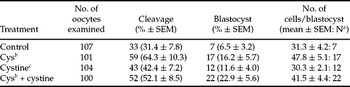
In Experiment 1, the cleavage rates in the control (31.4%), Cys (64.3%), cystine (42.4%) and Cys + cystine (52.1%) groups were not significantly different. The blastocyst rate (22.9%) in the Cys + cystine group was higher than that in the other three group (6.5–16.2%), but significant difference was not revealed among the four groups. The mean cell numbers per blastocyst were not significantly different among the four groups (Table 1).
Effects of cysteamine or cystine in a defined IVM medium on developmental competence of porcine oocytes following ICSI

a No. of blastocysts examined.
b Cys: 100 µM cysteamine.
c Cystine: 0.6 mM cystine.
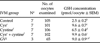
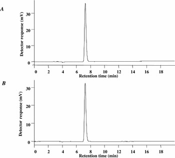
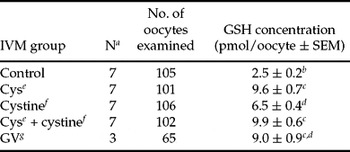
In Experiment 2, the peak shown in the Fig. 1A was assumed to represent intracellular GSH (Fig. 1) and the retention time was almost same as that of the standard GSH (Fig. 1B). The mean intracellular GSH concentrations of the Cys (9.6 pmol/oocyte) and Cys + cystine (9.9 pmol/oocyte) groups were significantly (p < 0.05) higher compared with those in the control (2.5 pmol/oocyte) and cystine (6.5 pmol/oocyte) groups (Table 2). Further, the GSH concentration of the cystine group was also significantly (p < 0.05) higher than that of the control group. However, the GSH concentration of the GV group (9.0 pmol/oocyte) was not significantly different compared with those in the Cys, cystine and Cys + cystine groups.
Intracellular glutathione (GSH) concentrations of porcine oocytes matured in defined maturation media
a The number of assays.
b –dWithin a column, the values with different letters are significantly different (p < 0.05).
e Cys: 100 µM cysteamine.
f Cystine: 0.6 mM cystine.
g GV: presumptive germinal vesicle oocytes (immature oocytes).
(A) Intracellular glutathione (GSH) concentration: 14 matured oocytes in the defined maturation medium supplemented with cysteamine (100 µM) and cystine (0.6 mM) were used for HPLC measurement. (B) A standard GSH: 125 pmol GSH (Sigma) was used for HPLC measurement.

In Experiment 3, the cleavage rates in the mPZM-3 group (56.1%), mPZM-4 group (54.0%), mPZM-4 + 5 ng/ml EGF group (43.0%) and mPZM-4 + 10 ng/ml EGF group (58.0%) were not significantly different. The blastocyst rates were also not significantly different among the four groups (14.0–23.6%). However, the rate resulting from the addition of 5 ng/ml EGF into the mPZM-4 (48.6%) was significantly (p < 0.05) higher than the other two defined groups (mPZM-4 and mPZM-4 + 10 ng/ml EGF; 23.4% and 23.1%, respectively) on the blastocyst rate per cleaved embryo and was similar to the mPZM-3 group (Table 3). The mean cell number per blastocyst in the mPZM-3 group (30.2 ± 2.2: 18; no. of cells/blastocyst ± SEM: no. of blastocysts examined), mPZM-4 group (32.5 ± 3.9: 11), mPZM-4 + 5 ng/ml EGF group (36.5 ± 4.6: 13) and mPZM-4 + 10 ng/ml EGF groups (34.4 ± 3.5: 13) were not significantly different among the four groups.
Effect of EGF in a defined IVC medium on developmental competence of porcine oocytes following ICSI

a mPZM, modified porcine zygote medium.
b Includes parthenogenetic development.
c Percentage of cleavage and blastocysts to the number of oocytes cultured. Cleavage and blastocyst formation were examined on days 2 and 6 (day of ICSI = day 0), respectively.
d Percentage of blastocysts to the number of cleaved embryos.
e,f Within a column, values (seven replicates) with different letters are different (p < 0.05).
Discussion
In the present study, the additional effects of Cys and/or cystine in the defined IVM medium, based on TCM199, were investigated on intracellular GSH concentration and subsequent embryonic development following ICSI. In general, it has been suggested that cumulus-free oocytes cannot uptake cystine, an oxidized dimer form of cysteine, but that they could uptake cysteine to synthesize GSH by the alanine–serine–cysteine (ASC) transport system during IVM (Nagai, Reference Nagai2001). In contrast, cumulus cells surrounding porcine oocytes can uptake both cystine and cysteine by system Xc– and system ASC, respectively, to synthesize GSH during IVM (de Matos et al., Reference de Matos, Furnus and Moses1997; Nagai, Reference Nagai2001). In addition, GSH synthesized in cumulus cells had also been shown to be transported to oocytes through a gap junction (Mori et al., Reference Mori, Amano and Shimizu2000; Nagai, Reference Nagai2001).
In the present study, the glutathione concentration in the control group was significantly low compared to that in the GV group (Table 2). However, the GSH concentrations of Cys and Cys + cystine groups were similar level of the GV oocytes. It was considered that GSH concentration of the control group was reduced because of the defined medium being without Cys during the IVM culture. The GSH concentration of the cystine group was also not significantly different from that of the GV oocytes. However, in the cystine group, the GSH concentration was significantly lower than those in the Cys or Cys + cystine group. Therefore, it appears that in the control group, cysteine was not fully provided to the oocytes and cumulus cells for GSH synthesis and that cysteine and cystine in the IVM medium (TCM199) were not sufficiently utilized by oocytes or cumulus cells. From these results, it was shown that the GSH concentration of the control group could not maintain comparable GSH levels as found in the GV oocytes.
The GSH concentration of in vitro matured oocytes was generally lower than that of in vivo matured oocytes (porcine, 36.3 pmol/oocyte; goat; 23.7 pmol/oocyte) (Brad et al., Reference Brad, Bormann, Swain, Durkin, Johnson, Clifford and Krisher2003; Gonzales et al., Reference Gonzales, Bejar, Mertens and Paramio2003). The developmental rates following IVF, ICSI or subzonal sperm injection and IVC of in vitro matured oocytes were also generally lower than the in vivo matured oocytes (Nagashima et al., Reference Nagashima, Grupen, Ashman and Nottle1996). In other studies using serum or follicular fluid, cysteine or cystine added to IVM media without Cys have been reported to result in significantly increased GSH concentrations (Brad et al., Reference Brad, Bormann, Swain, Durkin, Johnson, Clifford and Krisher2003) and enhanced developmental competence (de Matos & Furnus, 2000; Gasparrini et al., Reference Gasparrini, Boccia, Marchanidise, Palo, George, Donnay and Zicarelli2006). Therefore, it appeared that serum or follicular fluid contains some substances with the same antioxidant function as Cys (Rui-Hua et al., Reference Rui-Hua, Yong-Hai, Li-Hong, Xiao-Ning, Hong and Wei-Hua2002; Tatemoto et al., Reference Tatemoto, Muto, Sunagawa, Shinjo and Nakada2004). In vitro culture is generally performed with higher concentrations of O2 than those of in vivo conditions, resulting in increased production of reactive oxygen species (ROS). In vitro produced embryos are usually impaired by many ROS on the intracellular DNA, lipids and proteins (Guerin et al., Reference Guerin, El Mouatassim and Menézo2001; Kitagawa et al., Reference Kitagawa, Suzuki, Yoneda and Watanabe2004). GSH also plays an important role of antioxidation in oocytes against ROS. In the present study, the GSH concentration in the control oocytes matured in the defined IVM medium was lower than that of GV oocytes. These results were consistent with those of previous reports (Kishida et al., Reference Kishida, Lee and Fukui2004) stating that the GSH concentration in porcine oocytes matured in a defined IVM medium was lower than the GV oocytes. However, it has been reported that GSH concentration in oocytes increases during IVM (Sawai et al., Reference Sawai, Funahashi and Niwa1997; Zuelke et al., Reference Zuelke, Jeffay, Zucker and Perreault2003), reaching a peak level at the metaphase II stage (MII) (Luberda, Reference Luberda2005) and decreasing during IVC (Zuelke et al., Reference Zuelke, Jeffay, Zucker and Perreault2003). Gonzalez et al. (2003) and Brad et al. (Reference Brad, Bormann, Swain, Durkin, Johnson, Clifford and Krisher2003) reported that low intracellular GSH levels of IVM oocytes might be responsible, in part, for the lower developmental competence observed in in vitro matured goats or porcine oocytes. Therefore, the low GSH concentration in the oocytes after IVM without thiol compounds such as Cys would be one of the reasons for the low competence of embryonic development of porcine oocytes matured in a defined medium.
The GSH concentration in the oocytes matured in the IVM medium adding Cys was not significantly different from those of the Cys and cystine groups. Therefore, it was considered that TCM199 already contained enough cystine and cysteine to convert cystine to cysteine by adding 100 µM Cys. In the absence of Cys, as previously reported (de Matos et al., Reference de Matos, Furnus and Moses1997), the conversion of cystine into cysteine for GSH synthesis is mediated by cumulus cells. In the present study (Table 2), the GSH concentration of the oocytes matured in the medium adding cystine was significantly higher than that of the control oocytes. We believe that cumulus cells uptake, utilize cystine and then provide GSH to oocytes. However, the GSH concentration of the oocytes matured in the medium containing cystine was significantly lower than that of the Cys or Cys + cystine group. This suggested that the oocytes matured in TCM199 plus 0.6 mM cystine could not fully utilize cystine in the IVM medium. Furthermore, Bing et al. (Reference Bing, Hirao, Iga, Che, Takenouchi, Kuwayama, Fuchimoto, Rodriguez-Martinez and Nagai2002) reported that the GSH concentration of oocytes matured in an IVM medium containing 150 µM Cys was higher than that of the oocytes matured in the medium without Cys. These results indicate that uptake of cysteine by both oocytes and cumulus cells are more effective to synthesize GSH rather than uptake of cystine by only cumulus cells and then cumulus cells provide GSH to oocytes.
It has been shown that the addition of Cys to IVM media facilitates the increase of intracellular GSH concentration and developmental competence of embryos in various animal species (Takahashi et al., Reference Takahashi, Nagai, Hamano, Kuwayama, Okamura and Okano1993; Grupen et al., Reference Grupen, Nagashima and Nottle1995; Bing et al., Reference Bing, Hirao, Iga, Che, Takenouchi, Kuwayama, Fuchimoto, Rodriguez-Martinez and Nagai2002; de Matos et al., Reference de Matos, Gasparrini, Pasqualini and Thompson2002; Gasparrini et al., Reference Gasparrini, Boccia, Marchanidise, Palo, George, Donnay and Zicarelli2006). However, in the present study, although the addition of 100 µM Cys + 0.6 mM cystine resulted in a higher rate of blastocyst formation (22.9%) than found with the addition of Cys or cystine alone or, in the control medium, the rates of cleavage, blastocyst formation and the mean cell number per blastocyst were not significantly different among the control, Cys, cystine and Cys + cystine groups.
In Experiment 3, the efficacy of adding EGF to mPZM for IVC medium was investigated on embryonic development and the mean cell number of blastocysts following ICSI. Although the effects of EGF on IVP have been reported by several researchers, studies on the efficacy of adding EGF to IVC medium have been limited (Paria & Dey, Reference Paria and Dey1990; O'Neill, Reference O'Neill1997; Wei et al., Reference Wei, Park, Day and Prather2001) and there were conflicting results about the effects of EGF during preimplantation development. Abeydeera et al. (Reference Abeydeera, Wang, Cantley, Pieke, Prather and Day1998b) reported that the addition of EGF to the IVC medium for porcine embryos derived from oocytes matured in the presence of EGF was not positively effective on cleavage rates, blastocyst rates and the average cell number for blastocyst. On the other hand, it was reported that the EGF and EGF receptor (EGFr) mRNA are expressed in porcine IVF and porcine somatic cell nuclear transfer (SCNT) preimplantation embryos and that EGF increased the quality of blastocysts by increasing total cell numbers in porcine SCNT embryos (Lee et al., Reference Lee, Kang, Lee and Hwang2005).
In the present study, although the cleavage rates in the mPZM-4 + 5 ng/ml EGF group were somewhat lower compared to other three groups, the blastocyst formation to cleavage was significantly (p < 0.05) increased in the mPZM-4 + 5 ng/ml EGF group compared with other defined groups. Sirisathien et al. (Reference Sirisathien, Fonseca and Brackett2003) also found that a concentration of 5 ng/ml EGF, but not 1 or 25 ng/ml EGF, during bovine embryo culture improved percentages of 4-cell stage embryos reaching blastocysts compared to the control. The present results indicated that the addition of 5 ng/ml EGF to the mPZM-4 had a beneficial effect on blastocyst production using the defined IVC medium.
In contrast, Lee et al. (Reference Lee, Kim, Hyun, Jeon, Nam, Jeong, Kim, Kim, Kang, Lee and Hwang2005) reported that the 2-cell embryos, morulae and blastocysts derived from porcine IVF and SCNT had expressed EGFr mRNA, but 4-cell and 8-cell embryos did not express the EGFr mRNA. Lee et al. (Reference Lee, Kim, Hyun, Jeon, Nam, Jeong, Kim, Kim, Kang, Lee and Hwang2005) also found that the addition of EGF at the morula stage did not affect in vitro development to blastocysts or the cell number of porcine SCNT blastocysts. Wei et al. (Reference Wei, Park, Day and Prather2001) also reported that the addition of EGF at the morula stage did not influence blastocyst formation in porcine IVF embryos cultured in a defined IVC medium. Therefore, we consider that although the mechanism of the positive effect of EGF is not known, the addition of 5 ng/ml EGF would enhance 2-cell or 4-cell stage embryos to blastocyst development, as shown in our study. Lee & Fukui (Reference Lee and Fukui1995) suggested that EGF and fibroblast growth factor (FGF) might act synergistically on bovine embryonic development in vitro. Moreover, it was shown that the addition of 10 ng/ml EGF with 0.1% BSA significantly increased the rates of blastocyst formation, but there was no increase without BSA (Wei et al., Reference Wei, Park, Day and Prather2001). Therefore, it is possible that EGF has synergistically influenced on the preimplantation embryo development with some component(s) in the IVC medium used in the present study. Lee & Fukui (Reference Lee and Fukui1995) also reported that EGF increased the cell number of blastocyst with the addition of 10 ng/ml EGF to 2-cell stage bovine embryos, but blastocyst formation was not increased. However, in the present study, the mean cell number of blastocysts derived from the defined IVC medium adding EGF was not increased as compared with the negative control.
In the present study, the cleavage and developmental rates between mPZM-3 and mPZM-4 were not significantly different, and blastocysts (14–19%) were produced from embryos cultured in a defined IVC medium following defined IVM and ICSI. These results indicate that in vitro derived porcine embryos can develop to the blastocyst stage without BSA or serum, to a level similar to that achieved by adding BSA to IVC medium. To our best knowledge, this is the first report to produce porcine blastocysts by defined IVM and IVC media combined with ICSI. However, the rates of cleavage, blastocyst formation and the mean cell number in blastocysts were still low and further studies are needed to optimize a defined porcine IVP system to produce high-quality blastocysts in vitro.
In conclusion, the present results indicate that the addition of Cys to a defined maturation medium using TCM199 for IVM culture increases intracellular GSH concentration in porcine matured oocytes. Further addition of cystine may be not necessary and TCM199 plus Cys (100 µM) could be used as a defined IVM medium for porcine oocytes. The addition of 5 ng/ml EGF to a defined IVC medium has enhanced subsequent development after ICSI and porcine blastocysts can be produced using defined media throughout the steps of IVP (IVM, ICSI and IVC).
Acknowledgements
We thank the Hokkaido Meat Board in Obihiro for supplying porcine ovaries. The present study was partly supported by a Grant-in-Aid for Scientific Research (17580242) from the Japan Society for the Promotion of Science.