





Introduction
CHDs are the most common structural malformations of the heart or great vessels in the perinatal period and are one of the leading causes of neonatal mortality worldwide. These anomalies originate during embryonic development and can manifest from the fetal period. Their clinical spectrum is broad, ranging from mild defects that do not require treatment to moderate and complex life-threatening conditions requiring surgical intervention in the neonatal period. Reference Liu, Chen and Zühlke1
Globally, CHD is estimated to affect approximately 9 out of every 1,000 live births. According to a metaanalysis of 260 studies conducted between 1970 and 2017, the global prevalence at birth increased from 4,547 per 1,000 live births in the period 1970–1974 to 9,410 in the period 2010–2017. Reference Liu, Chen and Zühlke1 This increase is primarily attributed to improvements in prenatal and postnatal diagnostic techniques, rather than a true increase in incidence.
Despite diagnostic and therapeutic advances, CHD continues to be a significant cause of childhood morbidity and mortality. According to the 2017 Global Burden of Disease Study, CHD was responsible for approximately 261,247 deaths worldwide, 69% of which occurred in children under 1 year of age. 2 Furthermore, it is estimated to have generated more than 21 million years of life lost and 589,479 years lived with disability, reflecting a high global health burden. Although CHD mortality has decreased by 34.5% since 1990, most deaths occur in low- and middle-income countries, where access to early diagnosis and specialised surgery is limited. 2
Regarding their distribution, septal lesions predominate internationally, such as ventricular septal defects, atrial septal defects, and patent ductus arteriosus, representing approximately 58% of all CHD cases and accounting for more than 90% of the increase in prevalence observed in recent decades. Reference Liu, Chen and Zühlke1 In contrast, complex heart diseases such as transposition of the great arteries and hypoplastic left ventricular syndrome are less common, with prevalences of 0.29 and 0.18 per 1,000 live births, respectively. Reference Liu, Chen and Zühlke1,Reference Zambrano, Vaccaro and Jara3,Reference Carlos Smith, Millaray Brito, Manuel Álvarez, Alejandro Álvarez, Sergio De La Fuente and Juan G.Rodríguez4
In Chile, data from the Eastern Perinatal Reference Center (CERPO) show a distribution that is partially consistent with the international literature. Between 2003 and 2011, 568 cases were diagnosed prenatally, with ventricular septal defect being the most common, followed by hypoplastic left ventricle syndrome, atrioventricular canal defect, arrhythmias, coarctation or aortic stenosis, and transposition of the great arteries. Reference Mayorga, Rodríguez and Enríquez5 The high proportion of complex CHDs in this sample likely reflects CERPO’s status as a tertiary centre, which treats cases referred for suspected or confirmed CHD in our health system.
Prenatal diagnosis of CHD has been shown to have a significant impact on neonatal prognosis. Recent studies suggest that it allows for earlier timing of surgery and reduces both preoperative and postoperative mortality, especially in serious conditions such as transposition of the great arteries. Reference Li, Zhou, Fang, Wang, Hua and Mu6 It also makes it possible to plan delivery in centres with specialised support and anticipate interventions such as the administration of prostaglandins or urgent procedures such as Rashkind atrioseptostomy. Although variables such as birth weight and Apgar score do not show significant differences, fetal diagnosis provides critical time for the clinical, logistical, and emotional preparation of the treating team and family. Reference Li, Zhou, Fang, Wang, Hua and Mu6
Considering the high burden of CHD, its clinical heterogeneity, and the importance of prenatal diagnosis, it is essential to characterise its behaviour in the local context and share our experience in the prenatal diagnosis of CHD.
Objectives and methods
The main objective of this study was to retrospectively describe and characterise moderate and complex CHDs diagnosed by fetal echocardiography in the Fetal Examination Unit of the Guillermo Grant Benavente Regional Hospital, evaluating their distribution, the presence of chromosomal abnormalities and extracardiac malformations, as well as neonatal outcomes and their relationship with maternal comorbidities. Specifically, we sought to identify the subtypes of heart defects diagnosed prenatally during the study period, determine the frequency of chromosomal abnormalities and associated genetic syndromes, describe the main maternal comorbidities, and analyse postnatal outcomes, including the need for surgery, vital status, and short- and medium-term survival.
An observational, descriptive, and retrospective study was conducted. The population included all pregnant women whose fetuses were evaluated in the Fetal Examination Unit of the Guillermo Grant Benavente Regional Hospital as part of the fetal cardiology program. The period from January 1, 2018, to March 31, 2025, was considered.
Of the total fetal echocardiograms performed during the study period, cases with a confirmed diagnosis of moderate or complex CHD were selected, according to severity classification, excluding mild defects. All pregnancies, singleton or multiple, with a confirmed diagnosis of moderate or complex CHD at any gestational age were included.
The maternal variables analysed were age, comorbidities (pre-gestational and gestational diabetes, high blood pressure, hypothyroidism, bronchial asthma, systemic lupus erythematosus, and other less prevalent conditions), and relevant obstetric history. Regarding the fetuses, the subtype of CHD, the presence of extracardiac malformations, chromosomal abnormalities, or genetic syndromes, gestational age at diagnosis, sex of the newborn, and anthropometric data at birth were recorded.
Neonatal outcomes assessed included vital status at birth (live birth or stillbirth), early neonatal (≤7 days), late neonatal (8–28 days), postneonatal, and overall survival at the end of follow-up (March 31, 2025). The need for cardiac surgery was also recorded, and in cases where it was not performed, the reason for not being performed was stated as “out of scope.”
Data were obtained from anonymised institutional databases provided by the corresponding clinical services. Prenatal diagnoses were established using fetal echocardiography, considered the gold standard for the prenatal identification of cardiac malformations, and neonatal information was supplemented with hospital records and follow-up visits.
Statistical analysis was based on the description of variables using absolute and relative frequencies. Survival was assessed at different time points (7 days, 28 days, and follow-up at the end of the study) and graphically represented using Kaplan–Meier curves. The association between maternal comorbidities and outcomes was explored by comparing proportions between subgroups, without applying multivariate models due to the descriptive nature of the study and the limitations of the retrospective design. This work was approved by the scientific ethics committee of our hospital and the Concepción health service.
Results
During the study period, 2.260 fetal echocardiograms were performed, and 80 fetuses (3.5%) were diagnosed with moderate and complex CHD. One case corresponded to complete atrioventricular block treated with pacemaker implantation, while the remaining 79 presented structural heart disease.
Regarding sex distribution, 51 cases were male (63.8%) and 29 were female (36.3%). The average birth weight was 3,193 g (range: 1,855–5,309 g), and the average newborn length was 50 cm (range: 45–53 cm). According to weight-for-gestational age classification, 60 were adequate, 12 were small, and 8 were large. The maternal age at diagnosis was 31 years on average (range: 16–43 years). The median gestational age of the mothers at the time of diagnosis was 29 weeks of gestation (range: 22–37 weeks).
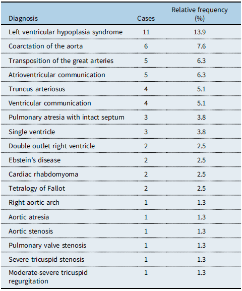
Hypoplastic left ventricle syndrome was the most common diagnosis, with 11 cases (14.0%). This was followed by coarctation of the aorta (6 cases; 7.6%), transposition of the great arteries (5 cases; 6.3%), common atrioventricular septal defect (5 cases; 6.3%), and truncus arteriosus (4 cases; 5.1%).
Large ventricular septal defect was also observed in 4 patients (5.1%), while pulmonary atresia with intact septum and single ventricle were diagnosed in 3 cases each (3.8%).
Other less prevalent diagnoses included double outlet right ventricle, Ebstein’s disease, cardiac rhabdomyoma, and tetralogy of Fallot (2 cases each; 2.5%). Isolated cases of right aortic arch, aortic atresia, aortic stenosis, pulmonary vein stenosis, severe tricuspid stenosis, and moderate-severe tricuspid regurgitation were also reported (1 case each; 1.3%). The complete distribution is presented in Table 1.
Distribution of isolated diagnoses of complex congenital heart disease

Table 1. Long description
A table showing the distribution of isolated diagnoses of complex congenital heart disease. The table has 18 rows and 3 columns. The columns are labeled Diagnosis, Cases, and Relative frequency (percent). The table lists various diagnoses along with the number of cases and their relative frequency in percent. Row 1: Left ventricular hypoplasia syndrome, 11, 13.9. Row 2: Coarctation of the aorta, 6, 7.6. Row 3: Transposition of the great arteries, 5, 6.3. Row 4: Atrioventricular communication, 5, 6.3. Row 5: Truncus arteriosus, 4, 5.1. Row 6: Ventricular communication, 4, 5.1. Row 7: Pulmonary atresia with intact septum, 3, 3.8. Row 8: Single ventricle, 3, 3.8. Row 9: Double outlet right ventricle, 2, 2.5. Row 10: Ebstein’s disease, 2, 2.5. Row 11: Cardiac rhabdomyoma, 2, 2.5. Row 12: Tetralogy of Fallot, 2, 2.5. Row 13: Right aortic arch, 1, 1.3. Row 14: Aortic atresia, 1, 1.3. Row 15: Aortic stenosis, 1, 1.3. Row 16: Pulmonary valve stenosis, 1, 1.3. Row 17: Severe tricuspid stenosis, 1, 1.3. Row 18: Moderate-severe tricuspid regurgitation, 1, 1.3.
Relative frequencies were calculated in relation to the total number of cases of structural CHD (n = 79). The percentages do not sum to 100%, as this table includes only isolated diagnoses.
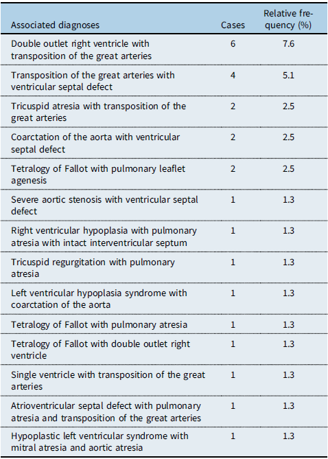
Twenty-one cases of diagnostic combinations were documented (26.6%). The most frequent association was double outlet right ventricle with transposition of the great arteries (6 cases; 7.6%), followed by transposition of the great arteries with ventricular septal defect (4 cases; 5.1%). Two cases of tricuspid atresia with transposition of the great arteries (2.5%) and two of tetralogy of Fallot with pulmonary leaflet agenesis (2.5%) were also recorded.
The remainder corresponded to highly complex unique combinations, such as coarctation of the aorta with ventricular septal defect, hypoplastic left ventricle syndrome with coarctation of the aorta, tetralogy of Fallot with pulmonary atresia, single ventricle with transposition of the great arteries, common atrioventricular septal defect with pulmonary atresia and transposition of the great arteries, and hypoplastic left ventricle syndrome with mitral and aortic atresia, all accounting for one case each (1.3%). The complete details are summarised in Table 2.
Distribution of diagnostic combinations of complex CHD

Table 2. Long description
A table with three columns and twelve rows. The columns are labeled ‘Associated diagnoses’, ‘Cases’, and ‘Relative frequency (%)’. The table lists various complex congenital heart disease (CHD) diagnoses along with the number of cases and their relative frequencies. Row 1: Double outlet right ventricle with transposition of the great arteries, 6 cases, 7.6 percent. Row 2: Transposition of the great arteries with ventricular septal defect, 4 cases, 5.1 percent. Row 3: Tricuspid atresia with transposition of the great arteries, 2 cases, 2.5 percent. Row 4: Coarctation of the aorta with ventricular septal defect, 2 cases, 2.5 percent. Row 5: Tetralogy of Fallot with pulmonary leaflet agenesis, 2 cases, 2.5 percent. Row 6: Severe aortic stenosis with ventricular septal defect, 1 case, 1.3 percent. Row 7: Right ventricular hypoplasia with pulmonary atresia with intact interventricular septum, 1 case, 1.3 percent. Row 8: Tricuspid regurgitation with pulmonary atresia, 1 case, 1.3 percent. Row 9: Left ventricular hypoplasia syndrome with coarctation of the aorta, 1 case, 1.3 percent. Row 10: Tetralogy of Fallot with pulmonary atresia, 1 case, 1.3 percent. Row 11: Tetralogy of Fallot with double outlet right ventricle, 1 case, 1.3 percent. Row 12: Single ventricle with transposition of the great arteries, 1 case, 1.3 percent. Row 13: Atrioventricular septal defect with pulmonary atresia and transposition of the great arteries, 1 case, 1.3 percent. Row 14: Hypoplastic left ventricular syndrome with mitral atresia and aortic atresia, 1 case, 1.3 percent.
Relative frequencies were calculated in relation to the total number of structural CHD cases (n = 79). The percentages do not sum to 100%, as this table only includes cases with diagnostic combinations.
When analysing the overall frequency by lesion, considering both isolated and combined diagnoses, transposition of the great arteries was observed in 19 patients (24.1%), hypoplastic left ventricle syndrome in 13 (16.5%), and coarctation of the aorta in 8 (10.1%), constituting the most prevalent entities in the study group.
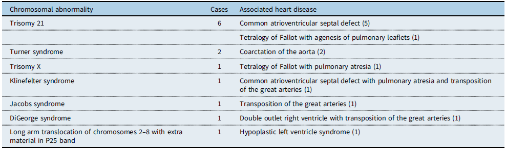
Genetic testing was performed in 39 of the 80 patients in the cohort (48.8%). Among those evaluated, 13 presented chromosomal abnormalities confirmed by karyotyping, corresponding to 16.3% of the total cases. The most frequent alteration was trisomy 21, identified in 6 patients, most of whom were associated with common atrioventricular septal defects, and one case with tetralogy of Fallot with agenesis of the pulmonary leaflets. Turner syndrome was documented in 2 patients, both with coarctation of the aorta. The remaining abnormalities corresponded to single cases, the details of which are presented in Table 3.
Chromosomal alterations and associated heart diseases

Table 3. Long description
A table listing chromosomal abnormalities, the number of cases, and associated heart diseases. The table has three columns: Chromosomal abnormality, Cases, and Associated heart disease. It contains eight rows, each detailing a specific chromosomal abnormality, the number of cases observed, and the corresponding heart disease. Row 1: Trisomy 21, 6 cases, Common atrioventricular septal defect (5), Tetralogy of Fallot with agenesis of pulmonary leaflets (1). Row 2: Turner syndrome, 2 cases, Coarctation of the aorta (2). Row 3: Trisomy X, 1 case, Tetralogy of Fallot with pulmonary atresia (1). Row 4: Klinefelter syndrome, 1 case, Common atrioventricular septal defect with pulmonary atresia and transposition of the great arteries (1). Row 5: Jacobs syndrome, 1 case, Transposition of the great arteries (1). Row 6: DiGeorge syndrome, 1 case, Double outlet right ventricle with transposition of the great arteries (1). Row 7: Long arm translocation of chromosomes 2-8 with extra material in P25 band, 1 case, Hypoplastic left ventricle syndrome (1).
The 13 cases with confirmed chromosomal abnormalities in the cohort (n = 80) are presented. The values correspond to absolute frequencies.
Additionally, a patient with a clinical phenotype compatible with trisomy 21 without genetic confirmation was reported.
According to the nutritional status of the 80 patients in the cohort, 50 (62.5%) were of normal weight, 10 (12.5%) were overweight, and 20 (25%) were obese. Maternal comorbidities were identified in 26 (32.5%) patients; of these, 14 mothers had obesity associated with another disease. The most frequent condition was diabetes, including gestational and pregestational forms, observed in 8 cases, followed by hypothyroidism in 7 cases. Hypertension was recorded in 3 patients, bronchial asthma in 2, and systemic lupus erythematosus in 2. Other less prevalent comorbidities, such as thrombophilia, mild mitral valve prolapse, chronic hepatitis, and ulcerative colitis, were grouped in the category of “other comorbidities.” The complete distribution of these conditions is presented in Figure 1.
Distribution of maternal comorbidities.The pie chart shows the proportion of mothers with each recorded comorbidity (n = 27) relative to the total cohort (n = 80). Categories include diabetes (gestational and pregestational), hypothyroidism, high blood pressure, bronchial asthma, systemic lupus erythematosus, and other less common comorbidities.

The pie chart shows the proportion of mothers with each recorded comorbidity (n = 26) relative to the total cohort (n = 80). Categories include diabetes (gestational and pregestational), hypothyroidism, high blood pressure, bronchial asthma, systemic lupus erythematosus, and other less common comorbidities.
Regarding postnatal management, 56 of the 80 patients (70%) underwent surgery, while 24 (30%) did not. Within this latter group, three cases were classified as “out of scope for surgery” due to the complexity of their injuries.
Regarding survival, 17 deaths were recorded (21.3%), of which 1 was a stillbirth at 35 weeks’ gestation (1.3%) and 16 occurred in the postnatal period (20.0%). The overall survival rate at the close of the study was 78.8% (63/80).
When analysing neonatal mortality (≤28 days), 13 deaths were documented (16.3% of the total cohort), representing 81.3% of postnatal deaths. Of these, 6 occurred in the first week of life (early neonatal mortality, 7.5%) and 7 between 8 and 28 days (late neonatal mortality, 8.8%). The remaining three deaths occurred at 30 days, 135 days, and 7 years and 2 months.
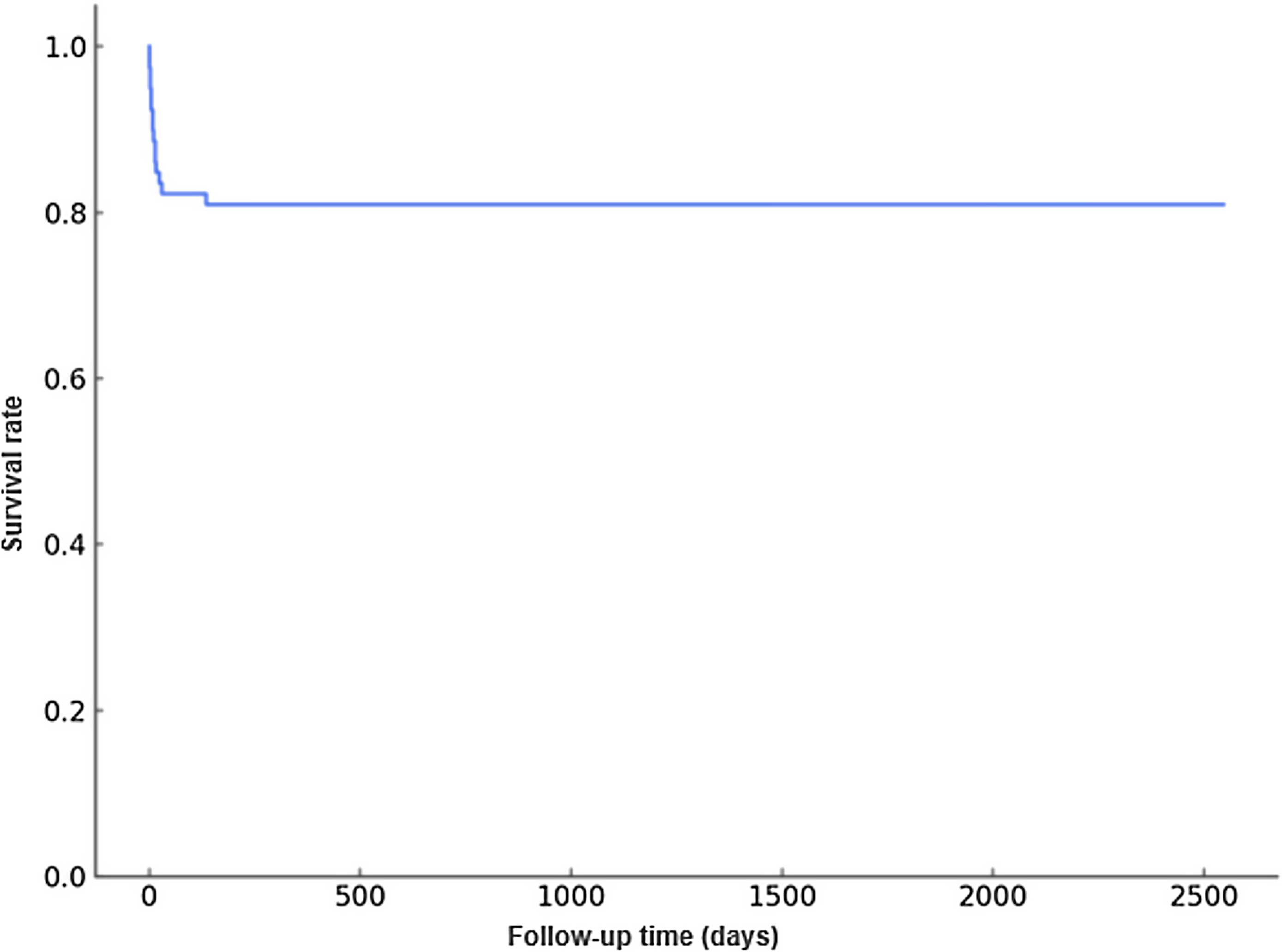
The Kaplan–Meier postnatal survival curve (Figure 2), which excludes stillbirths, shows a steep decline during the first month of life, followed by stabilisation at around 81% after the first year. The curve was truncated when the number at risk fell below five patients, with survival stability observed throughout the remainder of follow-up.
Kaplan–Meier curve of postnatal survival. Kaplan–Meier curve of postnatal survival in the group of 79 live-born patients. One stillbirth was excluded. The curve was truncated when the number at risk was <5. Surviving patients were censored at the end of the study (March 31, 2025).

Discussion
In our series of prenatally diagnosed CHDs, we found a distribution of diagnoses consistent with that described in the literature. Transposition of the great arteries, hypoplastic left ventricle syndrome, and coarctation of the aorta were the most prevalent entities, findings consistent with international reports identifying these malformations as having the greatest impact on neonatal morbidity and mortality. Reference Li, Zhou, Fang, Wang, Hua and Mu6 Transpositions of the great vessels not associated with another defect should be operated on within the first two weeks of life to achieve favourable results in the short and medium term. Reference Carlos Smith, Millaray Brito, Manuel Álvarez, Alejandro Álvarez, Sergio De La Fuente and Juan G.Rodríguez4,Reference Uricchio, Ghiselli and Marianeschi7,Reference Blyth, Howe, Gnanapragasam and Wellesley8
When compared with the national experience of CERPO, Reference Mayorga, Rodríguez and Enríquez5 where the most frequent diagnoses corresponded to interventricular communication, left ventricular hypoplasia, arrhythmias, and atrioventricular canal defect, our series showed a higher concentration of highly complex lesions and univentricular physiology. In this study, we present our experience with moderate and complex defects, leaving out minor defects that are not amenable to early surgical intervention and, in our opinion, do not benefit as much from antenatal diagnosis in their management after birth. This is unlike moderate and complex heart defects, which, with adequate diagnosis and management in the fetal period, can achieve significant improvements in mortality and in the quality of life of patients. The antenatal diagnosis of moderate and complex CHD in our setting presents many benefits for management in the neonatal period, early surgery, and survival of our patients, unlike publications that indicate that there would be no impact on the results. Reference Khoshnood, Lelong and Houyel9
The frequency of chromosomal abnormalities is within the range previously reported in the literature. Reference Mayorga, Rodríguez and Enríquez5,Reference Galindo, Gratacós and Martínez10 The most consistent association was between trisomy 21 and the common atrioventricular canal defect, which reaffirms the classic relationship between this chromosomal disorder and endocardial cushion defects. Reference Muñoz, Copado, Díaz, Muñoz, Enriquez and Aguilera11 The identification of less prevalent syndromes, such as Klinefelter or DiGeorge, underscores the need to move towards more detailed and less invasive genomic studies in clinical practice.
In our country, abortion has been legally permitted since 2017 under only three circumstances. One of these circumstances is lethal fetal inviability, when the embryo or fetus suffers from a congenital or genetic condition incompatible with independent life outside the womb. This procedure is available to mothers with affected fetuses who meet the legal requirements. The mother can choose to have an abortion or continue the pregnancy. Reference Sarria Cárcamo12
One-third of pregnant women had comorbidities, with diabetes being the most common condition, consistent with international evidence linking this condition with an increased risk of cardiovascular malformations. It is worth noting that, unlike other publications, hypothyroidism emerged as a relevant maternal comorbidity in our series, being very common in our study, consistent with other studies in our setting. Reference Mosso, Martínez and Rojas Mía13
Regarding prognosis, overall survival reached 78.8% at the end of follow-up. This figure contrasts with the 44% reported in the CERPO experience. Reference Mayorga, Rodríguez and Enríquez5 The differences could be explained by several factors: improvements in neonatal cardiovascular surgery over the last decade and, to a lesser extent, methodological variations in follow-up. The Kaplan–Meier curve showed a pronounced decline during the first month of life, followed by stability, which is consistent with the international literature, which indicates that most deaths are concentrated in the neonatal stage.
Although this study does not attempt to estimate population prevalence, the findings reinforce the burden represented by moderate and complex CHDs, precisely those with the highest mortality rates. The information obtained updates the national characterisation of CHDs, highlighting the importance of prenatal diagnosis to organise perinatal care and optimise the surgical and neurodevelopmental outcomes of our patients. Reference Peyvandi, De Santiago and Chakkarapani14 Ideally, all prenatal diagnostic units should have a paediatric and fetal cardiologist, which has improved the diagnosis of these heart defects, thus offering deliveries in a tertiary perinatal centre with a trained cardiosurgical team experienced in the surgical treatment of these moderate and complex cardiac malformations in the neonatal period. Reference Carvalho, Axt-Fliedner and Chaoui15–Reference Van Velzen, Clur and Rijlaarsdam17
We must mention the importance of creating a national registry in each country, including both paediatric and adult patients with CHD, and with a long-term focus. However, in Chile, there is still no law regulating the registration and follow-up of CHD.
Finally, the fact that a subgroup of patients remains outside the scope of surgery underscores the serious nature of fetal CHD and the need to strengthen screening and referral networks. Consolidating specialised regional programmes is emerging as a priority challenge to reduce survival and quality of life gaps in CHD.


 Open access

