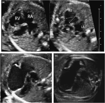
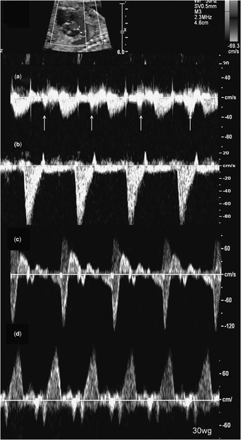
A 22-year-old woman was referred to our foetal cardiology unit for cardiomegaly at her 21st week of gestation. The initial echocardiogram showed dilatation of both the right atrium and ventricle and small muscular ventricular septal defects. The inter-ventricular septum was shifted towards the left ventricle with paradoxical movement. The tricuspid valve was difficult to assess. There was no tricuspid regurgitation but a swirling flow in the dilated right atrium. The tricuspid septal and posterior leaflets were thickened with insertion into a hypertrophic septal papillary muscle. This image was initially mistaken for an incomplete delamination of the septal leaflet (Fig 1a left). However, careful examination of the valve retrospectively demonstrated a normal insertion (Fig 1a right). The right ventricular inferior free wall below the tricuspid annulus was subtly thinner than normal (Fig 1a left). Flow in the ductus venosus and the arterial duct was normal (Fig 2b). Doppler interrogation in the main pulmonary artery showed diastolic ejection (Fig 2a).
( a ) At 21 WG, the right myocardium is thin with hypertrophied septal papillary muscle (arrow) mimicking septal attachment of the tricuspid septal leaflet (left) and normal insertion of the septal leaflet (right). ( b ) At 27 WG, the right ventricular free wall is thinner from the annulus to the apex. ( c ) At 30 WG, the anterior wall is thinner than the septum with a suggestive aspect of parchment and pericardial effusion. LA=left atrium; RA=right atrium; RV=right ventricle.

( a ) At 21 WG, diastolic ejection in the pulmonary artery (arrows) secondary to equalisation of right ventricular and right atrial pressures and poor right ventricular compliance. The atrial contraction is then transmitted to the pulmonary artery. ( b ) Normal flow in the ductus arteriosus. ( c ) At 27 WG, telesystolic and holodiastolic retrograde flow in the ductus arteriosus. ( d ) At 30 WG, trivial antegrade flow in protosystole and mid-diastole, the remaining flow through the ductus is retrograde from the aorta.

At 27 WG, the right ventricular free wall was clearly abnormally thin from the tricuspid annulus to the apex (Fig 1b), with decreased systolic function. There was mild-to-moderate tricuspid regurgitation. Flow in the ductus arteriosus was bidirectional in systole and diastole (Fig 2c).
At 30 WG, biventricular systolic function was severely impaired with sinus tachycardia, foetal hydrops, and circumferential pericardial effusion. The right ventricular free wall was homogeneously thin from the annulus to the apex, whereas septal thickness remained normal (Fig 1c). Antegrade pulmonary blood flow was almost absent with predominant retrograde flow in the ductus arteriosus and a pronounced retrograde A wave in the ductus venosus (Fig 2d). The foetus demised at 30+5 days. The parents refused autopsy.
Uhl’s anomaly was initially described as an almost total absence of the myocardium of the right ventricle, leading to global dilatation and a parchment-like appearance of the right ventricular free wall. 1 Only five foetal cases have been reported as of now with only one postnatal survivor.Reference Wager, Couser, Edwards, Gmach and Bessinger 2 – Reference Ikari, Azeka, Aiello, Atik, Barbero-Marcial and Ebaid 7 On the basis of our patient, changes described in Uhl’s anomaly occur progressively, as the abnormally thin aspect of the anterior wall was only mild at 20 weeks, making the initial diagnosis somewhat difficult. A progressive thinning of the anterior wall starting at the inlet portion and progressively reaching the apex was later observed. The thinning spared the inter-ventricular septum, but the latter showed an abnormal contractility with paradoxical movement. Simultaneously, the right ventricular haemodynamic changed with increased filling pressures, tricuspid regurgitation, and decreased output. Interestingly, at 21 weeks, a diastolic ejection wave was noticed on the pulmonary artery Doppler, which indicates an early equalisation of the right atrial and ventricular diastolic pressures.
Several diagnoses should be kept in mind in front of a dilated right ventricle in a foetus, the main differential diagnosis being Ebstein’s anomaly. In our patient, a hypertrophied papillary muscle was initially misdiagnosed as a displaced insertion of the septal leaflet. In addition, blood coming from the inferior caval vein had a swirling movement in the dilated right atrium, which could be mistaken for tricuspid regurgitation. This made the differential diagnosis between Uhl’s anomaly and Ebstein’s anomaly challenging, especially at the early stage when the right ventricular thinning was mild. Moreover, significant tricuspid regurgitation due to RV dilatation can appear later in the evolution of the disease and the tricuspid valve has been described as abnormal in Uhl’s anomaly, with redundant leaflets directly attached on thickened papillary muscles.Reference Wager, Couser, Edwards, Gmach and Bessinger 2 , Reference Cardaropoli, Russo and Paladini 6 Careful examination of the tricuspid insertions, focused on the septal leaflet, should help make the difference between these two diagnoses. The prenatal counselling will then be different, with Uhl’s anomaly having a worse prognosis. Some forms are probably less extensive as Uhl’s anomaly has been described in adults.Reference Hébert, Duthoit and Hidden-Lucet 8 , Reference Song 9 These partial forms could be underestimated because of their high tolerability, and most of the cases described in adults are diagnosed fortuitously on angiography or autopsy.Reference Hébert, Duthoit and Hidden-Lucet 8 , Reference Song 9
The pathophysiology of Uhl’s anomaly is still controversial and the border with entities showing a degenerative aspect of the right ventricular free wall-like arrhythmogenic right ventricular dysplasia remains difficult to establish. The typical histological description in Uhl’s anomaly is a transparent right ventricular free wall due to an aposition of the endocardium with the epicardium without intervening myocardium, infiltration, or inflammation. 1 , Reference Uhl 10 In arrhythmogenic right ventricular dysplasia, a fatty infiltrate in the myocardium is the classic pattern.
Several hypotheses have been stated to explain the absence of right ventricular myocardium. The description of our patient goes against failure of development during embryonic life.Reference Uhl 10 We clearly saw that the myocardium, initially only slightly abnormal, progressively disappeared during the evolution. The hypothesis of apoptosis has been proposed by James et alReference James, Nichols, Sapire, DiPatre and Lopez 5 . Apoptosis is characteristically a brief process in which the dying cell is rapidly engulfed by macrophages or adjacent cells without an inflammatory response. Uhl’s anomaly could be the result of an apoptotic process failing to stop and becoming extensive and destructive.
The parents refused autopsy for our patient. However, the echographic evolution with marked thinning of the right ventricular free wall over time is pathognomonic and leaves no doubt regarding the diagnostic.
Conclusion
This foetal patient extensively illustrates an extremely rare pathology, Uhl’s anomaly. This is an evolutive disease with progressive right ventricular free wall thinning leading to anterior wall myocardial dysfunction and terminal heart failure. The phenomenon of apoptosis could be an explanation for this rapid degeneration. Early diagnosis remains difficult to establish as the disease can be incomplete at first echocardiography.