Introduction
Takayasu arteritis is a rare disease characterised by the non-specific large vessels inflammation including the aorta, its main branches and pulmonary arteries. Takayasu arteritis is a global disease, but it occurs mainly in Southeast Asia. In China, the prevalence was 7.01 cases per million. Reference Sun, Yin and Ma1 Most patients are women of childbearing age. According to studies from Japan, the incidence of Takayasu arteritis involving the coronary artery varied from 9 to 10.5%. Reference Lupi-Herrem, Sanchez-Torres and Marcushamer2,3 The damage of Takayasu arteritis to the coronary artery can lead to coronary stenosis and occlusion, angina pectoris, and even acute myocardial infarction. With delayed diagnosis and the difficulty of treatment, Takayasu arteritis patients complicated with coronary abnormalities usually have poor prognosis. In this case report, we present a rare case of heart failure in a young woman due to total occlusion of the left main coronary artery, caused by Takayasu arteritis.
Case report
A 30-year-old female patient was admitted to our hospital for aggravated dyspnoea, accompanied by orthopnea. Five days before admission, She presented with paroxysmal dyspnoea at night, and intermittent chest pain that lasted for a few minutes and resolved spontaneously. The patient had experienced chest pain 2 years prior, which generally occurred after physical activity. She visited many hospitals and received no specific treatment.
Physical examination revealed a body temperature of 36°C, respiratory rate of 24 bpm, and blood pressure of 132/106 mmHg in the left upper limb, 176/100 mmHg in the right upper limb, 182/105 mmHg in the left lower limb, and 170/108 mmHg in the right lower limb. Moist rales were monitored during auscultation of both lungs. The apical beat was located in the area 0.2 cm medial to the left clavicular midline in the fifth intercostal space. The heart rate was 110 bpm with a regular rhythm. Electrocardiogram suggested sinus tachycardia and ST-segment depression and T-wave inversion in leads V4–6 (Supplement Figure 1). Upon hospital admission, the patient received chest CT angiography, which revealed obvious thickening in the aortic wall, rough internal wall, and local occlusion in the proximal end left subclavian artery (Fig 2). Laboratory parameters indicated a significant increase in the erythrocyte sedimentation rate (64 mm/h) and C-reactive protein (24 mg/l) level (Supplement Figure 2).
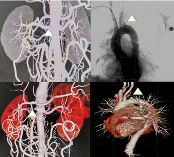
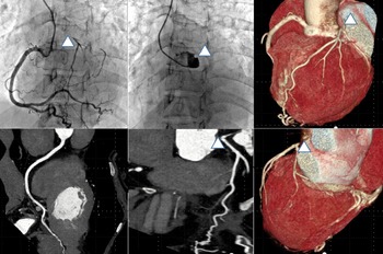
The patient underwent invasive aortic angiography, with the discovery of total occlusion at the proximal segment of the left subclavian artery (Fig 1b). Abdominal CT angiography indicated thickening of multiple vascular walls and lumen stenosis at the abdominal aorta, superior mesenteric artery, coeliac trunk, and origin of the right renal artery (Figure1 A and C). The patient underwent invasive coronary angiography and coronary CT angiography. They both indicated right-dominant type, occlusion the ostia of left main coronary artery and right coronary artery lumen enlargement with a secondary collateral branch to the left cronory artery (Fig 2) .The patient underwent echocardiography and was determined to have a slight enlargement in the left atrium and left ventricle, aortic regurgitation (mild-moderate), mitral regurgitation (mild-moderate), tricuspid regurgitation (mild-moderate) and pulmonary hypertension (mild-moderate). Also, left ventricular systolic dysfunction (EF = 45%) and pericardial effusion (a small amount) were identified. Vascular ultrasound was performed, which detected stenosis of multiple arteries and abnormal blood flow that covered the right renal artery and left upper extremity artery. Kawasaki disease was ruled out because of the absence of coronary artery aneurysm and typical childhood history. The patient was a young woman and lacked cardiovascular risk factors, which indicated a low possibility of premature atherosclerosis. Considering these clinical and imaging characteristics, the patient was diagnosed with Takayasu’s arteritis according to the Ishikawa criteria (1988). The initial drug treatment plan was as follows: (1) heart failure treatment: furosemide (20 mg, qd), spironolactone (20 mg, qd), β-blocker metoprolol succinate sustained-release tablets (23.75 mg, qd); (2) antiplatelete treatment and statin: aspirin enteric-coated tablets (100 mg, qd), atorvastatin (20 mg, qd); and (3) intravenous methylprednisolone (1000 mg/day for 5 days, followed by 40 mg, qd).
Right renal artery CT angiography and aortic angiography. Triangle showed the occlusion of the left subclavian artery and right renal artery.

CT angiography and invasive angiography for coronary. triangle showed the occlusion.

The patient’s symptoms began to resolve 1 week later. NT-proBNP decreased gradually. Meanwhile, the CRP and ESR levels gradually decreased to the normal range 2 weeks later (Supplement Figure 3). The patient was not a candidate for surgery because of the active stage of inflammation. Before discharge, we switched from intravenous methylprednisolone to oral methylprednisolone tablets, with simultaneous treatment using mycophenolate mofetil capsules (0.5 g, bid). Sacubitril valsartan sodium tablets (50 mg, bid) was added to prevent heart failure. Before discharge, her blood pressure was 131/70 mmHg with clear breath sounds in both lungs, no abnormal beat in the precordial area, a heart rate of 62 bpm, regular rhythm of the heart, no cardiac murmur, and no oedema of the lower limbs.
In the 2-year follow-up, the treatment improved the symptoms and cardiac function and maintained normal inflammatory markers (Supplement Figure 2, 3).
Discussion
Takayasu arteritis is a rare disease occurring primarily in young Asian women. There is no established specific biomarker and initial manifestations for the diagnosis of Takayasu arteritis. It poses a great diagnostic challenge, especially in early onset or stable stage. Cardiovascular symptoms are especially easy to miss. For chest pain symptoms, cardiologists mainly consider whether patients have coronary atherosclerotic disease and neglect myocardial ischaemia from other causes. And, coronary atherosclerotic heart disease is rare in these young women. Doctors prefer to attribute the chest pain of young woman to cardiac neurosis and microcirculation ischaemia. Therefore, no further examinations are given to them. Stenosis and occlusion developed in the coronary artery as the result of the long-lasting inflammation, which can lead to severe cardiovascular events, such as angina, myocardial infraction, and heart failure. In this case, the patient was admitted to the emergency because of heart failure with 2 years repeated chest tightness and chest pain. It is time to pay attention to the young women with chest distress or pain. We should at least give them equal attention, especially for patients with typical symptoms of myocardial ischaemia. In a retrospective analysis from Xiaofeng Zeng et al, Reference Li, Li and Sun4 angina pectoris (14/164, 8.5%) and myocardial infarction (3/164, 1.8%) were the most common clinical manifestations in coronary artery involvement group. No pulse or various types of ischaemic symptoms are clues to diagnosis. CT angiography and MRI of large artery are recognised as the gold standard for Takayasu arteritis’s diagnosis. Reference Hellmich, Agueda and Monti5 It is suggested that coronary angiography be performed immediately in the case of suspected Takayasu arteritis patients with coronary artery involvement.. A series of modern imaging methods might facilitate earlier diagnosis and play an important role in monitoring of arterial injury, including high-resolution colour doppler ultrasound, cardiovascular MRI, and 18F-fluorodeoxyglucose positron emission tomography. There are three keys can be used to distinguish Takayasu arteritis-involved coronary artery from Takayasu arteritis complicated with coronary artery disease. First, onset age of cardiac symptoms of the fomer was significantly younger, and most of the patients are women. Besides, the stenosis and occlusion of coronary of the former were commonly located in the ostial and proximal parts. Third, the latter often accompanied with risk factors for atherosclerosis, which are unusual in the former.
Takayasu arteritis is a chronic chronic inflammatory disease. However, systemic inflammatory response does not always show a positive correlation with inflammatory activity in the vessel wall. In the absence of systemic inflammation, Takayasu arteritis’s vascular injury continues to progress. The resulting myocardial ischaemia is one of the main causes of death related to Takayasu arteritis with coronary lesion. Considering the above, the diagnosis and treatment of coronary artery involvement in the early stage, especially before occlusion, are of great significance for reducing Takayasu arteritis patients' mortality. Once Takayasu arteritis is diagnosed, combined therapy of hormone and immunosuppression should be administered as soon as possible. The treatment not only control the progress of large vessel vasculitis but also the myocardial inflammatory infiltration. Antiplatelet treatment for Takayasu arteritis is controversial. European League against Rheumatism for managing Large Vessel Vasculitis (2018) Reference Hellmich, Agueda and Monti5 suggested that antiplatelet or anticoagulant therapy should not be routinely used for the treatment of Takayasu arteritis. But, it can be considered for coronary heart disease and cerebrovascular disease. Antiplatelets play a pivotal role in atherosclerotic coronary artery disease. However, a high-quality study is lacking on whether to use antiplatelet drugs for Takayasu arteritis patients with coronary artery involvement. Considering the side effects and low level of evidence, it should be used more carefully. In rheumatoid arthritis, for example a recent meta-analysis of the available studies showed that statin therapy may reduce disease activity and improve symptoms control by suppressing inflammatory response. Reference Li, Zhao and Li6 However, relapse-free survival was not significantly different in Takayasu arteritis patients treated with statins or aspirin. Reference Laurent, Prieto-González and Belnou7 And, cardiovascular risk factors in Takayasu arteritis could be associated with increased risk of ischaemic complications.
For Takayasu arteritis patients with heart failure, anti-heart failure therapy may alleviate or improve heart function and prevent the occurrence and development of heart failure. Except for emergency ischaemic events, surgical treatment for patients in the active stage is not recommended as it may increase the risk of complications and reduce the rate of vascular patency. Major intervention procedures include endovascular surgery and vascular bypass surgery to repair vascular abnormalities. To be specific, vascular bypass surgery is a traditional surgical choice suitable for patients with long-segment, complex, and severe stenosis/occlusion, and it exhibits a good long-term patency rate. The intravascular intervention has the advantage of being minimally invasive. However, stent placement during the percutaneous coronary intervention may stimulate intimal hyperplasia of the coronary artery, resulting in a relatively high long-term restenosis rate. Reference Furukawa, Tamura and Toma8 Considering the LM opening’s complete occlusion, vascular bypass surgery would be the first choice for this patient’s reperfusion treatment. However, given the surgery risk and the improvement of symptoms, the patient and her family ultimately refused operation. Monitoring disease activity and flow-up in Takayasu arteritis should be accomplished by the integrated use of noninvasive imaging methods. During the subsequent follow-up, the patient’s cardiac function had recovered well, and her left ventricular ejection fraction was improved, as indicated by a review using echocardiography. Over the 2-year follow-up, our case showed no new stenotic pathologic change of coronary evaluated by CT angiography.
TA involving the coronary artery has a more insidious onset is easily misdiagnosed and causes serious complications. We must pay more attention to the young women with chest pain. To coronary complications, large-scale randomised controlled trials are required in the future to evaluate the treatment strategy.
Supplementary material
To view supplementary material for this article, please visit https://doi.org/10.1017/S1047951123000835