Introduction
Zika virus (ZIKV) is an arthropod-borne virus in the genus Flavivirus and the family Flaviviridae. ZIKV is most closely related to the Spondweni virus and other mosquito-borne viruses, such as dengue (DENV), yellow fever (YFV), West Nile (WNV), Japanese encephalitis, and chikungunya virus (CHIKV).[ Reference Lanciotti, Kosoy, Laven, Velez, Lambert, Johnson, Stanfield and Duffy 1 ] ZIKV was first isolated in a Rhesus monkey that was used as a sentinel animal for yellow fever research in the Zika forest of Uganda. The second time the virus was isolated took place in early 1948 and involved an Aedes africanus mosquito in the same forest.[ Reference Dick, Kitchen and Haddow 2 ] The first case of ZIKV isolation in humans occurred in 1954 during an epidemic of jaundice in Nigeria.[ Reference Macnamara 3 ] According to the phylogenetic and geographical analysis, ZIKV can be categorized into three lineages, the East Africa lineage, the West Africa lineage, and the Asia lineage.[ Reference Haddow, Schuh, Yasuda, Kasper, Heang, Huy, Guzman, Tesh and Weaver 4 ] The introduction of ZIKV from Uganda to West Africa and Malaysia may have occurred in the 1940s.[ Reference Faye, Freire, Iamarino, Faye, de Oliveira, Diallo, Zanotto and Sall 5 ] Although ZIKV has been introduced across a wide geographical range, only sporadic cases of human infection have been reported in Africa (e.g., Senegal,[ Reference Monlun, Zeller, Le Guenno, Traoré-Lamizana, Hervy, Adam, Ferrara, Fontenille, Sylla, Mondo and Digoutte 6 , Reference Mccrae and Kirya 7 ] Sierra Leone,[ Reference Robin and Mouchet 8 ] Gabon,[ Reference Grard, Caron, Mombo, Nkoghe, Mboui Ondo, Jiolle, Fontenille, Paupy and Leroy 9 , Reference Jan, Languillat, Renaudet and Robin 10 ] Central African Republic,[ Reference Saluzzo, Gonzalez, Hervé and Georges 11 ] Ivory coast,[ Reference Akoua-Koffi, Diarrassouba, Bénié, Ngbichi, Bozoua, Bosson, Akran, Carnevale and Ehouman 12 ] and Egypt[ Reference Smithburn, Taylor, Rizk and Kader 13 ]) and Asia (e.g., Indonesia,[ Reference Olson, Ksiazek, Suhandiman and Triwibowo 14 ] Pakistan,[ Reference Darwish, Hoogstraal, Roberts, Ahmed and Omar 15 ] Malaysia,[ Reference Marchette, Garcia and Rudnick 16 ] India,[ Reference Smithburn, Kerr and Gatne 17 ] Thailand, North Vietnam,[ Reference Pond 18 ] and the Philippines[ Reference Hammon, Scurack and Sather 19 ]). For example, 6.1% of the residents in regions of Uganda were shown to possess specific anti-ZIKA antibodies.[ Reference Dick 20 ] Forty percent of Nigerians exhibited the ZIKV neutralizing antibody [ Reference Fagbami 21 ]; in Senegal, 10.1% and 2.8% of humans had IgM antibodies against ZIKV in 1988 and 1990,[ Reference Diallo, Sall, Diagne, Faye, Faye, Ba, Hanley, Buenemann, Weaver and Diallo 22 ] respectively. Underreporting or asymptomatic infection may account for the large difference between the number of reported infections and the number of individuals with antibodies against ZIKV. In 2007, an outbreak was reported on Yap Island in the Federated States of Micronesia.[ Reference Duffy, Chen, Hancock, Powers, Kool, Lanciotti, Pretrick, Marfel, Holzbauer, Dubray, Guillaumot, Griggs, Bel, Lambert, Laven, Kosoy, Panella, Biggerstaff, Fischer and Hayes 23 ] Approximately 73% of the residents were infected; 18% of those infected exhibited clinical illness. This outbreak represents the first transmission of ZIKV outside Africa and Asia. In October 2013, a second outbreak of ZIKV was reported in French Polynesia,[ Reference Cao-Lormeau, Roche, Teissier, Robin, Berry, Mallet, Sall and Musso 24 ] with 19,000 suspected cases. Large outbreaks in New Caledonia[ Reference Dupont-Rouzeyrol, O'Connor, Calvez, Daures, John, Grangeon and Gourinat 25 ] (1400 confirmed cases), the Cook Islands (over 900 cases) and Easter Island[ Reference Roth, Mercier, Lepers, Hoy, Duituturaga, Benyon, Guillaumot and Souares 26 ] have also been reported. In addition to autochthonous cases, cases associated with travel have been reported. Infections have been imported to Norway,[ Reference Wæhre, Maagard, Tappe, Cadar and Schmidt-Chanasit 27 ] Japan,[ Reference Kutsuna, Kato, Takasaki, Moi, Kotaki, Uemura, Matono, Fujiya, Mawatari, Takeshita, Hayakawa, Kanagawa and Ohmagari 28 ] and Italy[ Reference Zammarchi, Stella, Mantella, Bartolozzi, Tappe, Günther, Oestereich, Cadar, Munoz-Fontela, Bartoloni and Schmidt-Chanasit 29 ] from viremic people that traveled from French Polynesia. Infections have been imported to Canada[ Reference Fonseca, Meatherall, Zarra, Drebot, MacDonald, Pabbaraju, Wong, Webster, Lindsay and Tellier 30 ] and Germany[ Reference Tappe, Rissland, Gabriel, Emmerich, Günther, Held, Smola and Schmidt-Chanasit 31 ] from viremic people who traveled from Thailand. ZIKV may have been imported to the Americas by recreational travelers[ Reference Summers, Acosta and Acosta 32 ] to Brazil;[ Reference Musso 33 , Reference Zanluca, Melo, Mosimann, Santos, Santos and Luz 34 ] a plausible hypothesis for the arrival of ZIKV in Brazil involves travel associated with the 2014 World Cup. The lineage that emerged in the Pacific region and in Brazil were distinguished as the Asia lineage. The migration of humans and airline travel appear to facilitate the spread of ZIKV outside its typical geographical range.
ZIKV is a single-stranded RNA virus that exhibits positive sense. The RNA genome consists of 10,794 nucleotides,[ Reference Kuno and Chang 35 ] with a long open reading frame that is flanked by 5′ and 3′ non-coding regions. The open reading frame encodes three structural proteins (capsid, premembrane or membrane, and envelope) and five nonstructural proteins (NS1, NS2A, NS2B, NS3, NS4A, NS4B, and NS5).[ Reference Lindenbach and Rice 36 ] Most of the phylogenetic analysis on ZIKV has involved analysis of the envelope protein,[ Reference Lanciotti, Kosoy, Laven, Velez, Lambert, Johnson, Stanfield and Duffy 1 , Reference Dupont-Rouzeyrol, O'Connor, Calvez, Daures, John, Grangeon and Gourinat 25 , Reference Campos, Bandeira and Sardi 37 , Reference Faye, Faye, Dupressoir, Weidmann, Ndiaye and Alpha Sall 38 ] which is the major virion surface protein, or NS5,[ Reference Faye, Freire, Iamarino, Faye, de Oliveira, Diallo, Zanotto and Sall 5 , Reference Grard, Caron, Mombo, Nkoghe, Mboui Ondo, Jiolle, Fontenille, Paupy and Leroy 9 , Reference Buathong, Hermann, Thaisomboonsuk, Rutvisuttinunt, Klungthong, Chinnawirotpisan, Manasatienkij, Nisalak, Fernandez, Yoon, Akrasewi and Plipat 39 – Reference Balm, Lee, Lee, Chiu, Koay and Tang 41 ] which is the largest segment and is a well-conserved protein. NS5 contains an N-terminal component, which participates in synthesis of the 5′ cap and a C-terminal RNA-dependent RNA polymerase (RdRP).[ Reference Lindenbach and Rice 36 ]
Many types of sylvatic and peri-domestic mosquitos of the Aedes genus have been reported as major vectors for ZIKV, including Aedes africanus,[ Reference Dick, Kitchen and Haddow 2 ] Aedes aegypti,[ Reference Marchette, Garcia and Rudnick 16 ] Aedes polynesiensis,[ Reference Cao-Lormeau, Roche, Teissier, Robin, Berry, Mallet, Sall and Musso 24 ] Aedes luteocephalus, Aedes furcifer, Aedes taylori,[ Reference Diallo, Sall, Diagne, Faye, Faye, Ba, Hanley, Buenemann, Weaver and Diallo 22 ] Aedes apicoargenteus,[ Reference Mccrae and Kirya 7 ] Aedes albopictus,[ Reference Wong, Li, Chong, Ng and Tan 42 ] and Aedes hensilli.[ Reference Ledermann, Guillaumot, Yug, Saweyog, Tided, Machieng, Pretrick, Marfel, Griggs, Bel, Duffy, Hancock, Ho-Chen and Powers 43 ] The enzootic cycle of ZIKV and the broad range of hosts may facilitate recombination of ZIKV strains. Aedes africanus was the first species that was described as a vector for ZIKV; it has a wide dispersion among many animal species.[ Reference Faye, Freire, Iamarino, Faye, de Oliveira, Diallo, Zanotto and Sall 5 ] The main vectors in Asia were reported to be Ae. albopictus and Ae. aegypti; these species are widespread in tropical and subtropical regions. In addition, these species have a large presence in urban areas. In Europe, Ae. albopictus presents a major threat to public health.[ Reference Medlock, Hansford, Schaffner, Versteirt, Hendrickx, Zeller and Van Bortel 44 ] It is considered to be one of the most invasive mosquito species in the world; it is widely distributed in 20 European countries, particularly in the Mediterranean region. In Brazil, an where outbreak began in May 2015, both Ae. albopictus and Ae. aegypti are widely distributed.[ 45 ] Although Ae. hensilli and Ae. polynesiensis are the predominant species in the Federated States of Micronesia and French Polynesia, respectively, no direct evidence has been presented that they were associated with outbreaks; however, it is plausible that these species are vectors for ZIKV transmission.
Once the mosquitos take a blood meal from an affected host, the virus is transmitted from the midgut to salivary gland. The infection rate, dissemination rate, and transmission rate can be determined by the fraction of the mosquitos with infected midguts, by the number of infected salivary glands over the total number of mosquitos with infected midguts, and by the number of positive infected salivary glands over the number of infected salivary glands, respectively.[ Reference Wong, Li, Chong, Ng and Tan 42 ] When mosquitos take a blood meal from a human, transmission through skin immune cells, including dermal fibroblasts, epidermal keratinocytes, and immature dendritic cells has been demonstrated.[ Reference Hamel, Dejarnac, Wichit, Ekchariyawat, Neyret, Luplertlop, Perera-Lecoin, Surasombatpattana, Talignani, Thomas, Cao-Lormeau, Choumet, Briant, Despres, Amara, Yssel and Misse 46 ] Symptoms associated with ZIKV infection appear after an incubation period of a few days. The clinical symptoms associated with ZIKV infection include arthralgia, myalgia, headache, retro-orbital pain, conjunctivitis, cutaneous maculopapular rash, edema, and mild fever; severe fever has also been reported (>39° C).[ Reference Duffy, Chen, Hancock, Powers, Kool, Lanciotti, Pretrick, Marfel, Holzbauer, Dubray, Guillaumot, Griggs, Bel, Lambert, Laven, Kosoy, Panella, Biggerstaff, Fischer and Hayes 23 , Reference Zanluca, Melo, Mosimann, Santos, Santos and Luz 34 , Reference Foy, Kobylinski, Chilson Foy, Blitvich, Travassos da Rosa, Haddow, Lanciotti and Tesh 47 ] Some patients have also described symptoms such as malaise, anorexia, vomiting, diarrhea, and nausea.[ Reference Pinto, Luz, Parreira and Ferrinho 48 ] Most patients recover after 1 or 2 weeks; however, arthralgia can last for 1 month.[ Reference Foy, Kobylinski, Chilson Foy, Blitvich, Travassos da Rosa, Haddow, Lanciotti and Tesh 47 ] Although all of the symptoms mentioned above have been reported, a great portion of cases of infection are asymptomatic, which can lead to underdiagnosis and underreporting. ZIKV infection can be complicated by a neurological disorder, Guillain-Barré syndrome; this phenomenon was reported in French Polynesia.[ Reference Oehler, Watrin, Larre, Leparc-Goffart, Lastère, Valour, Baudouin, Mallet, Musso and Ghawche 49 ] Also, leukopenia and thrombocytopenia may be noted in laboratory measurements.[ Reference Gourinat, O'Connor, Calvez, Goarant and Dupont-Rouzeyrol 50 ] In addition to the human infections, infections of non-human primates, water buffalo, elephants, goats, hippos, impala, kongoni, lions, sheep, wildebeest, zebras, and rodents have been reported.[ Reference Darwish, Hoogstraal, Roberts, Ahmed and Omar 15 , Reference Henderson, Hewitt and Lule 51 – Reference Wolfe, Kilbourn, Karesh, Rahman, Bosi, Cropp, Andau, Spielman and Gubler 53 ] The symptoms associated with infection of various animals need further investigation.
Although the route of transmission is through mosquito bite, transmission of the ZIKV through perinatal transmission,[ Reference Besnard, Lastère, Teissier, Cao-Lormeau and Musso 54 ] sexual intercourse,[ Reference Foy, Kobylinski, Chilson Foy, Blitvich, Travassos da Rosa, Haddow, Lanciotti and Tesh 47 , Reference Musso, Roche, Robin, Nhan, Teissier and Cao-Lormeau 55 ] and blood transfusion[ Reference Musso, Nhan, Robin, Roche, Bierlaire, Zisou, Shan Yan, Cao-Lormeau and Broult 56 ] has been reported. The possible routes of perinatal transmission include transplacental, delivery, and breastfeeding routes. In the case of sexual intercourse, real-time reverse transcription PCR (rRT-PCR) results have indicated the presence of ZIKV in semen.[ Reference Musso, Roche, Robin, Nhan, Teissier and Cao-Lormeau 55 ] Transfusion-transmitted infection is theoretically possible with WNV[ Reference Pealer, Marfin, Petersen, Lanciotti, Page, Stramer, Stobierski, Signs, Newman, Kapoor, Goodman and Chamberland 57 ] and DENV[ Reference Stramer, Linnen, Carrick, Foster, Krysztof, Zou, Dodd, Tirado-Marrero, Hunsperger, Santiago, Munoz-Jordan and Tomashek 58 ] infections; the presence of CHIKV[ Reference Gallian, de Lamballerie, Salez, Piorkowski, Richard, Paturel, Djoudi, Leparc-Goffart, Tiberghien, Chiaroni and Charrel 59 ] in blood has also been suspected. Owing to the concurrent circulation of ZIKV with other arbovirus in a given area, co-infection with ZIKV and other arboviruses is a possibility. For example, co-infection of DENV and ZIKV has been reported in New Caledonia[ Reference Dupont-Rouzeyrol, O'Connor, Calvez, Daures, John, Grangeon and Gourinat 25 ]; however, the cross-reactivity and synergy between viruses are unknown and require additional study.
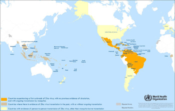
Owing to the recent outbreak of the ZIKV in multiple areas and the absence of specific antiviral therapy, a rapid and accurate characterization method for ZIKV is needed, particularly for regions that are undergoing outbreaks; a map of affected regions is shown in Fig. 1.[ 60 ] Novel materials may facilitate faster detection of ZIKV. For example, surface plasmon resonance and piezoelectric devices rely on advanced materials for sensitive detection of analytes. Novel materials for electrochemical sensing, laboratory on a chip-based sensing, lateral flow assay-based sensing, or optical sensing of ZIKV infection may facilitate more rapid detection and improved public health in affected regions. To date, detection of ZIKV has been based on viral isolation, serological identification, and molecular confirmation.[ 61 ] It is important to note that the similarity of ZIKV to other flaviviruses may hinder accurate virus identification (e.g., ZIKV may sometimes be misdiagnosed as DENV). In addition, the relationship between clinical symptoms and the presence of ZIKV can be more completely understood with the aid of more accurate detection methods.
Countries reporting Zika virus infection/outbreak, 2007–2016. Reprinted with permission from Ref. 60: World Health Organization (2016).

Viral isolation
Viral isolation in cell cultures has been recognized as the “gold standard” for viral detection and has been commonly performed since the 1960s.[ Reference Leland and Ginocchio 62 ] Viral isolation can be undertaken via intracerebral inoculation of mice, C6/36 cells (derived from Ae. albopictus), Vero cells (derived from the kidney epithelial cells of the African green monkey), or the AP 61 cell line (derived from Aedes pseudoscutellaris) with appropriate growth media and supplements. At the current time, researchers use Vero cell, C6/36 cell and AP 61 cell lines more frequently than intracerebral inoculation. Briefly, the samples are added to cell culture for incubation under controlled temperature and for an appropriate duration. Series passages can be performed to study the infectious capability of the virus. Microscopic identification of the unstained cell culture is the standard approach for examining viral proliferation. The presence of the virus causes degeneration in the cells; these changes are collectively called the cytopathic effect (CPE). However, there are some disadvantages to the viral isolation approach. Although viral isolation is not costly, it requires a long incubation period,[ Reference Faye, Faye, Diallo, Diallo, Weidmann and Sall 40 , Reference Leland and Ginocchio 62 ] which can be a few days; in addition, expertise may be needed to evaluate the CPE. It should be noted that the viremic stage is fleeting; it can cease at the time of or soon after the onset of symptoms; the viral load in the serum is often at a low level. Viral isolation with cell culture can be unsuccessful and is not practical for most clinical situations. As such, viral isolation is mainly utilized for research purposes.
Serological identification
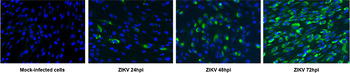
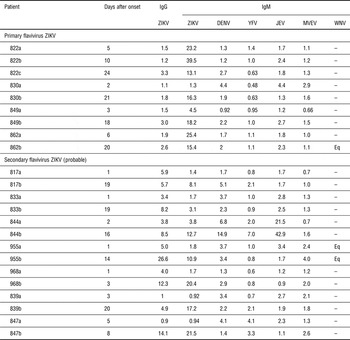
Serological identification is based on the interaction between antibodies and antigens. Enzyme-linked immunosorbent assay (ELISA) with plaque reduction neutralization test (PRNT), and immunofluorescence assay (IFA) are the most commonly used tests for ZIKV detection. An IFA for ZIKV-infected fibroblasts is shown as in Fig. 2.[ Reference Hamel, Dejarnac, Wichit, Ekchariyawat, Neyret, Luplertlop, Perera-Lecoin, Surasombatpattana, Talignani, Thomas, Cao-Lormeau, Choumet, Briant, Despres, Amara, Yssel and Misse 46 ] This type of assay was performed during the first outbreak on Yap Island. It should be noted that cross reactivity of antibodies between flaviviruses makes diagnosis using this approach challenging.[ Reference Lanciotti, Kosoy, Laven, Velez, Lambert, Johnson, Stanfield and Duffy 1 ] Data from patients at both the acute phase and the convalescent phase are shown in Table I. Patients with antibodies to more than one flavivirus show higher serological reactivity; this phenomenon is referred to as the “original antigenic sin.”[ Reference Lanciotti, Kosoy, Laven, Velez, Lambert, Johnson, Stanfield and Duffy 1 ] A complementary serological assay, PRNT, may needed for further confirmation of the ZIKV infection.[ Reference Fonseca, Meatherall, Zarra, Drebot, MacDonald, Pabbaraju, Wong, Webster, Lindsay and Tellier 30 , Reference Buathong, Hermann, Thaisomboonsuk, Rutvisuttinunt, Klungthong, Chinnawirotpisan, Manasatienkij, Nisalak, Fernandez, Yoon, Akrasewi and Plipat 39 , Reference Oehler, Watrin, Larre, Leparc-Goffart, Lastère, Valour, Baudouin, Mallet, Musso and Ghawche 49 ] Plaques can be counted and a plaque-forming unit (PFU) per unit volume can be calculated. Titers of neutralizing antibody to ZIKV and other flaviviruses with a cutoff value of 90% (PRNT90) can be determined. For confirmation of ZIKV infection, a titer of ZIKV greater than four times of the titer of other flaviviruses is recommended.[ Reference Duffy, Chen, Hancock, Powers, Kool, Lanciotti, Pretrick, Marfel, Holzbauer, Dubray, Guillaumot, Griggs, Bel, Lambert, Laven, Kosoy, Panella, Biggerstaff, Fischer and Hayes 23 ] Alternatively, ZIKV can be detected by indirect IFA using specific hyperimmune mouse ascitic fluid. A secondary antibody that binds to the primary antibody and causes an amplification of the signal can be used to provide enhanced sensitivity. It should be noted that low titers of immunoglobulin G (IgG) or immunoglobulin M (IgM) antibodies may be present during the early phase of infection.[ Reference Hayes 63 ] IgM antibodies may be found 3 days after the disease onset; IgG antibodies may be present in serum during the convalescent phase.[ Reference Pinto, Luz, Parreira and Ferrinho 48 ] In addition, PRNT and indirect IFA may also show cross reactivity associated with various flaviviruses.[ Reference Lanciotti, Kosoy, Laven, Velez, Lambert, Johnson, Stanfield and Duffy 1 ]
Primary human fibroblasts are susceptible to ZIKV. Primary fibroblasts infected with ZIKV (MOI =1) and mock-infected cells were analyzed at different times post-infection for the presence of the viral envelope protein by immunofluorescence with 4G2 MAb and FITC-conjugated anti-mouse IgG. Reprinted with permission from Ref. Reference Hamel, Dejarnac, Wichit, Ekchariyawat, Neyret, Luplertlop, Perera-Lecoin, Surasombatpattana, Talignani, Thomas, Cao-Lormeau, Choumet, Briant, Despres, Amara, Yssel and Misse46: American Society for Microbiology (2015).

Serological testing for heterologous flaviviruses of samples from ZIKV-positive patients, Yap State, Micronesia, 2007. Reprinted with permission from Ref. Reference Lanciotti, Kosoy, Laven, Velez, Lambert, Johnson, Stanfield and Duffy1: https://creativecommons.org/licenses/by/4.0/ (2008).

Ig, immunoglobulin; ZIKV, Zika virus; DENV, dengue virus type 1–4 mixture; YFV, yellow fever virus; JEV, Japanese encephalitis virus; MVEV, Murray Valley encephalitis virus; WNV, West Nile virus; -, negative. Eq, result in equivocal range of the assay. IgG and IgM testing was conducted by ELISA except for WNV, which was tested by microsphere assay; ELISA values are patient optical densities divided by negative control optical densities; <2, negative; 2–3 equivocal; >3 positive.
Molecular confirmation
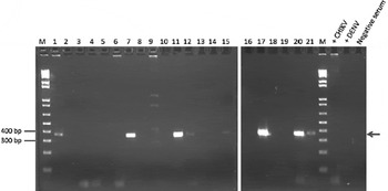
Since its development, PCR became a major tool for viral characterization. For ZIKV, RT–PCR can be performed to generate cDNA from RNA. RT–PCR can be categorized into traditional RT–PCR, real-time RT–PCR (quantitative), pan flavivirus RT–PCR, and nested RT–PCR. Full-ength sequences of ZIKV MR766[ Reference Kuno and Chang 35 ] and ZIKA EC 2007 were published in GenBank; these sequences were isolated from rhesus monkey in the Zika forest in 1947 and from an outbreak on Yap Island, respectively. The MR766 strain was most commonly used for RT–PCR; it is commonly compared with other strains since it was first isolated strain of ZIKV. For reverse transcription, the envelope or NS5 region are used as the target for primers. The primer is designed to be the counterpart to the nucleotides of the target segment. RNA is extracted from ZIKV, most commonly via the QIAamp viral RNA kit by means of centrifugation or a vacuum procedure. Viral RNA is then separated out using a QIAamp silica membrane. The mixture of ZIKV, primers, reverse transcriptase, DNA polymerase and buffer solution is added to the test tube and subject to thermocycling. The reverse transcriptase and DNA polymerase provide reverse transcription and DNA amplification functionalities, respectively. The amplicon size is determined by the forward primer and reverse primers. Rapid amplification of cDNA ends (RACE) kits, with a reverse primer for 5′-RACE and forward primer for 3′-RACE, can be used for amplification. Deoxynucleotide and buffer can be added for tailing to form a polynucleotide primer.[ Reference Kuno and Chang 35 ] The RT–PCR products can be analyzed using agarose gel electrophoresis. The pore size and strength of agarose gel make it an appropriate medium for DNA analysis. After applying voltage to the agarose gel, the nucleic acid migrates, with the velocity of migration being inversely proportional to the number of base pairs. The result can be visualized using colored dyes. ZIKV identification with a 364 bp amplicon is shown in Fig. 3.[ Reference Zanluca, Melo, Mosimann, Santos, Santos and Luz 34 ] Many groups utilize a one-step RT–PCR approach, which is suitable for a rapid diagnosis and is associated with less contamination than the two-step RT–PCR approach.
Agarose gel electrophoresis of RTPCR products for ZIKV detection. Reprinted with permission from Ref. Reference Zanluca, Melo, Mosimann, Santos, Santos and Luz34: Memorias do Instituto Oswaldo Cruz (2015).

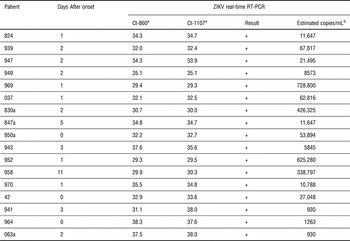
Real time RT–PCR (or quantitative RT–PCR) was considered to be the most powerful tool for molecular confirmation due to its high sensitivity and capability for quantitative analysis. During reverse transcription, a probe containing a reporter and a quencher is included with the primer. Emission of fluorescence from the probe occurs when the reporter is separated from the quencher by interaction with the primer; the optical density can subsequently be measured. In processing real time RT–PCR, the product of each PCR cycle is be recorded. The threshold cycle (Ct) versus the fluorescence is obtained during the course of the process; quantitative analysis can be obtained from a comparison of the Ct and a previously established standard curve. It is important to note that the Ct value is inversely proportional to the amount of RNA in the sample. Real-time RT–PCR results of samples from ZIKV-positive patients, Yap State, Micronesia, 2007 are shown in Table II.[ Reference Lanciotti, Kosoy, Laven, Velez, Lambert, Johnson, Stanfield and Duffy 1 ] In contrast, traditional RT–PCR is an end-point study that does not provide information during the process.
Threshold cycles and estimated copies/mL using real-time RT–PCR of samples from ZIKV-positive patients, Yap State, Micronesia, 2007. Reprinted with permission from Ref. Reference Lanciotti, Kosoy, Laven, Velez, Lambert, Johnson, Stanfield and Duffy1: https://creativecommons.org/licenses/by/4.0/ (2008).

RT–PCR, reverse transcription-PCR; ZIKV, Zika virus; Ct, crossing threshold; +, positive.
a Ct values with primer set 835/911c/860-FAM or 1086/1162c/1107-FAM. Values <38.5 are positive.
b Estimated by testing quantitated dilutions of ZIKV RNA transcripts and standard curve calculation generated by the iCycler instrument (Bio-Rad, Hercules CA, USA, see Methods).
A pan flavivirus RT–PCR or nested RT–PCR may sometimes be utilized. Pan flavivirus RT–PCR is performed with a degenerate primer. This degenerate primer can detect a range of flaviviruses; the notable trade-off in this approach is the loss of sensitivity. Nested RT–PCR requires an outer primer set, an inner primer set, and two successive PCRs. An outer primer or primary primer is targeted on a general area of the RNA, followed by the inner primer or nested primer that is targeted on a specific coding region. This approach can reduce the amplification of an unexpected binding site. Although RT–PCR is widely utilized and has many advantages, the highly specific primers and probes used in RT–PCR can pose a major drawback. It should be noted that there is a possibility of missing unanticipated sites, which may lead to failed amplification.
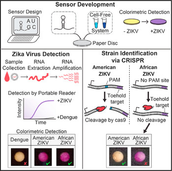
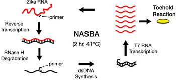
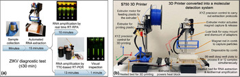
A molecular-based diagnostics method was recently proposed by Pardee et al., which offers a programmable sensor approach and paper-based detection (Fig. 4).[ Reference Pardee, Green, Takahashi, Braff, Lambert, Lee, Ferrante, Ma, Donghia, Fan, Daringer, Bosch, Dudley, O'Connor, Gehrke and Collins 64 ] The detection approach, which is anticipated to take 3 h, includes sample collection, RNA extraction, RNA amplification, and toehold reaction steps prior to colorimetric detection. A PCR approach was used to ligate the sensor to a LacZ reporter element. The sensor was embedded in paper, which was subsequently freeze dried. A reaction would occur in the presence of ZIKV trigger RNA, activating the LacZ gene for a colorimetric result. The result was associated with a change in the color of the paper from yellow (from chlorophenol red-β-D-galactopyranoside) to purple (from chlorophenol red). An electronic reader, which contained a 570 nm light-emitting diode source, was used to perform 29 readings from each sample; it provided low noise measurements of the color change. Boiling of the virus sample allowed for destruction of the ZIKV capsid and enabled a sufficient amount of RNA to be generated; this approach can be used in low resource envrionments and provides a straightforward alternative to traditional virus extraction. Use of nucleic acid sequence-based amplification (NASBA) with the paper-based system enabled Zika RNA to be distinguished from dengue RNA. NASBA-CRISPR cleavage (NASBACC), exploiting the Cas9 endonuclease enzyme, cleaves DNA if a specific protospacer adjacent motif (PAM) is present (Fig. 5).[ Reference Pardee, Green, Takahashi, Braff, Lambert, Lee, Ferrante, Ma, Donghia, Fan, Daringer, Bosch, Dudley, O'Connor, Gehrke and Collins 64 ] The clade (i.e., the branch of the viral family tree) that the ZIKV belongs to can be readily distinguished from the colorimetric results that are provided by the NASBACC approach. More recently, Chan et al. described use of magnetic particle (MP) and a consumer-grade three-dimensional (3D) printer for ZIKV RNA extraction and purification from ZIKV -spiked urine samples (Fig. 6).[ Reference Chan, Weaver, Wong, Lie, Wang, Guerbois, Vayugundla and Wong 65 ] Their device was shown to perform reverse transcription recombinase polymerase amplification and post-amplification detection (e.g., illumination by a light-emitting diode and analysis using a smartphone camera) of 8–12 urine samples in under 15 min. An absence of cross-reactivity with cross-reactivity with chikungunya and dengue was demonstrated using reverse transcription recombinase polymerase amplification assay and RT–PCR from the 3D printer-processed urine.
A schematic of sensor development, Zika virus detection, and strain identification via CRISPR. Reprinted with permission from Ref. Reference Pardee, Green, Takahashi, Braff, Lambert, Lee, Ferrante, Ma, Donghia, Fan, Daringer, Bosch, Dudley, O'Connor, Gehrke and Collins64: Elsevier (2016).

A schematic of NASBA (nucleic acid sequence based amplification)-mediated RNA amplification. Reprinted with permission from Ref. Reference Pardee, Green, Takahashi, Braff, Lambert, Lee, Ferrante, Ma, Donghia, Fan, Daringer, Bosch, Dudley, O'Connor, Gehrke and Collins64: Elsevier (2016).

(a) Rapid and low-cost diagnostics for ZIKV using a modified entry-level 3D printer. (b) Converting a low-cost 3D printer to perform rapid and automated nucleic acid isolation and real-time RPA amplification. Reprinted with permission from Ref. Reference Chan, Weaver, Wong, Lie, Wang, Guerbois, Vayugundla and Wong65: https://creativecommons.org/licenses/by/4.0/ (2016).

Discussion and conclusions
In comparing the three types of characterization methods, molecular confirmation is the most widely used method. Although viral isolation is the gold standard for detection, it is neither a time saving nor a labor saving approach. Serological identification can be hampered because of the cross-reactivity of flaviviruses. RT–PCR is a powerful tool for diagnosis of ZIKV infection and for genome sequencing analysis. The full sequence can be obtained using the genome walking approach. By Basic Local Alignment Search Tool (BLAST) analysis, a ZIKV strain can be compared with other flaviviruses or other ZIKV strains. A phylogenetic tree can be prepared, showing the clades of viruses; a potentially new strain of ZIKV can be identified as Asia lineage or Africa lineage. Real-time RT–PCR can provide quantitative analysis; it is less prone to contamination and error than agarose gel electrophoresis. However, the specificity of primer and probe should be taken into consideration. A finite mismatch between ZIKV MR766 strain and ZIKA EC 2007 strain led to a failed amplification of Yap Island virus during the initial analysis.[ Reference Lanciotti, Kosoy, Laven, Velez, Lambert, Johnson, Stanfield and Duffy 1 ] In addition to patient blood samples, saliva[ Reference Musso, Roche, Nhan, Robin, Teissier and Cao-Lormeau 66 ] and urine[ Reference Gourinat, O'Connor, Calvez, Goarant and Dupont-Rouzeyrol 50 ] samples were subjected to RT–PCR. ZIKV in both samples were detectable with higher loads; use of saliva samples did not increase the duration of detection window. ZIKV can be detected over 20 days after infection from collected urine samples. These two body fluids are useful if blood samples are difficult to collect (e.g., for potentially infected infants around the time of birth). In areas that lack medical facilities, non-invasive detection methods can are beneficial since they allow for collection of samples by patients. However, the onset of infection is sometimes difficult to define. A such, diagnosis with three kinds of samples is recommended.
In regions with concurrent circulation of ZIKV and other flaviviruses, multiple RT–PCR can be expensive and time consuming. Owing to the low availability of RT–PCR equipment and high cost of RT–PCR, Pardee et al.[ Reference Pardee, Green, Takahashi, Braff, Lambert, Lee, Ferrante, Ma, Donghia, Fan, Daringer, Bosch, Dudley, O'Connor, Gehrke and Collins 64 ] proposed a novel technology for molecular detection that enables rapid and low-cost diagnosis. This programmable and paper-based sensor does not require interpretation using laboratory equipment. With NASBA, a result with high sensitivity can be provided even in low resource regions. NASBACC can provide genetic information on the virus. As noted by Meagher et al., reverse transcription recombinase polymerase amplification and reverse transcription loop-mediated isothermal amplification can be used for highly sensitive detection of RNA with crude (minimally-processed) samples such as heat-treated blood.[ Reference Meagher, Negrete and Van Rompay 67 ] A combination of a liposome-based lateral flow assay and NASBA or a liposome-based immunoassay may also provide another avenue for rapid detection of ZIKV in clinical samples.[ Reference Bhalla, Jolly, Formisano and Estrela 68 ]
Owing to the ongoing ZIKV outbreak and the absence of either a vaccine or a curative treatment, a rapid mechanism for diagnosis of ZIKV infection is urgently needed. An inaccurate diagnosis may misguide individuals and may either underestimate or overestimate the incidence of infection in a given region. The cost and availability of various diagnostic methods should also be considered. Viral isolation is time consuming and serological identification may be hampered by cross reactivity with other viruses. Compared with viral isolation and serological identification, RT–PCR provides a rapid and sensitive approach to diagnose ZIKV infection; however, specific primers and probes may fail to provide appropriate amplification. A recently developed programmable and paper-based detection provides a low-cost and rapid detection method. A major challenge with current diagnostic approaches is that infection with another flavivirus (e.g., DENV) may be misdiagnosed as ZIKV infection.
More sensitive and accurate diagnosis methods are needed for faster treatment of affected individuals. With the development of novel biosensor materials, better detection accuracy, miniaturization of devices, increased stability, and a decrease in cost can be realized.[ Reference Bhalla, Jolly, Formisano and Estrela 68 , Reference Sin, Mach, Wong and Liao 69 ] The key features of a biosensor (e.g., low-power consumption, short assay time, selectivity, sensitivity, stability, and reproducibility) are primarily determined by the component materials.[ Reference Bhalla, Jolly, Formisano and Estrela 68 , Reference Sin, Mach, Wong and Liao 69 ] Use of nanomaterials may enable detection of ZIKV infection with smaller samples. As the analyte and the detector become closer in size, a reduction in non-specific binding and single molecule detection become possible.[ Reference Bhalla, Jolly, Formisano and Estrela 68 ] Nanoplasmonic sensors may enable group-specific antibodies in rapidly evolving viruses to be detected.[ Reference Sin, Mach, Wong and Liao 69 ] It should be noted that conversion of fluids obtained from a blood draw or a urine collection into biosensor samples may complicate use of new biosensor technologies.[ Reference Sin, Mach, Wong and Liao 69 ] Developing portable biosensors that are stable for at least 6 months and stable at high temperatures remains a significant challenge.