




No CrossRef data available.
Article contents
The SRA gene: the key to understanding the nature of Trypanosoma brucei rhodesiense
Published online by Cambridge University Press: 06 May 2005
Abstract
An abstract is not available for this content. As you have access to this content, full HTML content is provided on this page. A PDF of this content is also available in through the ‘Save PDF’ action button.
Information
- Type
- Review Article
- Information
- Copyright
- 2005 Cambridge University Press
INTRODUCTION
The discovery of the gene for human infectivity in Trypanosoma brucei rhodesiense (De Greef et al. 1989; Xong et al. 1998) ranks as one of the most significant landmarks in almost 100 years of research on this deadly parasite. The story of the discovery of that gene is as remarkable and as full of twists and turns, including the accidental infection of one of the investigators, as any at the dawn of tropical medicine, but here I want to focus on the impact of this important discovery on our understanding of the epidemiology of human trypanososomiasis.
Distinguishing Trypanosoma brucei rhodesiense from T. b. brucei
Trypanosoma brucei rhodesiense is the causative agent of rhodesian sleeping sickness, which is the acute form of human trypanosomiasis found in discrete foci throughout East Africa (Table 1). The prevalence of the disease is currently rated as moderate in Mozambique, Tanzania and Uganda, and there are also endemic foci in Kenya, Malawi, Zambia, and possibly also in Botswana, Burundi, Ethiopia and Rwanda (http://www.who.int/mediacentre/factsheets/fs259/en/). Trypanosoma brucei rhodesiense was first recognized in Northern Rhodesia (now Zambia) in 1910 (Stephens & Fantham, 1910) and the course of the disease was reported to be different from that caused by T. b. gambiense, the only human infective trypanosome known in Africa at the time (Yorke, 1910). The role of wild animals as reservoir hosts of T. b. rhodesiense was clear from the start, because rhodesian sleeping sickness occurred as an occupational hazard of hunters and other visitors to areas devoid of human habitation (Ashcroft, 1959; Apted, 1962). Indeed, this is still the case today, with sporadic cases reported in tourists on wildlife safaris in East Africa (Jelinek et al. 2002; Faust et al. 2004). Coupled with the fact that T. b. rhodesiense is morphologically indistinguishable from sympatric T. b. brucei, a trypanosome not infective to humans, the occurrence of both parasites in the same range of wild hosts led to a seemingly endless debate on the nature of the 2 trypanosomes. Some, including David Bruce, argued that T. b. rhodesiense and T. b. brucei were one and the same, while others contended that they were distinct species (Ashcroft, 1959). Taute & Huber (1919) tried to prove the existence of T. b. brucei by inoculating trypanosomes obtained from naturally-infected horses and mules into themselves and over a hundred other volunteers, fortunately without successful infection in any case.

In the famous Tinde experiment, T. b. rhodesiense was passaged through a series of experimental hosts including sheep over a period of about 18 years, and its infectivity periodically re-tested in human volunteers (Willett & Fairbairn, 1955). In this way it was shown that although infectivity of T. b. rhodesiense varied over time, it was never lost. Subsequent in vitro experiments using the Blood Incubation Infectivity Test (BIIT), developed by Rickman & Robson (1970), confirmed that human infectivity, as judged by resistance to the lytic effects of human serum, was an unstable character, which varied with strain passage (Targett & Wilson, 1973). Surprisingly, human serum resistance was also found to vary with antigenic type and it was shown that cloned antigenic variants of exactly the same T. b. rhodesiense strain could be either resistant or sensitive to human serum (Van Meirvenne, Magnus & Janssens, 1976; Rickman, 1977). In consequence, it was realised that the BIIT could not distinguish between human serum sensitive T. b. rhodesiense and T. b. brucei, and that sensitivity to human serum was unfortunately not a reliable guide to the potential human infectivity of a trypanosome strain.
Multiple genotypes in T. b. rhodesiense
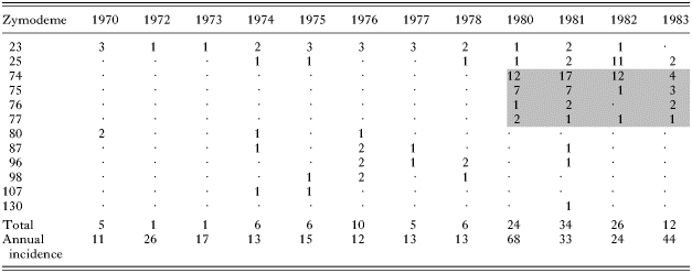
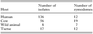
Nor was any satisfactory end to the debate on the identity of T. b. rhodesiense and T. b. brucei provided by the advent of biochemical methods for strain characterization from the late 1970's. Instead these studies showed that both subspecies comprised many different genotypes (e.g. Gibson, Marshall & Godfrey, 1980; Tait et al. 1985; Godfrey et al. 1990; Hide et al. 1990; MacLeod, Turner & Tait, 1999; Biteau et al. 2000), and that several T. b. rhodesiense genotypes were often circulating in a single outbreak of rhodesian sleeping sickness (Gibson & Gashumba, 1983; Gibson & Wellde, 1985; Komba et al. 1997). Results from a brief epidemic of rhodesian sleeping sickness in the Lambwe valley in south-western Kenya serve to illustrate this point. The Lambwe valley occupies a comparatively small area, with a wildlife reserve on the valley floor and settlements of subsistence farmers on the slopes (Allsopp & Baldry, 1972). In the early 1980's the valley contained an isolated but large population of tsetse, Glossina pallidipes, infesting thickets of vegetation on the valley floor and slopes (Wellde et al. 1989b). Close and frequent contact between tsetse, wildlife, domestic stock and humans led to outbreaks of both human and livestock trypanosomiases (Wellde et al. 1989a). Despite the small geographical area affected and the short duration, many different genotypes of T. b. rhodesiense were present (Table 2). Similar levels of genetic variation among T. b. rhodesiense isolates have been found in other foci when these have been studied in detail (Gibson & Gashumba, 1983; Komba et al. 1997). The conclusion is clear: T. b. rhodesiense is not one genetic entity. In the Lambwe Valley, the total number of genotypes of T. brucei sspp. recovered from all hosts was even greater (Table 3), reflecting the fact that T. b. brucei is also comprised of a great range of genotypes, some quite divergent, e.g. sindo and kiboko genotypes (Gibson, Fase-Fowler & Borst, 1985; Godfrey et al. 1990). The extent of genetic diversity needs to be borne in mind when interpreting the results of comparative studies of T. b. rhodesiense and T. b. brucei, for example, Hide et al. (1994), Coleman & Welburn (2004).
Discovery of the SRA gene in T. b. rhodesiense
Meanwhile research was in progress to find out why T. b. rhodesiense, but not T. b. brucei, was able to infect humans. The finding that different antigenic variants of exactly the same cloned line of T. b. rhodesiense could be either human serum sensitive or resistant (Van Meirvenne et al. 1976), led to the identification of the serum-resistance-associated (SRA) gene by comparison of cDNA from sensitive and resistant clones expressing the same variant surface glycoprotein (VSG) gene (De Greef et al. 1989). This work revealed that the difference between serum sensitive and resistant clones lay in the expression of a single gene, SRA (De Greef et al. 1989). However, when the SRA gene was found to be similar to VSG genes by DNA sequence analysis (De Greef & Hamers, 1994), many disbelieved that the SRA gene was more than a somewhat odd VSG gene.
But the evidence was building up. Independently Milner & Hajduk (1999) isolated the same gene from another strain of T. b. rhodesiense using a similar approach, and crucially Xong et al. (1998) showed that the SRA gene alone was able to confer human serum resistance on T. b. brucei after transfection. An accidental laboratory infection, with a T. b. brucei strain transfected with the SRA gene, proved the case beyond all doubt (E. Pays, personal communication). Xong et al. (1998) found that the SRA gene was located in a VSG expression site in the T. b. rhodesiense strain investigated (Fig. 1), with the important corollary that the gene was only expressed when that particular expression site was active. This finding nicely explained why resistance to human serum changed with antigenic variation (Van Meirvenne et al. 1976; Rickman, 1977).

Fig. 1. Diagram of (A) a typical VSG expression site of Trypanosoma brucei sspp. and (B) the VSG expression site containing the SRA gene, as described by Xong et al. (1998). The arrows indicate the position of the promotor, which drives transcription of all downstream genes. Genes are indicated by shaded blocks: black, expression site associated genes or ESAGs; hatched, serum resistance associated gene, SRA; grey, variant surface glycoprotein gene, VSG.
The SRA gene as a molecular marker for T. b. rhodesiense
Research on T. b. rhodesiense could now move forward. In terms of understanding the nature of T. b. rhodesiense, this discovery was potentially the ‘holy grail’ sought for so long – a single gene capable of identifying T. b. rhodesiense. However, before this marker could be used for epidemiological studies, it was necessary to demonstrate that all T. b. rhodesiense strains had the gene. To date this has been proved for each and every T. b. rhodesiense strain isolated from human patients in Ethiopia, Uganda, Rwanda, Kenya, Tanzania, Zambia, Malawi and Botswana, a total of over 120 isolates (De Greef et al. 1989, 1992; Welburn et al. 2001; Gibson, Backhouse & Griffiths, 2002; Radwanska et al. 2002; MacLean et al. 2004; Njiru et al. 2004) (Fig. 2). To prove the converse – that the SRA gene is not found in T. b. brucei – is more difficult, since this begs the question of how this trypanosome can be identified without recourse to human volunteers. Such information is available for only a handful of old isolates (e.g. Onyango, Van Hoeve & De Raadt, 1966), but it can be assumed that other T. brucei ssp. strains commonly used in the laboratory, e.g. 427, are also not infective to humans. Other lines of evidence suggesting that a T. brucei ssp. isolate is not human-infective originate from an area of Africa where sleeping sickness has never been reported, or sensitivity to human serum in vitro (although such results are not definitive – see the cautionary note above). No isolate satisfying one or more of these criteria has yet been shown to possess the SRA gene (De Greef et al. 1989, 1992; Welburn et al. 2001; Gibson et al. 2002; Radwanska et al. 2002; Njiru et al. 2004), although SRA-like gene sequences have been demonstrated in some T. b. brucei strains, most notably the genome strain TREU 927/4 (Campillo & Carrington, 2003; Vanhamme et al. 2004). However, the SRA-like gene in this strain does not have the deletion characteristic of the SRA gene (Vanhamme et al. 2004), nor does TREU 927/4 manifest full resistance to lysis by human serum in vitro (Turner et al. 2004; Vanhamme et al. 2004).
The SRA gene therefore appears to be a robust, single-gene marker for T. b. rhodesiense throughout its area of distribution, and can be used with confidence to identify T. b. rhodesiense in tsetse or putative reservoir hosts. In this way, cattle have been shown to be important reservoir hosts of T. b. rhodesiense in Uganda (Welburn et al. 2001), and cattle and a number of other domestic animal species were found to harbour T. b. rhodesiense in Kenya and Uganda (Njiru et al. 2004). Whereas in the past wild animals were considered to constitute the major reservoir of rhodesian sleeping sickness (Ashcroft, 1959), their decrease in numbers and distribution, and the concomitant increase in the human population and associated livestock, mean that domestic animals now pose the greatest risk. This is a major shift in the epidemiology of rhodesian sleeping sickness and may have unforeseen consequences, for example, the spread of rhodesian sleeping sickness into new areas with movement of livestock (Fevre et al. 2001; Hutchinson et al. 2003).

Fig. 2. Sketch map of East Africa showing the foci (stars) from which Trypanosoma brucei rhodesiense isolates from patients were derived. All T. b. rhodesiense isolates examined had the SRA gene by PCR analysis (data from De Greef et al. 1989; De Greef et al. 1992; Welburn et al. 2001; Gibson, Backhouse & Griffiths, 2002; Radwanska et al. 2002; MacLean et al. 2004; Njiru et al. 2004).
Origins and divergence of the SRA gene
Not only is the SRA gene found exclusively in T. b. rhodesiense, but its sequence is also conserved. Among 10 T. b. rhodesiense isolates from 5 disease foci (Ethiopia, Kenya, Tanzania, Uganda, Zambia) variation in the sequence of the SRA gene was less than 3%, and sequences from several different isolates were found to be identical (Gibson et al. 2002). There are limited sequence data available from T. b. rhodesiense for comparison, but several copies of the ESAG6 and ESAG7 genes, which encode variants of the transferrin receptor and are also located in VSG expression sites, have been sequenced from T. b. rhodesiense EATRO 3 (ETar 1) and WRATat 1. However, the comparison is a bit inconclusive, as levels of identity between homologous genes from the two T. b. rhodesiense strains were typically 93–94%, while one ESAG6 sequence from WRATat 1 was identical to that of EATRO 3.
SRA gene sequences fall into 2 groups: the northern variant found in Uganda, Kenya and Tanzania, and the southern variant found in Ethiopia, Malawi and Zambia (De Greef & Hamers, 1994; Milner & Hajduk, 1999; Gibson et al. 2002; MacLean et al. 2004). The gene appears to have originated from a VSG gene with a deletion of 378 bp (De Greef & Hamers, 1994; Campillo & Carrington, 2003). The genomic location of the gene is also conserved; in the Ugandan strain analysed by Xong et al. (1998), the SRA gene was found in a telomeric VSG expression site, lacking most of the typical ESAGs, except ESAG5, 6, and 7 (Fig. 1). Gibson & Ferris (2003) similarly found that the SRA gene was located downstream of ESAG5 in a range of T. b. rhodesiense isolates, and in addition showed that the northern and southern SRA variants have slightly different genomic environments, reinforcing the idea that there are 2 major genetic lineages of T. b. rhodesiense (Gibson et al. 1980; Godfrey et al. 1990; Hide et al. 1991; MacLeod et al. 2000). This echoes clinical observations of a milder form of rhodesian sleeping sickness towards the south of the range (Ormerod, 1967), recently confirmed in a comparison of the clinical course of trypanosomiasis in patients from Uganda and Malawi and correlation with parasite genotype (MacLean et al. 2004). The reasons for the divergence of northern and southern parasite lineages remain unknown, but may relate to isolation of human or vector populations, for example during climatic upheavals of the last few thousand years.
The degree of conservation of the SRA gene was unexpected considering the extent of genetic variation among T. b. rhodesiense isolates revealed by previous biochemical characterization studies, even in a single outbreak. The simplest explanation is that the SRA gene arose once by deletion of the central portion of a VSG gene (Campillo & Carrington, 2003), and spread into different T. b. brucei genetic backgrounds by genetic exchange (Jenni et al. 1986). In support of this hypothesis, Campillo & Carrington (2003) noted that the putative breakpoint regions for the deletion are remarkably conserved in all SRA sequences on database, suggesting a single origin of the gene. Genetic exchange between T. b. rhodesiense and T. b. brucei has been demonstrated in the laboratory; in a genetic cross of T. b. rhodesiense and T. b. brucei, the trait for human serum resistance was shown to be heritable by testing the human serum resistance of the parents and progeny (Gibson, 1989).
Mechanism of human serum resistance
The detailed mechanism of human serum resistance has been controversial for some time. Early on, the trypanolytic factor in human serum was recognized to be a component of high density lipoprotein (HDL) particles (Rifkin, 1978), but there may be more than one factor (Raper et al. 2001). The most recent and convincing evidence has identified apolipoprotein L-1 (apoL-1) in the HDL particle as the active factor, and demonstrated a molecular interaction between apoL-1 and the SRA protein (Vanhamme et al. 2003). ApoL-1 belongs to a family of 6 proteins, with genes clustered on human chromosome 22; it is expressed in many tissues, but its function is unknown (Page et al. 2001; Duchateau et al. 2001). Intriguingly, the apolipoprotein L gene cluster appears to have evolved recently in primates (Monajemi et al. 2002), and the apoL-1 protein was detected only in the serum of humans and the gorilla by specific binding to antibody and the SRA protein when a range of primate sera were tested (Poelvoorde et al. 2004). Chimpanzees, although considered the nearest living relative of humans, can be infected with T. b. brucei (Baker, 1962; Baker & Taylor, 1971) and have no trypanolytic activity or apoL-1 in their sera (Seed, Sechelski & Loomis, 1990; Poelvoorde et al. 2004). However, while baboons also had no detectable apoL-1 (Poelvoorde et al. 2004), baboon plasma has strong trypanolytic activity (Hawking, Ramsden & Whytock, 1973; Seed et al. 1990), suggesting the existence of another trypanolytic factor in primates. A potential candidate is haptoglobin-related protein, another component of HDL particles that appeared recently in primate evolution and has demonstrable trypanolytic activity (reviewed by Raper et al. 2001).
Implications for evolution and taxonomy of human pathogenic trypanosomes
While the discovery of the SRA gene has solved the mystery of human infectivity in T. b. rhodesiense, a different mechanism must operate in T. b. gambiense, because the SRA gene is not present (De Greef et al. 1992; Gibson et al. 2002; Radwanska et al. 2002). Both the avirulent and virulent types of T. b. gambiense [Group 1 and Group 2 T. b. gambiense respectively (Gibson, 1986)] show resistance to the trypanolytic activity of human serum (Hawking, 1976a,b; Mehlitz et al. 1982), but whereas the classical avirulent type, Group 1 T. b. gambiense, shows solid resistance (Mehlitz et al. 1982; Zillmann, Mehlitz & Sachs, 1984), the less common and more virulent Group 2 T. b. gambiense shows variable resistance to human serum like T. b. rhodesiense (Mehlitz et al. 1982). This suggests that resistance in Group 2 T. b. gambiense may work by a similar mechanism to that in T. b. rhodesiense, with the serum resistance gene located in a VSG expression site, giving rise to a variable human serum resistance phenotype (Xong et al. 1998). In contrast, an equivalent human serum resistance gene in Group 1 T. b. gambiense would be expected to be constitutively expressed, leading to the observed solid resistance to lysis by human serum (Mehlitz et al. 1982; Zillmann et al. 1984).
The working definition of T. brucei sspp. relies on host range. Thus the variability in expression of the SRA gene in T. b. rhodesiense leads to an intriguing taxonomic dilemma: does T. b. rhodesiense become T. b. brucei when the SRA gene is switched off? The pragmatic answer is to define T. b. rhodesiense by possession rather than expression of the SRA gene. Presumably, the SRA gene is always expressed by some trypanosomes in the human bloodstream, to avoid complete elimination of the population by the trypanolytic factor in the serum. Since the SRA gene was found in a VSG expression site (Xong et al. 1998), a genomic location which appears to be conserved in different strains of T. b. rhodesiense (Gibson & Ferris, 2003), this implies strong selection of this VSG expression site in the human host. Upstream of the SRA gene are ESAG6 and ESAG7, and one could further speculate that the variant of the transferrin receptor that these two genes encode should be suitable for uptake of human transferrin (Bitter et al. 1998). Intriguingly one ESAG6 copy from T. b. rhodesiense WRATat 1 was identical to the ESAG6 gene from the SRA expression site of EATRO 3, while the other copies were only 93–94% identical (see above); is the identical copy located in the SRA expression site of WRATat 1?
If there is a single expression site with the SRA gene, then only trypanosomes that undergo antigenic variation by switching the VSG gene in situ would survive. Given this scenario, there would surely be a selective drive for human-adapted trypanosomes with extra expression sites containing the SRA gene, but there is as yet no evidence for this. However, a set of identical or closely similar expression sites containing the SRA gene might be difficult to distinguish.
Metacyclics, which are the infective stages inoculated into the human host in tsetse saliva, transcribe a different set of VSG expression sites from established bloodstream forms. The metacyclic VSG expression sites are short and lack any ESAG's (Rudenko, Cross & Borst, 1998), presumably including the SRA gene. How then can metacyclics withstand the trypanolytic effect of human serum? Brun & Jenni (1987) have argued that metacyclics are not exposed to the trypanolytic effects of human serum at the site of the fly bite, where they multiply initially in the tissues, causing localized inflammation – the chancre. All metacyclics of T. b. brucei and a proportion of metacyclics of T. b. rhodesiense are susceptible to the trypanolytic effect of human serum in vitro, dying within a few hours of incubation (Jenni & Brun, 1982; Brun & Jenni, 1987). This suggests that metacyclics are fully capable of taking up the trypanolytic factor, unlike procyclics, which are resistant to lysis by human serum and fail to bind or endocytose the trypanolytic factor (Moore et al. 1995). Bloodstream forms of T. b. brucei rapidly develop resistance to human serum as they transform to procyclics in vitro (Moore et al. 1995), giving rise to possible confusion with T. b. rhodesiense and potential confounding effects in the design of in vitro assays of human serum resistance (e.g. Turner et al. 2004).
CONCLUSION
The SRA gene is a single gene marker for T. b. rhodesiense, which is already proving invaluable for epidemiological tracking of reservoir hosts and vectors. While the discovery of this gene has illuminated important aspects of the biology of T. b. rhodesiense, many questions about the human infectivity trait remain, perhaps most importantly what the equivalent mechanism in T. b. gambiense might be.
References
REFERENCES
ALLSOPP, R. &
BALDRY, D. A. T. (
1972).
A general description of the Lambwe valley area of South Nyanza District, Kenya.
Bulletin of the World Health Organization 47,
691–697.Google Scholar
APTED, F. I. C. (
1962).
Sleeping sickness in Tanganyika, past, present, and future.
Transactions of the Royal Society of Tropical Medicine and Hygiene 56,
15–29.CrossRefGoogle Scholar
ASHCROFT, M. T. (
1959).
A critical review of the epidemiology of human trypanosomiasis in Africa.
Tropical Diseases Bulletin 56,
1073–1093.Google Scholar
BAKER, J. R. (
1962).
Infection of the chimpanzee (Pan troglodytes verus) with Trypanosoma rhodesiense and T. brucei.
Annals of Tropical Medicine and Parasitology 56,
216–217.CrossRefGoogle Scholar
BAKER, J. R. &
TAYLOR, A. E. R. (
1971).
Experimental infections of the chimpanzee (Pan troglodytes) with Trypanosoma brucei brucei and Trypanosoma brucei rhodesiense.
Annals of Tropical Medicine and Parasitology 65,
471–485.CrossRefGoogle Scholar
BITEAU, N.,
BRINGAUD, F.,
GIBSON, W.,
TRUC, P. &
BALTZ, T. (
2000).
Characterization of Trypanozoon isolates using a repeated coding sequence and microsatellite markers.
Molecular and Biochemical Parasitology 105,
185–201.CrossRefGoogle Scholar
BITTER, W.,
GERRITS, H.,
KIEFT, R. &
BORST, P. (
1998).
The role of transferrin-receptor variation in the host range of Trypanosoma brucei.
Nature, London 391,
499–502.CrossRefGoogle Scholar
BRUN, R. &
JENNI, L. (
1987).
Human serum resistance of metacyclic forms of Trypanosoma brucei brucei, T. brucei rhodesiense and T. brucei gambiense.
Parasitology Research 73,
218–223.Google Scholar
CAMPILLO, N. &
CARRINGTON, M. (
2003).
The origin of the serum resistance associated (SRA) gene and a model of the structure of the SRA polypeptide from Trypanosoma brucei rhodesiense.
Molecular and Biochemical Parasitology 127,
79–84.CrossRefGoogle Scholar
COLEMAN, P. G. &
WELBURN, S. C. (
2004).
Are fitness costs associated with resistance to human serum in Trypanosoma brucei rhodesiense?
Trends in Parasitology 20,
311–315.Google Scholar
DE GREEF, C.,
CHIMFWEMBE, E.,
KIHANG'A WABACHA, J.,
BAJYANA SONGA, E. &
HAMERS, R. (
1992).
Only the serum-resistant bloodstream forms of Trypanosoma brucei rhodesiense express the serum resistance associated (SRA) protein.
Annales de la Société Belge de Medecine Tropicale 72,
13–21.Google Scholar
DE GREEF, C. &
HAMERS, R. (
1994).
The serum resistance associated (SRA) gene of Trypanosoma brucei rhodesiense encodes a VSG-like protein.
Molecular and Biochemical Parasitology 68,
277–284.CrossRefGoogle Scholar
DE GREEF, C.,
IMBERECHTS, H.,
MATTHYSSONS, G.,
VAN MEIRVENNE, N. &
HAMERS, R. (
1989).
A gene expressed only in serum-resistant variants of Trypanosoma brucei rhodesiense.
Molecular and Biochemical Parasitology 36,
169–176.CrossRefGoogle Scholar
DUCHATEAU, P. N.,
PULLINGER, C. R.,
CHO, M. H.,
ENG, C. &
KANE, J. P. (
2001).
Apolipoprotein L gene family: tissue-specific expression, splicing, promoter regions; discovery of a new gene.
Journal of Lipid Research 42,
620–630.Google Scholar
FAUST, S. N.,
WOODROW, C. J.,
PATEL, S.,
SNAPE, M.,
CHIODINI, P. L.,
TUDOR-WILLIAMS, G. &
LYALL, E. G. H. (
2004).
Sleeping sickness in brothers in London.
Pediatric Infectious Disease Journal 23,
879–881.CrossRefGoogle Scholar
FEVRE, E. M.,
COLEMAN, P. G.,
ODIIT, M.,
MAGONA, J. W.,
WELBURN, S. C. &
WOOLHOUSE, M. E. J. (
2001).
The origins of a new Trypanosoma brucei rhodesiense sleeping sickness outbreak in eastern Uganda.
Lancet 358,
625–628.CrossRefGoogle Scholar
GIBSON, W.,
BACKHOUSE, T. &
GRIFFITHS, A. (
2002).
The human serum resistance associated gene is ubiquitous and conserved in Trypanosoma brucei rhodesiense throughout East Africa.
Infection, Genetics and Evolution 1,
207–214.CrossRefGoogle Scholar
GIBSON, W. &
FERRIS, V. (
2003).
Conservation of the genomic location of the human serum resistance associated gene in Trypanosoma brucei rhodesiense.
Molecular and Biochemical Parasitology 130,
159–162.CrossRefGoogle Scholar
GIBSON, W. C. (
1986).
Will the real Trypanosoma brucei gambiense please stand up?
Parasitology Today 2,
255–257.Google Scholar
GIBSON, W. C. (
1989).
Analysis of a genetic cross between Trypanosoma brucei rhodesiense and T. b. brucei.
Parasitology 99,
391–402.CrossRefGoogle Scholar
GIBSON, W. C.,
FASE-FOWLER, F. &
BORST, P. (
1985).
Further analysis of intraspecific variation in Trypanosoma brucei using restriction site polymorphisms in the maxi-circle of kinetoplast DNA.
Molecular and Biochemical Parasitology 15,
21–36.CrossRefGoogle Scholar
GIBSON, W. C. &
GASHUMBA, J. K. (
1983).
Isoenzyme characterisation of some Trypanozoon stocks from a recent trypanosomiasis epidemic in Uganda.
Transactions of the Royal Society of Tropical Medicine and Hygiene 77,
114–118.CrossRefGoogle Scholar
GIBSON, W. C.,
MARSHALL, T. F. D. C. &
GODFREY, D. G. (
1980).
Numerical analysis of enzyme polymorphism: a new approach to the epidemiology and taxonomy of trypanosomes of the subgenus Trypanozoon.
Advances in Parasitology 18,
175–246.CrossRefGoogle Scholar
GIBSON, W. C. &
WELLDE, B. T. (
1985).
Characterisation of Trypanozoon stocks from the South Nyanza sleeping sickness focus in Western Kenya.
Transactions of the Royal Society of Tropical Medicine and Hygiene 79,
671–676.CrossRefGoogle Scholar
GODFREY, D. G.,
BAKER, R. D.,
RICKMAN, L. R. &
MEHLITZ, D. (
1990).
The distribution, relationships and identification of enzymic variants within the subgenus Trypanozoon.
Advances in Parasitology 29,
1–74.CrossRefGoogle Scholar
HAWKING, F. (
1976 a).
The resistance to human plasma of Trypanosoma brucei, T. rhodesiense and T. gambiense. I. Analysis of the composition of trypanosome strains.
Transactions of the Royal Society of Tropical Medicine and Hygiene 70,
504–520.Google Scholar
HAWKING, F. (
1976 b).
The resistance to human plasma of Trypanosoma brucei, T. rhodesiense and T. gambiense. II. Survey of strains from East Africa and Nigeria.
Transactions of the Royal Society of Tropical Medicine and Hygiene 70,
513–520.Google Scholar
HAWKING, F.,
RAMSDEN, D. B. &
WHYTOCK, S. (
1973).
The trypanocidal action of human serum and of baboon plasma.
Transactions of the Royal Society of Tropical Medicine and Hygiene 67,
501–516.CrossRefGoogle Scholar
HIDE, G.,
BUCHANAN, N.,
WELBURN, S.,
MAUDLIN, I.,
BARRY, J. D. &
TAIT, A. (
1991).
Trypanosoma brucei rhodesiense: characterisation of stocks from Zambia, Kenya, and Uganda using repetitive DNA probes.
Experimental Parasitology 72,
430–439.CrossRefGoogle Scholar
HIDE, G.,
CATTAND, P.,
LE RAY, D.,
BARRY, D. J. &
TAIT, A. (
1990).
The identification of Trypanosoma brucei subspecies using repetitive DNA sequences.
Molecular and Biochemical Parasitology 39,
213–226.CrossRefGoogle Scholar
HIDE, G.,
WELBURN, S. C.,
TAIT, A. &
MAUDLIN, I. (
1994).
Epidemiological relationships of Trypanosoma brucei stocks from South East Uganda: evidence for different population structures in human infective and non-infective isolates.
Parasitology 109,
95–111.CrossRefGoogle Scholar
HUTCHINSON, O. C.,
FEVRE, E. M.,
CARRINGTON, M. &
WELBURN, S. C. (
2003).
Lessons learned from the emergence of a new Trypanosoma brucei rhodesiense sleeping sickness focus in Uganda.
Lancet Infectious Diseases 3,
42–45.CrossRefGoogle Scholar
JELINEK, T.,
BISOFFI, Z.,
BONAZZI, L.,
VAN THIEL, P.,
BRONNER, U.,
DE FREY, A.,
GUNDERSEN, S. G.,
McWHINNEY, P. &
RIPAMONTI, D. (
2002).
Cluster of African trypanosomiasis in travelers to Tanzanian national parks.
Emerging Infectious Diseases 8,
634–635.CrossRefGoogle Scholar
JENNI, L. &
BRUN, R. (
1982).
A new in vitro test for human serum resistance of Trypanosoma (T.) brucei.
Acta Tropica 39,
281–284.Google Scholar
JENNI, L.,
MARTI, S.,
SCHWEIZER, J.,
BETSCHART, B.,
LEPAGE, R. W. F.,
WELLS, J. M.,
TAIT, A.,
PAINDAVOINE, P.,
PAYS, E. &
STEINERT, M. (
1986).
Hybrid formation between African trypanosomes during cyclical transmission.
Nature, London 322,
173–175.CrossRefGoogle Scholar
KOMBA, E. K.,
KIBONA, S. N.,
AMBWENE, A. K.,
STEVENS, J. R. &
GIBSON, W. C. (
1997).
Genetic diversity among Trypanosoma brucei rhodesiense isolates from Tanzania.
Parasitology 115,
571–579.CrossRefGoogle Scholar
MacLEAN, L.,
CHISI, J. E.,
ODIIT, M.,
GIBSON, W. C.,
FERRIS, V.,
PICOZZI, K. &
STERNBERG, J. M. (
2004).
Severity of Human African Trypanosomiasis in East Africa is associated with geographic location, parasite genotype and host-inflammatory cytokine response profile.
Infection and Immunity 72,
7040–7044.CrossRefGoogle Scholar
MacLEOD, A.,
TURNER, C. M. R. &
TAIT, A. (
1999).
A high level of mixed Trypanosoma brucei infections in tsetse flies detected by three hypervariable minisatellites.
Molecular and Biochemical Parasitology 102,
237–248.CrossRefGoogle Scholar
MacLEOD, A.,
TWEEDIE, A.,
WELBURN, S. C.,
MAUDLIN, I.,
TURNER, C. M. R. &
TAIT, A. (
2000).
Minisatellite marker analysis of Trypanosoma brucei: Reconciliation of clonal, panmictic, and epidemic population genetic structures.
Proceedings of the National Academy of Sciences, USA 97,
13442–13447.CrossRefGoogle Scholar
MEHLITZ, D.,
ZILLMANN, U.,
SCOTT, C. M. &
GODFREY, D. G. (
1982).
Epidemiological studies on the animal reservoir of gambiense sleeping sickness. III. Characterisation of Trypanozoon stocks by isoenzymes and sensitivity to human serum.
Tropenmedizin und Parasitologie 33,
113–118.Google Scholar
MILNER, J. D. &
HAJDUK, S. L. (
1999).
Expression and localization of serum resistance associated protein in Trypanosoma brucei rhodesiense.
Molecular and Biochemical Parasitology 104,
271–283.CrossRefGoogle Scholar
MONAJEMI, H.,
FONTIJN, R. D.,
PANNEKOEK, H. &
HORREVOETS, A. J. G. (
2002).
The apolipoprotein L gene cluster has emerged recently in evolution and is expressed in human vascular tissue.
Genomics 79,
539–546.CrossRefGoogle Scholar
MOORE, D. R.,
SMITH, A.,
HAGER, K. M.,
WALDON, R.,
ESKO, J. D. &
HAJDUK, S. L. (
1995).
Developmentally-regulated sensitivity of Trypanosoma brucei brucei to the cytotoxic effects of human high density lipoprotein.
Experimental Parasitology 81,
216–226.CrossRefGoogle Scholar
NJIRU, Z. K.,
NDUNG'U, K.,
MATETE, G.,
NDUNGU, J. M. &
GIBSON, W. C. (
2004).
Detection of Trypanosoma brucei rhodesiense in animals from sleeping sickness foci in East Africa using the serum resistance associated (SRA) gene.
Acta Tropica 90,
249–254.CrossRefGoogle Scholar
ONYANGO, R. J.,
VAN HOEVE, K. &
DE RAADT, P. (
1966).
The epidemiology of Trypanosoma rhodesiense sleeping sickness in Alego location, Central Nyanza, Kenya. I. Evidence that cattle may act as reservoir hosts of trypanosomes infective to man.
Transactions of the Royal Society of Tropical Medicine and Hygiene 60,
175–182.Google Scholar
ORMEROD, W. E. (
1967).
Taxonomy of the sleeping sickness trypanosomes.
Journal of Parasitology 53,
824–830.CrossRefGoogle Scholar
PAGE, N. M.,
BUTLIN, D. J.,
LOMTHAISONG, K. &
LOWRY, P. J. (
2001).
The human apolipoprotein L gene cluster: Identification, classification, and sites of distribution.
Genomics 74,
71–78.CrossRefGoogle Scholar
POELVOORDE, P.,
VANHAMME, L.,
VAN DEN ABBEELE, J.,
SWITZER, W. M. &
PAYS, E. (
2004).
Distribution of apolipoprotein L-I and trypanosome lytic activity among primate sera.
Molecular and Biochemical Parasitology 134,
155–157.CrossRefGoogle Scholar
RADWANSKA, M.,
CHAMEKH, M.,
VANHAMME, L.,
CLAES, F.,
MAGEZ, S.,
MAGNUS, E.,
DE BAETSELIER, P.,
BUSCHER, P. &
PAYS, E. (
2002).
The serum resistance-associated gene as a diagnostic tool for the detection of Trypanosoma brucei rhodesiense.
American Journal of Tropical Medicine and Hygiene 67,
684–690.CrossRefGoogle Scholar
RAPER, J.,
PORTELA, M. P. M.,
LUGLI, E.,
FREVERT, U. &
TOMLINSON, S. (
2001).
Trypanosome lytic factors: novel mediators of human innate immunity.
Current Opinion in Microbiology 4,
402–408.CrossRefGoogle Scholar
RICKMAN, L. R. (
1977).
Variation in the test response of clone-derived Trypanosoma (Trypanozoon) brucei brucei and T. (T.) b. rhodesiense relapse antigenic variants, examined by a modified blood incubation infectivity test and its possible significance in rhodesian sleeping sickness transmission.
Medical Journal of Zambia 11,
31–41.Google Scholar
RICKMAN, L. R. &
ROBSON, J. (
1970).
The testing of proven Trypanosoma brucei and T. rhodesiense strains by the blood incubation infectivity test.
Bulletin of the World Health Organization 42,
911–916.Google Scholar
RIFKIN, M. R. (
1978).
Identification of the trypanocidal factor in normal human serum: high density lipoprotein.
Proceedings of the National Academy of Sciences, USA 75,
3450–3454.CrossRefGoogle Scholar
RUDENKO, G.,
CROSS, M. &
BORST, P. (
1998).
Changing the end: antigenic variation orchestrated at the telomeres of African trypanosomes.
Trends in Microbiology 6,
113–117.CrossRefGoogle Scholar
SEED, J. R.,
SECHELSKI, J. B. &
LOOMIS, M. R. (
1990).
A survey for a trypanocidal factor in primate sera.
Journal of Protozoology 37,
393–400.CrossRefGoogle Scholar
STEPHENS, J. &
FANTHAM, H. (
1910).
On the peculiar morphology of a trypanosome from a case of sleeping sickness and the possibility of its being a new species (T. rhodesiense).
Annals of Tropical Medicine and Parasitology 4,
343–350.CrossRefGoogle Scholar
TAIT, A.,
BARRY, J. D.,
WINK, R.,
SANDERSON, A. &
CROWE, J. S. (
1985).
Enzyme variation in Trypanosoma brucei spp. II. Evidence for T. b. rhodesiense being a set of variants of T. b. brucei.
Parasitology 90,
89–100.Google Scholar
TARGETT, G. A. T. &
WILSON, V. C. L. C. (
1973).
The blood incubation infectivity test as a means of distinguishing between Trypanosoma brucei brucei and T. brucei rhodesiense.
International Journal for Parasitology 3,
5–11.CrossRefGoogle Scholar
TAUTE, M. &
HUBER, F. (
1919).
Die Unterscheidung des Trypanosoma rhodesiense vom Trypanosoma brucei. Beobachtungen und Experimente aus dem Kriege in Ostafrika.
Archiv für Schiffs- und Tropen-Hygiene 23,
212–234.Google Scholar
TURNER, C. M. R.,
McLELLAN, S.,
LINDERGARD, L. A. G.,
BISONI, L.,
TAIT, A. &
MacLEOD, A. (
2004).
Human infectivity trait in Trypanosoma brucei: stability, heritability and relationship to sra expression.
Parasitology,
129,
445–454.CrossRefGoogle Scholar
VAN MEIRVENNE, N.,
MAGNUS, E. &
JANSSENS, P. G. (
1976).
The effect of normal human serum on trypanosomes of distinct antigenic type (ETat 1–12) isolated from a strain of Trypanosoma brucei rhodesiense.
Annales de la Société Belge de Medecine Tropicale 56,
55–63.Google Scholar
VANHAMME, L.,
PATURIAUX-HANOCQ, F.,
POELVOORDE, P.,
NOLAN, D.,
LINS, L.,
VAN DEN ABBEELE, J.,
PAYS, A.,
TEBABI, P.,
XONG, H.,
JACQUET, A.,
MOGUILEVSKY, N.,
DIEU, M.,
KANE, J. P.,
DE BAETSELIER, P.,
BRASSEUR, R. &
PAYS, E. (
2003).
Apolipoprotein L-1 is the trypanosome lytic factor of human serum.
Nature, London 422,
83–87.CrossRefGoogle Scholar
VANHAMME, L.,
RENAULD, H.,
LECORDIER, L.,
POELVOORDE, P.,
VAN DEN ABBEELE, J. &
PAYS, E. (
2004).
The Trypanosoma brucei reference strain TREU927/4 contains T. brucei rhodesiense-specific SRA sequences, but displays a distinct phenotype of relative resistance to human serum.
Molecular and Biochemical Parasitology 135,
39–47.Google Scholar
WELBURN, S. C.,
PICOZZI, K.,
FEVRE, E. M.,
COLEMAN, P. G.,
ODIIT, M.,
CARRINGTON, M. &
MAUDLIN, I. (
2001).
Identification of human-infective trypanosomes in animal reservoir of sleeping sickness in Uganda by means of serum-resistance-associated (SRA) gene.
Lancet 358,
2017–2019.CrossRefGoogle Scholar
WELLDE, B. T.,
CHUMO, D. A.,
REARDON, M. J.,
WAEMA, D.,
SMITH, D. H.,
GIBSON, W. C.,
WANYAMA, L. &
SIONGOK, T. A. (
1989 a).
Epidemiology of Rhodesian sleeping sickness in the Lambwe Valley, Kenya.
Annals of Tropical Medicine and Parasitology 83,
43–62.Google Scholar
WELLDE, B. T.,
WAEMA, D.,
CHUMO, D. A.,
REARDON, M. J.,
OLOO, F.,
NJOGU, A. R.,
OPIYO, E. A. &
MUGUTU, S. (
1989 b).
Review of tsetse control measures taken in the Lambwe Valley in 1980–1984.
Annals of Tropical Medicine and Parasitology 83,
119–125.Google Scholar
WILLETT, K. &
FAIRBAIRN, H. (
1955).
The Tinde experiment: A study of Trypanosoma rhodesiense during eighteen years of cyclical transmission.
Annals of Tropical Medicine and Parasitology 49,
278–292.CrossRefGoogle Scholar
XONG, V. H.,
VANHAMME, L.,
CHAMEKH, M.,
CHIMFWEMBE, C. E.,
VAN DEN ABBEELE, J.,
PAYS, A.,
VAN MEIRVENNE, N.,
HAMERS, R.,
DE BAETSELIER, P. &
PAYS, E. (
1998).
A VSG expression site-associated gene confers resistance to human serum in Trypanosoma rhodesiense.
Cell 95,
839–846.CrossRefGoogle Scholar
YORKE, W. (
1910).
On the pathogenicity of a trypanosome (T. rhodesiense, Stephens and Fantham) from a case of sleeping sickness contracted in Rhodesia.
Annals of Tropical Medicine and Parasitology 4,
351–368.Google Scholar
ZILLMANN, U.,
MEHLITZ, D. &
SACHS, R. (
1984).
Identity of Trypanozoon stocks isolated from man and domestic dog in Liberia.
Tropenmedizin und Parasitologie 35,
105–108.Google Scholar
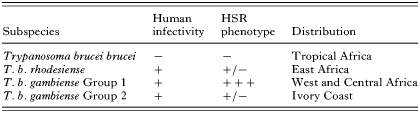
Table 1. Characteristics of subspecies within the Trypanosoma brucei group
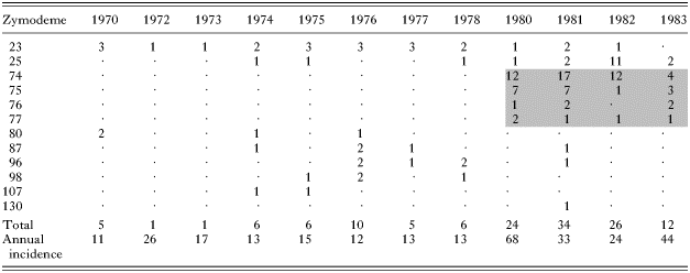
Table 2. Trypanosoma brucei rhodesiense in Lambwe Valley, Kenya, 1970–1983
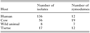
Table 3. Trypanosoma brucei rhodesiense zymodemes in Lambwe Valley, Kenya, 1969–1983
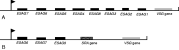
Fig. 1. Diagram of (A) a typical VSG expression site of Trypanosoma brucei sspp. and (B) the VSG expression site containing the SRA gene, as described by Xong et al. (1998). The arrows indicate the position of the promotor, which drives transcription of all downstream genes. Genes are indicated by shaded blocks: black, expression site associated genes or ESAGs; hatched, serum resistance associated gene, SRA; grey, variant surface glycoprotein gene, VSG.
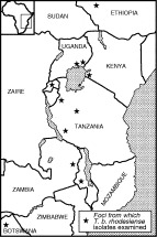
Fig. 2. Sketch map of East Africa showing the foci (stars) from which Trypanosoma brucei rhodesiense isolates from patients were derived. All T. b. rhodesiense isolates examined had the SRA gene by PCR analysis (data from De Greef et al. 1989; De Greef et al. 1992; Welburn et al. 2001; Gibson, Backhouse & Griffiths, 2002; Radwanska et al. 2002; MacLean et al. 2004; Njiru et al. 2004).
 You have
Access
- 25
- Cited by