




No CrossRef data available.
Neonatal hemodynamics in patients with hypoplastic left heart syndrome
Published online by Cambridge University Press: 21 September 2005
Extract
The neonate with hypoplastic left heart syndrome presents a challenge for clinical diagnosis and management. Three diagnostic goals must be met. First, it is necessary to make an etiologic cardiac diagnosis so as to rule out any genetic abnormality. Second, the anatomic cardiac diagnosis is made by segmental echocardiographic analysis, including details of the atrial arrangement, venous return, the patency of the arterial duct, atrial anatomy, and the arrangement of the aortic arch. Finally, the physiologic cardiac diagnosis is made by Doppler evaluation. In some patients, the diagnosis of hypoplastic left heart syndrome is not synonymous with functionally univentricular physiology, and a bi-ventricular repair can be achieved.1
Information
- Type
- Clinical Aspects
- Information
- Copyright
- © 2004 Cambridge University Press
Prenatal assessment
Optimal care of the neonate is achieved by prenatal diagnosis.2 Fetal echocardiography will reveal left-to-right shunting at the oval fossa (Fig. 1), and the other anatomic findings consistent with the diagnosis. Such prenatal diagnosis facilitates monitoring of the growth and normal development of the fetus and the vital organs. During fetal life, the pattern of growth should be monitored by the perinatal team, and transfer to the cardiac surgical center should be planned. The difficult decision about univentricular as opposed to biventricular repair may be aided by fetal observations.3, 4 A vaginal delivery is preferable for infants with hypoplastic left heart syndrome, and avoids unnecessary risk for the mother as long as the fetus has no signs of cardiac failure. We use the cardiovascular profile score to select those fetuses requiring special attention, and we have shown that this cardiovascular score correlates with the Tei index.5 Medications in pregnancy should not include any known constrictor of the fetal arterial duct, such as ibuprofen. The goal of all prenatal interventions is to prevent premature delivery. Such an occurrence will change the prognosis, and completely alter the postnatal plan. Evidence that prenatal diagnosis can improve the care of the neonate with hypoplastic left heart syndrome comes from a recent study by Mahle et al.6 showing a decrease in neurological events in the group of patients diagnosed prenatally.

Figure 1. Fetal aortic stenosis, with left to right shunting at the oval fossa (arrow in upper panel) AS: aortic stenosis; Ao: aorta; LA: left atrium; LV: left ventricle; MR: mitral regurgitation; RA: right atrium.
Neonatal Doppler evaluation
After birth, pulsed and continuous wave Doppler techniques can be used to assess the functional state of the heart and cardiovascular system, evaluating all sites for intra-cardiac and extra-cardiac shunting. Thus, attention should be directed to the patency of the arterial duct, the integrity of the atrial septum, the function of the right ventricle, tricuspid and pulmonary valves, the flows through the aortic arch and pulmonary arteries, and the peripheral arterial Doppler findings in critical sites of the body such as the middle cerebral and the superior mesenteric arteries.
The patency of the arterial duct must be confirmed by echocardiography, and it should be maintained widely patent. Prostaglandin E1 at doses from 0.0125 to 0.050 μg/kg body weight per minute should accomplish this objective. If the arterial duct is constricted in a shocked neonate presenting with hypoplastic left heart syndrome, the pathway from the right ventricle to the aorta is obstructed. Doppler interrogation will then confirm the presence of a right-to-left gradient across the constricted arterial duct (Fig. 2).

Figure 2. Continuous wave Doppler in a neonate with hypoplastic left heart syndrome with constriction of the arterial duct. Right to left velocity acceleration to a maximum of 2.9 m/s is present.
The atrial septum is interrogated to evaluate the left-to-right atrial gradient, and to assess the pattern of flow in the pulmonary veins so as to detect any interatrial obstruction. Large, or increasing, atrial reversal of flow in the pulmonary veins suggests significant obstruction. Right ventricular function is assessed by a careful evaluation of the global and segmental function of the portions of the right ventricular myocardium. Tricuspid valvar regurgitation can be assessed by color Doppler, and may be due to an intrinsic anatomic abnormality of the valve, be secondary to right ventricular dysfunction, or be the consequence of increased work load of the right ventricle (Fig. 3). The tricuspid valvar function is assessed to detect significant regurgitation. If present, the first derivative of the pressure change, the so-called dP/dt, can be calculated. This should be greater than 800 mmHg/s.

Figure 3. Neonate with hypoplastic left heart syndrome and regurgitation across the tricuspid valve.
Flows across the arterial valves are evaluated with Doppler to detect any regurgitation, which is always an abnormal finding, and to ensure an unobstructed pathway to the body for cardiac output. The flow through the aortic arch may be reversed when there is aortic atresia. The flow in the right and left pulmonary arteries can be used to assess the magnitude of the flow of blood to the lungs, and to detect any difference in the flow to the right and left lungs. Diastolic flow should be present in the superior mesenteric and middle cerebral arteries, but ductal steal can compromise the circulation (see below).
Management in the delivery room
The neonate with hypoplastic left heart syndrome undergoes a dramatic transitional change after birth. Assuming that the gestation is from 38 to 40 weeks, that the neonate is mature, the arterial duct is widely patent, and that there are normal Apgar scores after birth, the hemodynamics will change after birth primarily due to decreases in the pulmonary vascular resistance. After the first breath, there is a sudden decrease in the resistance of the pulmonary bed, to less than two-fifths of the fetal resistance. From this point, the balance of the circulation is governed by the ratio between the pulmonary and systemic vascular resistances. This ratio determines how much flow will go to the body, and how much to the lungs. The ratio of these flows changes with alterations in the pulmonary and systemic resistances. For example, if the newborn with hypoplastic left heart syndrome is stressed by cold, then the systemic resistance is elevated, and the ratio of pulmonary to systemic flows is increased.
The attending physician in the delivery room should be aware of the correct management of ventilation in a neonate with hypoplastic left heart syndrome. This means that the primary determinant of the pulmonary resistance, namely the arterial partial pressure of carbon dioxide, should be maintained in a eucapnic range, namely from 35 to 45 Torr. The use of oxygen is usually contraindicated, but may be necessary in the delivery room if there is respiratory depression, pulmonary dysfunction, or airway obstruction. Use of the pulse oximeter is mandatory, and the saturation of oxygen should be maintained in the range of 80–85%, and not allowed to go higher. Higher saturations indicate a higher than normal ratio of pulmonary to systemic flows, and should be avoided.
To date, no quantitative measurements have been made of the preoperative ratio of pulmonary to systemic flows in the neonate with hypoplastic left heart syndrome. Estimates are based on assumptions about the pulmonary venous saturations, and use the arterial level of oxygenation, coupled with the Fick principle, to estimate the flow. For example, an umbilical arterial saturation of 85% in room air correlates approximately with a ratio of flows in the range from 1.5 or 2 to one. The impact on systemic flow of a rising ratio can be seen by estimating the decrease in effective cardiac output to the neonate. For example, an increase of the arterial saturation from 85 to 92% could be associated with a decrease of systemic flow of half, due entirely to redistribution of cardiac output to the lungs. The total combined cardiac output pumped by the right ventricle then is primarily affected by the flow to the lungs. At high values of flow to the lungs, the stroke volume of the right ventricle can be huge, and the work of the heart is significantly increased. Under such conditions, the right ventricle may dilate, and result in significant tricuspid valvar regurgitation. The echocardiographer must assess the situation. One feature that can help in this regard is the appearance of the pulsed Doppler tracing assessed in the patent arterial duct. Rychik et al.7 showed that the area ratio of forward to reversed flow in this site correlates with estimates of the ratio of pulmonary to systemic flows as made using the Fick principle. Increased flow of blood to the lungs also affects the diastolic blood pressure and, in time, also decreases the systolic pressure. This decrease in diastolic flow to the body is known as the “diastolic steal”.
Preoperative assessment and stabilization
Doppler examination can aid in the diagnosis of the “diastolic steal.” In diastole, the arterial valves are closed, and the circulation redistributes according to the various resistances that are connected to the aorta. These resistances are in parallel, and include the pulmonary resistance, which is connected to the aorta by the patent arterial duct. Reversal of flow of blood in the descending aorta below the arterial duct in diastole indicates that the pulmonary resistance is significantly less than the systemic resistance. As this resistance drops, the diastolic flow into the pulmonary arteries increases as a sign of the larger left-to-right shunt (Fig. 4). This reversal of flow from the lower body, and accentuation of diastolic flow from the upper body and head, can lead to abnormal diastolic perfusion of vital organs. These include the heart, the brain, and the gut (Fig. 5).

Figure 4. Doppler interrogation of the left pulmonary artery, showing evidence of a large left-to-right shunt. Note the large diastolic velocity.

Figure 5. Pulsed Doppler recording in the superior mesenteric artery (SMA) in a neonate with hypoplastic left heart syndrome with good diastolic flow.
Coronary arterial flow has been assessed in patients prior to the Norwood operation with an aortopulmonary shunt via the patent arterial duct, and diastolic coronary flow is known to be abnormal. If untreated, this “diastolic steal” from the myocardium can lead rapidly to low cardiac output. Patterns of flow of cerebral blood are known to be affected by the level of pulmonary vascular resistance in neonates with patent arterial ducts. For example, after administration of surfactant, the pulmonary resistance is decreased immediately, and the perfusion is altered through the carotid and middle cerebral arteries. The cerebral perfusion, therefore, can be abnormal in the neonate with hypoplastic left heart syndrome, and is a sign of abnormal hemodynamics requiring attention. The middle cerebral artery can be evaluated easily during cardiac evaluation, and there should always be diastolic flow in this vessel. Absence of diastolic flow suggests that the steal phenomenon is affecting the flow of blood to the brain. With treatment, this steal should diminish, and diastolic flow should reappear in the middle cerebral artery. The superior mesenteric artery supplies the gut, and flow through this vessel can be used to monitor the perfusion of this vascular bed. Some presumed stable neonates with hypoplastic left heart syndrome will have absence of diastolic flow in this artery, but this is not a normal finding, and should always raise the issue of the advisability of preoperative feeding in these patients. The goal of management is to balance the pulmonary and systemic circulations. The rule of “40s” states that the arterial partial pressures of oxygen and carbon dioxide should both be about 40 Torr. By achieving this goal, there is usually a modest degree of overcirculation of the lungs, with a ratio of pulmonary to systemic flow in the range of 2 to 1.
Measures used to stabilize the neonate with hypoplastic left heart syndrome and “diastolic steal” syndrome are well established, but there is little data to support one approach over another. The peripheral resistance should be maintained low by keeping the newborn warm and dry. Use of overhead warmers is mandatory, and maintenance of a neutral thermal environment is the goal. When the warmer is used to maintain temperature, the core temperature should be normal. If shock occurs, the core temperature will rise, and there is a temptation to turn the warmer off. This should be avoided, because the desired result is peripheral vasodilation, which requires external warming as part of the therapy. Proper management requires frequent assessment of lower body perfusion and temperature. If the feet are warm, with less than 3 s capillary refill time, then the hemodynamics are usually good. Replacement of volume is always indicated during the phase of shifting of circulating volume from the body to the lung compartment as the pulmonary vascular resistance falls after birth. We usually give 5% albumin intravenously, at a dose of 10 ml/kg body weight. Prostaglandin E1 will keep the arterial duct open, and should be started within 3 to 4 hours after birth. We use a dose of 0.025 μg/kg body weight per minute as an intravenous infusion. Higher doses produce apnea, and also cause pulmonary vasodilation, as well as side effects involving the central nervous system, such as agitation or fever. Catacholamine-containing medications for inotropic support should be avoided in doses that constrict the peripheral vascular bed and raise the systemic resistance. For example, Dopamine is good for renal perfusion, and has significant inotropic effect in neonates up to a dose of 3 μg/kg/min, but at higher doses may cause systemic vasoconstriction. Digoxin at a dose of 5μg/kg given intravenously twice each day is indicated to support the dilating myocardium, and to reduce the central release of catacholamine. A diuretic such as furosemide at 1 mg/kg, given intravenously twice a day, is used to clear free water, so as to try to avoid pulmonary congestion. The best way to accomplish this goal, however, is to provide good nutrition to maintain the plasma oncotic pressure. Originally, hypoventilation and sedation were used to raise the pulmonary vascular resistance, but difficulty with atelectasis was frequently encountered. Now, use of inspired carbon dioxide, given at a partial pressure of 7 to 14 Torr, or inspired nitrogen added to decrease the fixed inspiratory oxygen from 0.18 to 0.20, both given to raise the pulmonary vascular resistance, allows adequate ventilation volumes to prevent collapse of the lungs.
In general, the systemic vascular resistance should be maintained as low as possible to be compatible with a normal diastolic blood pressure. Afterload reduction is the best way to balance the circulation, and is done with agents that decrease systemic vascular tone. Milrinone at an intravenous infusion dose of 0.5μg/kg/min is an excellent vasodilator, and provides some intropic support for neonates who are manifesting less than ideal right ventricular function. If needed, nitroprusside can be given as an intravenous infusion further to lower the systemic resistance. Muscular paralysis may be indicated rarely if there is severe acidosis or hypoxemia, and will lower the consumption of oxygen by the whole body, but we try to avoid this approach when the neonates are stable.
Intraoperative and postoperative management
After the Norwood operation, echocardiographic attention should be focused in the same areas as were the case preoperatively. The aortic arch deserves special attention to exclude the possibility of obstruction.8 An unobstructed pathway for the pulmonary venous return is confirmed at each examination.
In the postoperative unit, it is always useful to monitor the myocardial function. The global assessment of function will guide decisions concerning closure of the chest, weaning from ventilation, and inotropic medications. Echocardiography may be useful in predicting the outcome of a neonate early after the first stage of the Norwood sequence. In a small study in our center, we evaluated the right ventricular function using a nongeometric approach after a classical first stage procedure.9 A combination of the Tei myocardial performance index and dP/dt proved to be useful in predicting the outcome. We evaluated 20 neonates within one week of the first stage procedure, and 17 of them survived to discharge. Assessment of right ventricular function was done by measuring the Tei index. This is the ratio of the isovolemic time divided by the ejection time of the right ventricle. The isovolemic time can be determined by measuring the interval from closing to opening of the tricuspid valve and subtracting the ejection time. In our experience, the normal range for patients with hypoplastic left heart syndrome is from 0.37 to 0.46. The Tei index was then combined with dP/dt. The latter value is calculated from the jet of tricuspid regurgitation by measuring the time interval in milliseconds between the velocities of 1 and 3 m/s as seen on the upstroke of the waveform. The dP/dt is 32 Torr divided by this time interval, and has units of mmHg/s. A normal value is greater than 900 mmHg/s. The results of our study are shown in Table 1. Non-survivors had a higher Tei index, and lower dP/dt, than did those who survived. We concluded that non-geometric measurements of right ventricular function may be useful in predicting hospital survival after the first stage of the Norwood sequence. Newer concepts of providing flow to the lungs after the Norwood operation include the Sano modification. A conduit is connected from the right ventricle to the pulmonary arteries (Fig. 6). This creates a physiology similar to tetralogy of Fallot, and eliminates the problem of diastolic steal.10 Whether this technique leads to improvement in overall results and late functional class has yet to be determined.
Table 1. Tei index and dP/dt measurements as assessed in 20 patients in the first week after Norwood operation as a measure of survival to hospital discharge.


Figure 6. A conduit placed from the right ventricle to the pulmonary arteries in the modified Norwood operation. Note the small area of the conduit anastomosed to the right ventricle.
References
Nurozler F, Bradley SM. Considerations in biventricular repair after the Norwood procedure. Eur J Cardiothorac Surg 2000; 18: 540–544.Google Scholar
Brackley KJ, Kilby MD, Wright JG, et al. Outcome after prenatal diagnosis of hypoplastic left-heart syndrome: a case series. Lancet 2000; 356: 1143–1147.Google Scholar
Daebritz SH, Tiete AR, Rassoulian D, et al. Borderline hypoplastic left heart malformations: Norwood palliation or two-ventricle repair? Thorac Cardiovasc Surg 2002; 50: 266–270.Google Scholar
Schwartz ML, Gauvreau K, Geva T. Predictors of outcome of biventricular repair in infants with multiple left heart obstructive lesions. Circulation 2001; 104: 682–687.Google Scholar
Falkensammer CB, Paul J, Huhta JC. Fetal congestive heart failure: Correlation of Tei-Index and Cardiovascular-Score. J Perinat Med 2001; 29: 390–398.Google Scholar
Mahle WT, Clancy RR, McGaurn SP, Goin JE, Clark BJ. Impact of prenatal diagnosis on survival and early neurologic morbidity in neonates with the hypoplastic left heart syndrome. Pediatrics 2001; 107: 1277–1282.Google Scholar
Rychik J, Bush DM, Spray TL, Gaynor JW, Wernovsky G. Assessment of pulmonary/systemic blood flow ratio after first-stage palliation for hypoplastic left heart syndrome: development of a new index with the use of Doppler echocardiography. J Thorac Cardiovasc Surg 2000; 120: 81–87.Google Scholar
Lemler MS, Zellers TM, Harris KA, Ramaciotti C. Coarctation index: identification of recurrent coarctation in infants with hypoplastic left heart syndrome after the Norwood procedure. Am J Cardiol 2000; 86: 697–699, A9.Google Scholar
Paul JJ H.G., Morell V, Cook JR, Huhta JC. Non-geometric RV Function Assessment After Stage I Norwood for HLHS. Abstract presented at the International Meeting of the Latin American Cardiology Society, Miami, September 2000.
Drinkwater DC Jr, Aharon AS, Quisling SV, et al. Modified Norwood operation for hypoplastic left heart syndrome. Ann Thorac Surg 2001; 72: 2081–2087.Google Scholar

Fetal aortic stenosis, with left to right shunting at the oval fossa (arrow in upper panel) AS: aortic stenosis; Ao: aorta; LA: left atrium; LV: left ventricle; MR: mitral regurgitation; RA: right atrium.

Continuous wave Doppler in a neonate with hypoplastic left heart syndrome with constriction of the arterial duct. Right to left velocity acceleration to a maximum of 2.9 m/s is present.

Neonate with hypoplastic left heart syndrome and regurgitation across the tricuspid valve.

Doppler interrogation of the left pulmonary artery, showing evidence of a large left-to-right shunt. Note the large diastolic velocity.

Pulsed Doppler recording in the superior mesenteric artery (SMA) in a neonate with hypoplastic left heart syndrome with good diastolic flow.
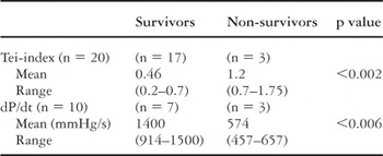
Table 1.

A conduit placed from the right ventricle to the pulmonary arteries in the modified Norwood operation. Note the small area of the conduit anastomosed to the right ventricle.
 You have Access