Introduction
Ventricular septal defect (VSD) is the most common form of congenital heart disease (CHD) accounting for about 40% of all forms of heart defects. Reference Penny and Vick1 Peri-membranous, subaortic, and doubly committed sub-arterial ventricular septal defects are found to be associated with aortic valve prolapse. Reference Monsefi, Zierer and Risteski2 Long-term follow-up studies have shown that transcatheter closure of ventricular septal defect is a feasible alternative of surgery. Reference Fu3 However, most of the published literatures have excluded ventricular septal defect with aortic cusp prolapse with or without aortic regurgitation from device closure. Main concern is risk of aggravation of aortic regurgitation after device closure. In this article, we summarised short-term follow-up results of transcatheter closure of ventricular septal defect in the presence of aortic cusp prolapse with or without mild aortic regurgitation.
Objective
To evaluate feasibility, safety, and short-term outcome of transcatheter closure of ventricular septal defect (peri-membranous or outlet muscular) with aortic cusp prolapse with or without mild aortic regurgitation.
Materials and methods
This prospective observational study was done in the Department of Pediatric Cardiology in NH-Rabindranath Tagore International Institute of Cardiac Sciences, Kolkata, India. Total 68 patients who had ventricular septal defect with aortic valve prolapse with or without mild aortic regurgitation and underwent ventricular septal defect closure by transcatheter route from January 2018 to December 2023 had been included in this study.
Inclusion criteria
(1) Peri-membranous or outlet muscular ventricular septal defect with aortic cusp prolapse with or without trivial/mild aortic regurgitation who were attempted for device closure and (2) presence of subaortic rim 2 mm or more.
Exclusion criteria
(1) Ventricular septal defects other than peri-membranous or outlet muscular (inlet /doubly committed ventricular septal defects); (2) ventricular septal defect diameter >20 mm by trans-thoracic echocardiography; (3) ventricular septal defect with aortic cusp prolapse if associated with more than mild aortic regurgitation; (4) gross aortic cusp prolapse; (5) absent or less than 2 mm subaortic rim; (6) severe pulmonary arterial hypertension that is not reversible after oxygen (with pulmonary vascular resistance index >8 WU.m2); (7) ventricular septal defect with right to left shunt; (8) associated abnormalities (e.g. mitral valve involvement) requiring open heart surgery.
Methods
Medical records, echocardiography recordings and reports, device closure reports, procedure (angiography) recordings, discharge summary, and follow-up data were analysed.
Patient selection was done in out-patient department as per inclusion criteria on the day before elective transcatheter device closure. Clinical evaluation (including gender, age, and body weight of the patient), chest X-ray, 12-lead electrocardiogram, and echocardiography were done.
Pre-intervention echocardiographic assessment
Suitability of device closure was meticulously evaluated measuring size and type of ventricular septal defect, rim size, its relationship with adjacent aortic and tricuspid valves, degree of aortic valve prolapse, presence or absence of aortic regurgitation, and its severity by transthoracic echocardiography. In case of adult or adolescent patient with limited echocardiographic views, transesophageal echocardiography was done. If it appeared to be suitable for transcatheter closure, then we moved forward.
The cut-off value for subaortic rim (distance between aortic valve and ventricular septal defect) was ≥2 mm. Defects with right ventricular exit-to-left ventricular entry ratio (right ventricular side/left ventricular side) ≤ 0.5 were classified as conical defects, whereas defects with right ventricular exit-to-left ventricular entry ratio >0.5 were classified as tubular defects. Selected peri-membranous defects (PM ventricular septal defects) were either linear without septal aneurysm formation or conical defects with septal aneurysm. Exit(s) on right ventricular side was meticulously evaluated. Selected outlet muscular defects were away from aortic valve cusps with sufficient subaortic rim (≥2 mm).
Interventional procedural technique
Written informed consent was signed by the parents of the children or patients. One dose of 3rd generation cephalosporin was given intravenously as antibiotic prophylaxis 30 minutes before the procedure and continued for 24 hour post-procedure. Most of the procedures were done under local anaesthesia and intravenous sedation without endotracheal intubation in the catheterisation laboratory. 2 patients under 1 year of age required endotracheal intubation. Mostly one femoral venous and one femoral arterial access were placed. Some patients needed internal jugular venous access. Sheath size was chosen according to the predicted device size. Sometimes, smaller initial sheath size was upgraded before taking the device. After that, intravenous heparin (100 IU/kg, maximum 5000 IU) was administered to all patients with regular monitoring of activated clotting time with the target of longer than 200 s.
We started the procedure by doing standard right and left heart catheterisation, and the haemodynamic data were recorded. Left ventriculogram with appropriate-sized pigtail catheter was performed. In case of PM ventricular septal defect, standard left anterior oblique 300 cranial 300 and left anterior oblique 600 cranial 200 views were used, whereas in case of outlet muscular ventricular septal defect, right anterior oblique 300 caudal 200 view was used. In case of multiple right ventricular exits, the largest one was chosen for measurement. The left ventricular entry side was measured on angiography at diastolic phase. Ventricular septal defect was crossed from retrograde route in maximum cases and rest of the cases from antegrade route using Judkins Right Coronary Catheter (JR) over J-tip glide wire (Terumo wire). In some cases, arteriovenous loop was formed by snaring the wire into the inferior vena cava. Device deployment was done mostly from retrograde approach, in others from antegrade approach (venous side).
In case of retrograde approach, we injected dye by hand injection though a long Touhy-Borst (Y connector) attached to the end of right coronary artery guiding catheter and position of the left ventricular disk in relation to the aortic valve cusp was assessed before releasing the device. Here, 2nd arterial access was not required. In case of antegrade approach, left ventriculogram or aortic root angiogram was done by pigtail catheter for the same purpose. After device deployment, thorough assessment was done to see the position of the device, residual defect, any change in geometry of aortic valve, impingement of aortic valve cusp by the disk of the device, appearance of new onset aortic regurgitation, and any increment in pre-existing aortic regurgitation by trans-thoracic echocardiography or transesophageal echocardiography. It was repeated after releasing the device. Patients were shifted to paediatric ICU and received aspirin.
Device and technique
We used AmplatzerTM Duct Occluder II (St. Jude Medical, St Paul, Minnesota) (ADO-II) in the majority of cases followed by Konar-MF occluder (MFO) and then CeraTM muscular ventricular septal defect occluder devices in these patients. In one case, AmplatzerTM Duct Occluder (ADO I) device was used.
ADO II was chosen depending on the following criteria:
(1) If the defect diameter <5 mm with a subaortic rim≥2 mm in non-aneurysmal type (tubular) defects, (2) for aneurysmal type, the right ventricular exit should be <5 mm with a left ventricular entry diameter <12 mm, and (3) the diameter of the device waist in ADO II was chosen to be 1–2 mm greater than the largest ventricular septal defect diameter.
MFO device was found suitable for (1) Peri-membranous conical defects with septal aneurysm where left waist diameter (D2) was same or just 1 mm larger than the defect dimension on the left ventricular side and (2) peri-membranous defects without septal aneurysm and outlet muscular defects with sufficient subaortic rim (≥2 mm) where D2 was chosen 1–2 mm larger than the defect diameter on the left ventricular side.
The Cera muscular ventricular septal defect occluder device was chosen according to the left ventricular entry diameter without any need to oversize.
Here, ADO I device was used in one case where aneurysm was large enough and to avoid touching the aortic valve by larger retention skirt of aortic disk, it was deployed in reverse fashion. The right disk (pulmonary end) of ADO I was kept in left ventricular side and left disk (aortic end) with retention skirt on right ventricular side (in 1 out of 68 patients).
Follow up
Tablet aspirin was given at a dose of 3–5 mg/kg/day to all the patients after ventricular septal defect device closure for 6 months. Clinical examination, 12 lead electrocardiogram and trans-thoracic echocardiography were performed on the next day of procedure, on the day of discharge (second post-intervention day), on 7th day, 3 months, 6 months and 1 year after device closure and yearly after that/SOS basis.
Statistical analysis
Categorical variables were expressed as number and percentages. Continuous variables were summarised as mean with standard deviation or median with range as appropriate. The statistical analyses were computed using the Statistical Package for the Social Sciences (SPSS Statistics), version 25.
Results
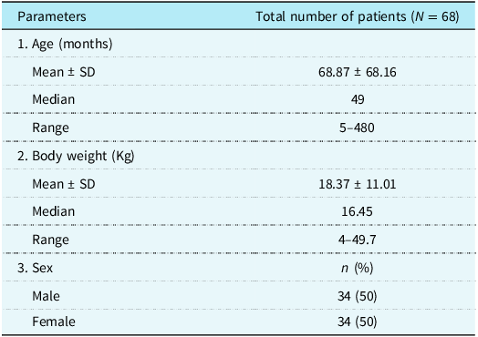
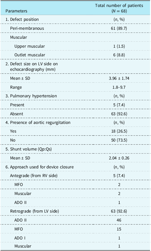
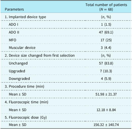
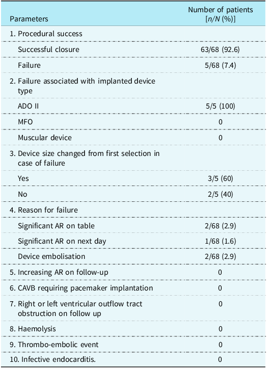
In this study, total 68 patients (as per inclusion and exclusion criteria) were included. Mean age of the study population was 68.87 months (standard deviation±68.16) with minimum age being 5 months and maximum age being 480 months (Table 1). The lowest body weight included in this study was 4 kg with mean of 18.37 kg (standard deviation±11.01) (Table 1). Male and female were equally distributed (50% each) in this study population. Ventricular septal defect was in the peri-membranous region in 61 (89.7%) patients, upper muscular region in one patient (1.5%) and outlet muscular region in 6 (8.8%) patients (Table 2). Mean defect size on left ventricular side was 3.96 mm (standard deviation±1.74) on echocardiography (Table 2). 7.4% patients had pulmonary arterial hypertension. Among all, 18 (26.5%) patients had trivial to mild aortic regurgitation, others had no aortic regurgitation (pre-procedure) (Table 2). Transcatheter ventricular septal defect closure was done with ADO II in 47 (69.1%) cases, 17 (25%) cases with MFO, another 3 (4.4%) cases with Muscular ventricular septal defect Occluder, and 1 (1.5%) case with ADO I (Table 3). Only in 5 cases (7.4%), device closure was done from antegrade approach. Among them, we used MFO device in 2 patients, muscular device in another 2 cases, and ADO II in 1 case. Remaining 63 (92.6%) cases, ventricular septal defect device deployment was done from left ventricular side (retrograde approach) and was tried to avoid device touching the aortic valve as much as possible. Among them, we used ADO II devices in 46 patients, MFO devices in 15 patients, muscular device in 1 patient and ADO I device in 1 patient (Table 2). In 63 out of 68 (92.6%) cases, ventricular septal defect device closure was successful and were not associated with appearance of new onset aortic regurgitation or aggravation of existing trivial to mild AR (Table 4). In 2 patients (2.9%), ventricular septal defect occluder appeared to be touching the aortic valve and aggravating aortic regurgitation even after repositioning and re-deploying the device from venous side and ultimately that devices were taken out and sent for surgical closure (Table 4). In one (1.6%) patient, device position appeared to be perfect on table before release and aortic regurgitation was also same as before and aortic root angiogram was also satisfactory. But re-evaluation by echocardiography on the day after procedure revealed aggravating aortic regurgitation. The patient was sent for device retrieval and surgical ventricular septal defect closure (Table 4). Another immediate major complication occurred in 2 patients (2.9%) in the form of device embolisation into right pulmonary artery few hours after releasing the device and the patients were sent for device retrieval and surgical ventricular septal defect closure (Table 4). All procedural failure were associated with ADO II devices. In 60% of failure cases, final device size was either upgraded or downgraded from the first device size used (Table 4). During the perioperative period, no death, major bleeding, pericardial tamponade, residual shunt, arrhythmia occurred. Mean follow up period of this study was 27 months (12–49 month). None of the children had shown any increase in severity of aortic regurgitation on follow up. No one developed atrioventricular block requiring permanent pacemaker implantation on short-term follow up and till the last out-patient department visit. None of the child had right ventricular or left ventricular outflow tract obstruction due to the device during follow-up. None of them had haemolysis, thrombo-embolic event or infective endocarditis (Table 4).
Demographic data

Procedural data

Procedural data continued

Outcome analysis

Discussion
Aortic valve prolapse and aortic regurgitation are well known to be associated with ventricular septal defect. Reference Monsefi, Zierer and Risteski2 Incidence of aortic valve prolapse is 5–8% in case of peri-membranous ventricular septal defect and as high as 30% in doubly committed subarterial ventricular septal defect. Reference Perloff4 In case of peri-membranous ventricular septal defect, aortic valve prolapse occurs due to the Venturi effect produced by the blood flowing from the left ventricle to the right ventricle through the ventricular septal defect during systole. Later, prolapse occurs due to the damaged aortic valve which is unable to withstand the high aortic pressure. In case of doubly committed subarterial ventricular septal defect, aortic valve prolapse occurs due to the lack of supporting structure in the subaortic area as there is complete deficiency of conal septum. The development of aortic valve prolapse is a risk factor for increasing aortic regurgitation. Reference Tomita, Arakaki, Ono, Yamada, Yagihara and Echigo5 Subaortic ventricular septal defect will enhance progression of aortic regurgitation with the incidence of 18%. Reference Pan, Xing and Cao6 So, early closure of such defects can prevent further damage of the aortic valve and can prevent progression of aortic regurgitation. Traditionally, surgery has been the treatment of choice for ventricular septal defect with aortic valve prolapse with or without aortic regurgitation. But, it does have some potential risks of complications, including heart block in 1–5% of the cases, Reference Ross-Hesselink, Mejiboom and Spitaels7–Reference Bol-Raap, Weerheim, Kappetein, Witsenburg and Bogers10 significant residual ventricular septal defect in 1–10% of the cases, Reference Mavroudis, Backer, Jacobs, Mavroudis and Backer8,Reference Kitagawa, Durham, Mosca and Bove11–Reference Backer, Winters and Zales14 the necessity for re-operation in 2% of the patients, Reference Mavroudis, Backer, Jacobs, Mavroudis and Backer8 and even death in 0.6–5% of the cases. Reference Mavroudis, Backer, Jacobs, Mavroudis and Backer8,Reference Hobbins, Izukawa, Radford, Williams and Trusler9,Reference Wollenek, Wyse, Sullivan, Elliot, de Leval and Stark12,Reference Serraf, Lacour-Gayet and Bruniaux13 Furthermore, infections, tachyarrhythmias, and neurological complications may occur after surgery. Reference Mavroudis, Backer, Jacobs, Mavroudis and Backer8 However, confronted with this obstacle, considering that transcatheter closure is less invasive than surgery and early occlusion can prevent development of aortic regurgitation and reduce the need for surgery, we proceeded with transcatheter closure for these patients and manifested a satisfactory result. However, there are limited data in the literature to provide clinical information on device closure of ventricular septal defects in patients with aortic valve prolapse and mild aortic regurgitation. The diameter of the defects and a limited aortic rim can pose a significant challenge to the operator. If the left ventricular disk of the occluder impairs movement of aortic valve leaflet, procedure may need to be stopped because of significant aortic regurgitation. This tends to spontaneously resolve with time but not always. Therefore, whether to perform the closure or not remains controversial.
ACC/AHA Guideline also reminded that patients with a small ventricular septal defect and aortic valve prolapse may not recommended as the primary indication for closure and high possibility of developing progressive aortic regurgitation. Reference Warnes, Williams and Bashore15
Our result is comparable with the study done by Guan-Liang Chen et al. Reference Chen, Li and Li16 and study by S. Ghosh et al. Reference Ghosh, Sridhar, Solomon and Sivaprakasham17 where ventricular septal defect device closure was done in ventricular septal defect with aortic valve prolapse.
In our study, mean age of the patients was 68.87 months (ranged from 5 to 480 months). In the study by Chen G-L, et al. Reference Chen, Li and Li16 and study by Ghosh S, et al., Reference Ghosh, Sridhar, Solomon and Sivaprakasham17 mean age was 72 months and 96 months, respectively. Here, number of male and female patients were equal, whereas males were slightly more in number in other study described by Chen G-L, et al. Reference Chen, Li and Li16 In this study, body weight was not considered as an inclusion criterion. Any patient who developed progressive aortic valve prolapse and trivial or mild aortic regurgitation needing immediate treatment were included for transcatheter intervention if the anatomy was suitable. Here, the lowest body weight included was 4 kg with mean of 18.37 kg. As this baby developed aortic valve prolapse with mild aortic regurgitation, we attempted for ventricular septal defect device closure keeping the body weight and risks of surgery in mind and ventricular septal defect was closed successfully without any complications. In the study by Ghosh S, et al., Reference Ghosh, Sridhar, Solomon and Sivaprakasham17 body weight of <5 kg was considered as an exclusion criterion, and the lowest body weight included was 9 kg with mean of 26.03 kg. Chen G-L, et al. Reference Chen, Li and Li16 described lowest body weight as 13.2 kg with mean of 24.9 kg in his study. In our study, the majority (89.7 %) had peri-membranous ventricular septal defect followed by outlet muscular ventricular septal defect (8.8%) similar to other studies by Ghosh S, et al. Reference Ghosh, Sridhar, Solomon and Sivaprakasham17 and Chen G-L, et al. Reference Chen, Li and Li16 Among all patients, 26.5% had trivial to mild pre-procedural aortic regurgitation, others had no aortic regurgitation. Transcatheter closure was done with ADO-II in 69.1% cases, 25% cases with MFO, another 4.4% cases with muscular ventricular septal defect occluder, and 1.5% cases with ADO I. Here, device closure was successful and was not associated with appearance of new-onset aortic regurgitation or aggravation of existing trivial to mild aortic regurgitation in 92.6% patients which was comparable to the study by Ghosh S, et al. Reference Ghosh, Sridhar, Solomon and Sivaprakasham17 and Chen G-L, et al. Reference Chen, Li and Li16 where immediate success rate was 89% and 96.9%, respectively. In 2 (2.9%) cases, ventricular septal defect device appears to be touching the aortic valve and aggravating aortic regurgitation even after repositioning and re-deploying the device from venous side and ultimately that devices were taken out and the patients were sent for surgical closure. In this study, 1 (1.6%) patient’s device position appeared to be perfect on table before release and aortic regurgitation was also same as before, but re-evaluation by echocardiography on the day after procedure revealed aggravating aortic regurgitation. This patient was sent for surgical removal of the device and ventricular septal defect closure. In the study by Chen G-L, et al. Reference Chen, Li and Li16 and study by Ghosh S, et al., Reference Ghosh, Sridhar, Solomon and Sivaprakasham17 3% and 5% cases were referred for surgery because of significant aggravation of device related aortic regurgitation. Here, 2 patients (2.9%) had embolisation of device into right pulmonary artery few hours after releasing the device and it was comparable to the study by Ghosh S, et al. Reference Ghosh, Sridhar, Solomon and Sivaprakasham17 where 1 (5.2%) patient had device embolisation. None of the children had shown any increase in severity of aortic regurgitation on follow-up, but in the study by Ghosh S, et al., Reference Ghosh, Sridhar, Solomon and Sivaprakasham17 one (5.2%) patient had immediate new-onset mild aortic regurgitation which progressed to moderate aortic regurgitation over 18 months of follow-up and required partial device amputation and surgical ventricular septal defect closure. Complete atrio-ventricular block is one of the most dangerous complications associated with either transcatheter device closure or surgical closure of PM ventricular septal defect because of mechanical stimulation or compression of the conduction system. In recent years, complete atrio-ventricular block has been deemed a rare complication after transcatheter closure of PM ventricular septal defect. In general, the incidence of late presenting complete atrio-ventricular block after surgical ventricular septal defect closure was described between 0.3 and 0.7% in most of the reported paediatric studies. Reference Lin, Mahle and Frias18 However, a few reports described higher incidence up to 1–2%. Reference Laurens, Gavelle, Piwnica, Farge, Dubost and Maurice19 In our study, no one developed complete atrio-ventricular block requiring pacemaker implantation on follow-up, whereas in the study by Azab S, et al. Reference Azab, Shahawy, Samy and Mahdy20 3.5% and in the study by Siehr SL, et al. Reference Siehr, Hanley, Reddy, Miyake and Dubin21 1.9% patients developed permanent complete atrio-ventricular block requiring pacemaker implantation following ventricular septal defect surgical closure. None of the children had infective endocarditis (IE) on follow-up. Although, it was seen that 2.7% patients developed IE after surgical ventricular septal defect closure on 25 years of follow-up, Reference Morris, Reller and Menashe22 in case of transcatheter ventricular septal defect closure incidence of infective endocarditis is very rare. In our experience, the proper selection of patients was the most important factor for the success of the procedure. Pre intervention profiling of ventricular septal defect by trans-thoracic echocardiography or transesophageal echocardiography included: (1) Location—PM, upper muscular, outlet muscular, subaortic; (2) proximity to aortic valve, right coronary cusp prolapse, non-coronary cusp prolapse; (3) amount of aortic regurgitation – no, mild, more than mild; (4) distortion of aortic valve; and (5) size of the defects.
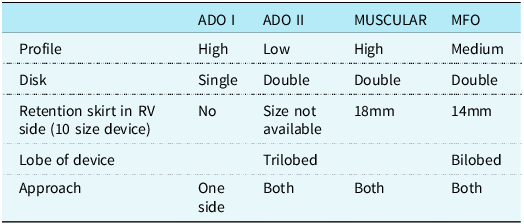
With the presence of adequate aortic rim, and not more than mild aortic regurgitation, we successfully closed the ventricular septal defect with aortic cusp prolapse (Figure 1 & 2). We did not try for transcatheter closure of ventricular septal defect with gross aortic cusp prolapse, distorted cusps, more than mild aortic regurgitation (Figure 3). Intra-operative profiling of ventricular septal defect during left ventricular angiography according to the location of ventricular septal defect is important (Figure 4 & 5). Proper device selection is also important (Table 5).
TTE in A5C view showing PM VSD who are suitable for device closure. TTE = trans-thoracic echocardiography; PM VSD = Peri-membranous ventricular septal defect.

TTE in PLAX view showing outlet muscular VSD who are suitable for device closure. TTE = Trans-thoracic echocardiography; VSD = ventricular septal defect.

TTE in PLAX view showing outlet VSD with gross aortic cusp prolapse who are not suitable for device closure. TTE = Trans-thoracic echocardiography; VSD = ventricular septal defect.

LV angiogram in LAO 600 cranial 200 view with pigtail catheter showing PM VSD. LV= left ventricle; PM VSD = Peri-membranous ventricular septal defect.

LV angiogram in RAO 300 caudal 200 view with pigtail catheter showing outlet muscular VSD without any aortic cusp prolapse. LV = left ventricle; VSD = ventricular septal defect.

ADO II: Low profile, self-centering device sits away from cusp without much manoeuvre (touch the part of device to keep away from aortic cusps). 4–5 Fr ADO II delivery system and 5–6 Fr guiding catheter (e.g. guiding JR coronary catheter) can be used for the largest device. Limiting factor is only small defects measuring up to 4–5 mm can be closed (Figure 6).
PLAX view on TTE showing ADO II device perfectly positioned after releasing the device without any residual shunt. TTE = Trans-thoracic echocardiography.

MFO: Wide spectrum of size from 5/3 to 14/12 mm are available. Medium profile (more than ADO II, but less than muscular) device and can be approached from both venous side and aortic side. The device needs manoeuvre to properly place as it is not self-centering. In our experience, some amount of manipulation was needed to achieve final desired position. If part of the device was still inside aortic cusps with or without causing significant aortic regurgitation, pushing the left ventricular disk with cable during release of the device helped to keep the device away from aortic cusps. The secured device position was documented after releasing the device by doing aortic root angiogram (Figure 7 & 8).
Fluoroscopy in RAO 300 caudal 200 view showing Konar-MFO device deployment from retrograde approach to close outlet muscular VSD. VSD = ventricular septal defect.

Aortic root angiogram in RAO 300 caudal 200 view after releasing MFO device showing device is positioned perfectly away from aortic cusps without any AR. AR = aortic regurgitation.

Muscular device: High profile, double disk device; wide range of sizes are available (waist size 4 mm to 24 mm), length of the device is same (7 mm), can be deployed from both antegrade and retrograde route. This device is mostly used for defect in muscular septum, however can also be used in peri-membranous defects. Quite stout device that are suitable for thick interventricular septum in older kids or defect with septal aneurysm formation.
ADO I: In one case. we used ADO I, and it was done before widespread use of MFO device in our unit. To avoid touching of the larger aortic disk of ADO I to aortic cusp, we put it in reverse position (Figure 9).
Fluoroscopy showing deployment of ADO I device from retrograde approach in reverse fashion in LAO 600 cranial 200 view.

Proper technique also plays an important role, whether it is antegrade approach or retrograde approach (Figure 7, 10 & 11). Post-deployment aortogram was done in every case to assess whether device was away from aortic cusp or restriction of movement of the same and aortic regurgitation (Figure 12). Touhy-Borst adapter was attached to the hub of guiding catheter. It was helpful where larger (6/7 Fr) guiding catheters being used. Aortogram avoids cumbersome process and long radiation exposure associated with right ventricular angiogram with levophase. Aortic root angiogram after releasing the device was also done in every case (Figure 8). Do not release the device in the presence of new onset or aggravation of previous aortic regurgitation (Figure 13).
Fluoroscopy showing arteriovenous loop formation by snaring the tip of the wire through IVC. IVC = inferior vena cava.

Fluoroscopy showing device deployment from antegrade approach in LAO 600 cranial 200 view.

Post device deployment aortogram in LAO 600 cranial 200 showing MFO device is positioned perfectly away from aortic cusps.

TTE in A5C view after releasing the device showing development of aortic regurgitation. TTE = trans-thoracic echocardiography.

Conclusion
Transcatheter closure of ventricular septal defect with aortic cusps prolapse with or without mild aortic regurgitation in selected patients is technically feasible and safe with high procedural success rate. However, thorough understanding of anatomy and operator’s expertise along with meticulous case selection, device selection, deployment route, and technique is necessary for desired outcome. If aortic regurgitation appears to be increasing in nature on table after device deployment, then taking out the device is highly recommended. Increasing aortic regurgitation is due to the mechanical hindrance of coaptation of valve leaflets or distortion of valve, which will progress with time (adherence /fibrosis). Spontaneous recovery of increasing degree of aortic regurgitation after device release is not expected. Long-term safety and efficacy need to be assessed to draw conclusion.
Study limitation
Single centre-based study. Study population was small. It was observational study; long-term follow-up was not done for all patients. Study population was heterogeneous in terms of age, type of defect, and devices used.
Properties of different devices used for ventricular septal defect closure

Acknowledgements
None.
Financial support
This research received no specific grant from any funding agency, commercial, or not-for-profit sectors.
Competing interests
None.
Ethical standard
Not applicable.