





Non-technical Summary
The Late Ordovician (ca. 458 to 443 million years ago) shales of the Cincinnati Arch region are well known as the “butter shales” for their lithological characteristics. They contain well-preserved fossils. Among the diverse taxonomic groups recovered from the shales, ostracods (small bivalved crustaceans) have been neglected, despite their great potential for reconstructing diversity histories and distributional patterns of the past. Here we examine the taxonomy of ostracods from the butter shale for the first time. Twenty-nine species of 24 genera are systematically described, including two new species: Milleratia binodosa n. sp. and Daleiella? cincinnatiensis n. sp. Results indicate that ostracods also participated in the process of the “Richmondian Invasion” (a regional Biotic Immigration Event whereby organisms migrated into the Cincinnati Basin) during the Late Ordovician. Furthermore, strong biogeographical connections between the present fauna and faunas within Laurentia (an ancient continent generally consisting of present-day North America and Greenland) as well as faunas from Baltica (an ancient continent generally consisting of present-day Northern and northeastern Europe) are also found.
Introduction
The Upper Ordovician (Katian) from the Cincinnati Arch region of North America is well known for its continuous sedimentary successions with great fossil diversities and has been studied extensively for over 150 years (Orton, Reference Orton, Newberry, Orton, Andrews, Wormley and Meek1873; Nickles, Reference Nickles1902; Shideler, Reference Shideler1914; McFarlan, Reference McFarlan and Jillson1931; Fox, Reference Fox1962; Holland, Reference Holland1993; Holland and Patzkowsky, Reference Holland and Patzkowsky1996, Reference Holland, Patzkowsky, McLaughlin, Brett, Holland and Storrs2008; McLaughlin et al., Reference McLaughlin, Brett, Holland and Storrs2008; Brett et al., Reference Brett, Cramer, Dattilo, McLaughlin, Schramm, Thomka, Brett, Cramer and Gerke2012a, Reference Brett, McLaughlin, Schramm, Sullivan, Thomka, Brett, Cramer and Gerke2012b, Reference Brett, Hartshorn, Wald, McLaughlin, Bulinski, Thomka, Paton, Freeman, Dattilo and Flora2018a, Reference Brett, Hartshorn, Young, Schwalbach and Stigall2018b, Reference Brett, Aucoin, Dattilo, Freeman, Hartshorn, McLaughlin and Schwalbach2020). Within the region’s predominant carbonates are the conspicuously recurring Lagerstätten, informally referred to as “butter shales,” which have yielded an abundance of articulated trilobites (Schumacher and Shrake, Reference Schumacher, Shrake, Brett and Baird1997; Hunda et al., Reference Hunda, Hughes and Flessa2006; Hunda and Hughes, Reference Hunda and Hughes2007; Hunda, Reference Hunda, McLaughlin, Brett, Holland and Storrs2008), as well as well-preserved brachiopods, echinoderms, bivalves, bryozoans, and many other fossil groups (Foerste, Reference Foerste1916; Frey, Reference Frey1987a, Reference Frey1987b; Holland and Patzkowsky, Reference Holland and Patzkowsky2007), making them critical layers for sequence stratigraphy and taphonomy studies (e.g., Brett et al., Reference Brett, Baird, Speyer, Brett and Baird1997, Reference Brett, Aucoin, Dattilo, Freeman, Hartshorn, McLaughlin and Schwalbach2020; Aucoin et al., Reference Aucoin, Brett, Dattilo and Thomka2016; Aucoin and Brett, Reference Aucoin and Brett2016). Yet microfossils, a very good proxy for biogeographical, paleoecological, and macroevolutionary analyses (Williams et al., Reference Williams, Floyd, Salas, Siveter, Stone and Vannier2003; Meidla et al., Reference Meidla, Tinn, Salas, Williams, Siveter, Vandenbroucke, Sabbe, Harper and Servais2013; Yasuhara et al., Reference Yasuhara, Tittensor, Hillebrand and Worm2017, Reference Yasuhara, Hunt and Jordan2022; Yasuhara, Reference Yasuhara2019), have received limited attention in this region, with focus mainly on conodonts for biostratigraphical correlations (Bergström and Sweet, Reference Bergström and Sweet1966; Sweet, Reference Sweet1979, Reference Sweet1984).
Ostracods range from the Tremadocian (Early Ordovician) to the present and are one of the most species-diverse clades of crustaceans (Horne, Reference Horne, Holmes and Chivas2002; Williams et al., Reference Williams, Siveter, Salas, Vannier, Popov and Ghobadi Pour2008). As a fundamental component of marine benthic communities during the Great Ordovician Biodiversification Event (Braddy et al., Reference Braddy, Tollerton, Racheboeuf, Schallreuter, Webby, Paris, Droser and Percival2004; Harper et al., Reference Harper, Cascales-Miñana and Servais2020), ostracods offer great potential for biodiversity and biogeographical analyses due to their remarkable abundance and wide distribution. Early studies on ostracods from the Upper Ordovician of the Cincinnati Arch region were initiated by Hall (Reference Hall1871), Hall and Whitfield (Reference Hall and Whitfield1875), and Miller (Reference Miller1874a– Reference Millerd, Reference Miller1875), who included limited descriptions of a few species in their geological survey reports. Building on this foundation, Ulrich (Reference Ulrich1879, Reference Ulrich1890) documented more than 40 species from the Upper Ordovician sporadically distributed in Ohio, Kentucky, Tennessee, and Minnesota. However, these species were described with hand-drawn sketches and imprecise stratigraphical information. Later, Shideler (Reference Shideler1927) listed 26 ostracod species from the late Katian Arnheim, Waynesville, Liberty, and Whitewater formations of the Cincinnati Arch region, yet provided no taxonomy. Subsequent effort was carried out by Warshauer and Berdan (Reference Warshauer and Berdan1982), who systematically described 53 species from the early Katian Lexington and Clays Ferry formations of central Kentucky, representing the most comprehensive work in the region thus far.
Here we describe newly recovered, well-preserved ostracods from the butter-shale Lagerstätten, accompanied by taxonomic revisions. Faunal composition, paleoenvironmental, and biogeographical implications are being discussed accordingly.
Geologic setting
During the Late Ordovician, the region now known as Cincinnati in southern Ohio was paleogeographically situated in southern Laurentia in a region of shallow epicontinental sea (Holland, Reference Holland1993; Holland and Patzkowsky, Reference Holland and Patzkowsky2007; Meyer and Davis, Reference Meyer and Davis2009; Brett et al., Reference Brett, Malgieri, Thomka, Aucoin, Dattilo and Schwalbach2015). The depositional environment of the Cincinnati Arch exhibits mixed carbonate–siliciclastic facies that lead to the notable Cincinnatian Series, a carbonate-dominated succession interspersed with shales, siltstones, and argillaceous dolostones (Brett et al., Reference Brett, Aucoin, Dattilo, Freeman, Hartshorn, McLaughlin and Schwalbach2020). Stratigraphically, the Cincinnatian is further divided into a trio of regional stages: the Edenian, Maysvillian, and Richmondian stages, each comprising numerous formations and members (Fig. 1) (Brett et al., Reference Brett, Aucoin, Dattilo, Freeman, Hartshorn, McLaughlin and Schwalbach2020). These stages range from the Katian Amorphognathus superbus to A. ordovicicus conodont zones (Bergström and Sweet, Reference Bergström and Sweet1966; Sweet, Reference Sweet1979, Reference Sweet1984). Within sequence stratigraphic contexts, the Cincinnatian Series is described as the “C” sequence with eight further subdivisions and is underlain by the Mohawkian Series “M” sequence (Fig. 1).
(1, 2) Location map of the sampled area (indicated by orange diamond) and sections (indicated by green and blue diamonds). (3) Stratigraphical framework of the Cincinnatian Series (Holland and Patzkowsky, Reference Holland and Patzkowsky1996; Brett et al., Reference Brett, Aucoin, Dattilo, Freeman, Hartshorn, McLaughlin and Schwalbach2020) with butter-shales positions (Aucoin et al., Reference Aucoin, Brett, Dattilo and Thomka2016) and sequence stratigraphy (Brett et al., Reference Brett, Aucoin, Dattilo, Freeman, Hartshorn, McLaughlin and Schwalbach2020). Chr. Str. = Chronostratigraphy; Gl. = Global; N.A. = North American; Seq. Str. = Sequence Stratigraphy. Samples collected from two sections (indicated by green and blue shading) are categorized from “Shale 1” (S1) to “Shale 7” (S7). Gray shading indicates sequences containing Richmondian Invasion stages (Stigall, Reference Stigall2023).

Figure 1. Long description
Panel 1 is a regional map of Indiana, Ohio, and Kentucky. An orange diamond marks Cincinnati at the intersection of the three states. Panel 2 is a local map showing Caesar Creek Section marked with a green diamond in the North and Flat Run Section marked with a blue diamond in the Southeast, near Mt Orab. Major highways I 75, I 71, and I 275 are labeled.
Panel 3 is a stratigraphic table with seven columns. From left to right:
1. Global Chronostratigraphy: Upper Ordovician Series, Katian Stage.
2. North American Chronostratigraphy: Cincinnatian Series, divided into Edenian, Maysvillian, and Richmondian stages.
3. Formation: Lists units from bottom to top including Lexington, Kope, Fairview, Grant Lake, Arnheim, Waynesville, Liberty, Whitewater, and Elkhorn.
4. Member: Specific subdivisions like Point Pleasant, Economy, Southgate, McMicken, Mt. Hope, Fairmount, Miamitown, Bellevue, Corryville, Mt. Auburn, Sunset, Oregonia, Fort Ancient, Clarksville, and Blanchester.
5. Butter Shales: Identifies seven specific shale layers. S 1 (Dent Trilobite Shale) is shaded blue. S 2 (Mt. Orab Shale) is shaded blue. S 3 through S 7 are shaded green, including lowest Ft. Ancient (S 3), Harpers Run (S 4), Oldenburg (S 5), Roaring Brook (S 6), and F. minuens Shale (S 7).
6. Sequence Stratigraphy: Labeled M, then C 1 through C 8. Gray shading highlights sequences C 4 B, C 5, and C 6, indicating Richmondian Invasion stages.
Although a wide range of carbonate lithologies form the Upper Ordovician of the region, siliciclastic facies also comprise a part, representing a sedimentary cycle that reflects sea-level fluctuations (e.g., Holland, Reference Holland1993; Holland and Patzkowsky, Reference Holland and Patzkowsky1996; Brett et al., Reference Brett, Hartshorn, Wald, McLaughlin, Bulinski, Thomka, Paton, Freeman, Dattilo and Flora2018a, Reference Brett, Aucoin, Dattilo, Freeman, Hartshorn, McLaughlin and Schwalbach2020). The butter-shale Lagerstätten, which corresponds to mudstone-rich facies where fine-grained mud deposition outpaced skeletal accumulation (Aucoin et al., Reference Aucoin, Dattilo, Brett and Cooper2015, Reference Aucoin, Brett, Dattilo and Thomka2016; Brett et al., Reference Brett, Aucoin, Dattilo, Freeman, Hartshorn, McLaughlin and Schwalbach2020), are typically highly fossiliferous. These layers are composed primarily of clays largely undiluted by carbonate material, resulting in a soft, butter-like consistency (Aucoin et al., Reference Aucoin, Dattilo, Brett and Cooper2015, Reference Aucoin, Brett, Dattilo and Thomka2016). In this paper, we adopt the stratigraphical framework outlined by Aucoin et al. (Reference Aucoin, Brett, Dattilo and Thomka2016) and Brett et al. (Reference Brett, Aucoin, Dattilo, Freeman, Hartshorn, McLaughlin and Schwalbach2020) (Fig. 1).
There are eight layers of butter shales interbedded within the Cincinnatian Series (Fig. 1), including the Moranburg shale, Western Hills Trilobite shale, Dent Trilobite shale, Mt. Orab shale, Harpers Run submember, Oldenburg submember, Roaring Brook submember, and Flexicalymene minuens shale, which form parts of the Kope to the Liberty formations in an ascending order.
Materials and methods
Seven ostracod samples were newly obtained from two sections in the Cincinnati Arch region, the Flat Run section (privately owned) at Frye Road, Mt. Orab, Ohio, and the Caesar Creek section at the Caesar Creek State Park, Waynesville, Ohio (Figs. 1, 2). The Flat Run section was thoroughly described and studied previously for its well-preserved trilobites (Hunda et al., Reference Hunda, Hughes and Flessa2006), while the Caesar Creek section is a classic site for studying the Richmondian Stage in the region (Shrake, Reference Shrake1992; Aucoin et al., Reference Aucoin, Dattilo, Brett and Cooper2015, Reference Aucoin, Brett, Dattilo and Thomka2016; Aucoin and Brett, Reference Aucoin and Brett2016; Brett et al., Reference Brett, Aucoin, Dattilo, Freeman, Hartshorn, McLaughlin and Schwalbach2020, Reference Brett, Forsythe, Dattilo, Farnam, Hartshorn and Little2024). Specifically, two samples were collected from the Arnheim Formation of the Flat Run section, including the Dent Trilobite shale (S1) of the Sunset Member and the Mt. Orab shale (S2) of the Oregonia Member (Figs. 1, 2). We further gathered five samples from the Waynesville and Liberty formations of the Caesar Creek section, comprising the lowest shale (S3) and the Harpers Run submember (S4) of the Fort Ancient Member, the Oldenburg (S5) and Roaring Brook (S6) submembers of the Blanchester Member, and last, the Flexicalymene minuens shale (S7) of the Liberty Formation (Figs. 1, 2). Each sample weighs 2 kg and is processed with water only. Samples were washed through a 63-μm mesh sieve, and a total of 6,743 specimens were preserved as calcified valves and carapaces and manually picked out of residues using a binocular microscope. Uncoated specimens were digitally imaged using a HITACHI S3400N variable pressure scanning electronic microscope in low-vacuum mode at the Electron Microscope Unit of the University of Hong Kong.
Stratigraphical range of ostracods from the butter shales of the Cincinnati Arch region. Width of the bars represents relative abundance (%) of each species in each sample from the two sections (indicated by green and blue shading). See Figure 1 for age determinations of each stratigraphic formation and member. Note that 7 m of lithology in the Clarksville Member are omitted because no samples were collected from this interval.

Figure 2. Long description
The chart is divided into two main horizontal panels.
Top Panel: Caesar Creek Section. The vertical axis on the left shows stratigraphic members from bottom to top: Fort Ancient, Clarksville (with a 7 m gap), Blanchester, and Liberty. A lithology column uses patterns for Limestone, Shale, and Shale with carbonate nodules. Arrows labeled S 3 through S 7 indicate specific sample points.
Bottom Panel: Flat Run Section. The vertical axis shows the Sunset and Oregonia members. Arrows S 1 and S 2 mark sample points.
Horizontal Axis: Lists 28 ostracod species names including Macronotella sp., Saccelatia? sp., Ctenobolbina ventrispinifera, and others ending with Euprimitia cf. sanctipauli.
Data Representation: Vertical bars of varying widths represent relative abundance in percent. A legend in the center-right defines the widths: a dot for less than 1 percent, and increasingly thick rectangles for 10, 20, 40, and 60 percent.
Key Observations:
- Daleiella? cincinnatiensis n. sp. shows high abundance (40 to 60 percent) consistently across most samples in both sections.
- Warthinia nodosa is prominent in the lower sections (Sunset, Oregonia, and Fort Ancient).
- Species like Milleratia cincinnatiensis and Americoncha punctata appear more frequently in the Oregonia and Blanchester members.
- Several species, such as Bollia regularis and Krausella brevicornis, appear only sporadically in specific samples.
The classification scheme used in this study follows Vannier et al. (Reference Vannier, Siveter and Schallreuter1989), Mohibullah et al. (Reference Mohibullah, Williams and Zalasiewicz2014), and Siveter et al. (Reference Siveter, Briggs, Siveter and Sutton2024), as well as Scott (Reference Scott and Moore1961). The morphological terminology is from Jaanusson (Reference Jaanusson1957), Scott (Reference Scott and Moore1961), and Henningsmoen (Reference Henningsmoen1965).
Repository and institutional abbreviation
Type and figured specimens are housed in the Cincinnati Museum Center (catalog numbers CMC IP103873–IP103969).
Results
In total, 29 species of 24 genera are identified from the shales (Figs. 2, 3). The butter-shale ostracod fauna is characterized by the dominance of beyrichiocopids (15 species of 12 genera), among which the suborder Binodicopina (seven species of six genera) accounts for the most (Fig. 3). The most abundant components of the fauna at the species level are the podocopid Daleiella? cincinnatiensis n. sp. and beyrichiocopid Warthinia nodosa (Ulrich, Reference Ulrich1890) (Fig. 2). The podocopids Longiscula subcylindrica (Ulrich, Reference Ulrich and Ulrich1889), Phelobythocypris cylindrica (Hall, Reference Hall1871), and Rectella cf. R. derosforma Taha, Reference Taha2018, and beyrichiocopids Quasibollia persulcata (Ulrich, Reference Ulrich1879) and Euprimitia minuta Keenan, Reference Keenan1951 are less abundant, while the remaining species exhibit rather low abundances (Fig. 2). In general, the fauna is characterized by relatively low diversity but high abundance.
General composition of the butter-shale ostracod fauna from the Cincinnati Arch region at the standpoints of taxonomy (orders and suborders) and endemicity (species and genus levels).

Figure 3. Long description
The chart uses a vertical Y-axis labeled Percentage % ranging from 0 to 100 in increments of 25.
Column 1, Faunal composition by Order:
* Bottom section (orange): Beyrichiocopida at approximately 52%.
* Middle section (dark gray): Podocopida from 52% to 76%.
* Top section (blue): Platycopida from 76% to 100%.
Column 2, Faunal composition by Suborder:
* Bottom section (black): Binodicopina at 24%.
* Second section (dark charcoal): Palaeocopina from 24% to 42%.
* Third section (medium-dark gray): Eridostracina from 42% to 48%.
* Fourth section (medium gray): Leiocopina from 48% to 52%.
* Fifth section (light-medium gray): Cytherelliformes from 52% to 76%.
* Top section (light gray): Metacopina from 76% to 100%.
Column 3, Endemicity at Species level:
* Bottom section (blue-gray): Recorded elsewhere at 55%.
* Top section (olive green): Endemic at 45%.
Column 4, Endemicity at Genus level:
* Single solid section (blue-gray): Recorded elsewhere at 100%.
Discussion
Richmondian Invasion
The late Katian “Richmondian Invasion” is interpreted as a regional Biotic Immigration Event (BIME), during which the inter-basinal migration of organisms from multiple source regions into the Cincinnati Basin led to a drastic increase in diversity (Holland, Reference Holland, Brett and Baird1997; Holland and Patzkowsky, Reference Holland and Patzkowsky2007; Stigall, Reference Stigall2010, Reference Stigall2023; Stigall et al., Reference Stigall, Bauer, Lam and Wright2017). Characterized by niche partitioning and the restructuring of shallow marine communities, the invasion records the influx of mainly cephalopods, brachiopods, cnidaria, and mollusks (Stigall et al., Reference Stigall, Bauer, Lam and Wright2017; Stigall, Reference Stigall2019, Reference Stigall2023). Ostracods also participated in this process as evidenced by the presence of both native and migrated elements, forming a merging butter-shale fauna (Fig. 4).
Ranges of butter-shale ostracod fauna of the Cincinnati Arch region against the Richmondian Invasion stages of Stigall (Reference Stigall2023), based on the sequence stratigraphical framework (Holland and Patzkowsky, Reference Holland and Patzkowsky1996; Brett et al., Reference Brett, Aucoin, Dattilo, Freeman, Hartshorn, McLaughlin and Schwalbach2020). Basins modified from Stigall (Reference Stigall2023), with data from Cincinnati Basin (Warshauer and Berdan, Reference Warshauer and Berdan1982), Eastern Midcontinent (Keenan, Reference Keenan1951; Swain, Reference Swain and Sloan1987; Swain and Cornell, Reference Swain, Cornell and Sloan1987), and Appalachian Basin (A. B.) (Swain, Reference Swain1957, 1962; Kraft, Reference Kraft1962; Copeland, Reference Copeland1982; Zhang et al., Reference Zhang, Yasuhara, Tian and Hopkins2025). Due to lack of study on ostracods, data are missing from C1 to C3 sequences (marked with dashed line).

Figure 4. Long description
The chart is organized into three vertical groups of genera. The horizontal axis lists four stratigraphic stages: M (before), C 1 dash 3 (marked with dashed lines indicating missing data), C 4 (early), and C 5, C 6 (main).
* Native elements (orange bars): Warthinia, Quasibollia, Americoncha, Milleratia, Schmidtella, and Daleiella? span all stages. Phelobythocypris spans all stages. Ctenobolbina, Macronotella, and Laccoprimitia end after C 4. Elliptocyprites and Krausella are present in M and C 1 dash 3, absent in C 4, and reappear in C 5, C 6.
* From Eastern Midcontinent (blue bars): Bollia, Aechmina, Tetradella, Euprimitia, and Primitiella? begin at C 1 dash 3 and continue through C 5, C 6. Saccelatia? begins at C 1 dash 3 and ends after C 4. Pseudulrichia, Hastatellina, and Primitia appear only in the C 5, C 6 stage.
* From A. B. (Appalachian Basin) (gray bars): Longiscula and Rectella begin at C 1 dash 3 and continue through C 5, C 6. Macrocyproides appears only in the C 5, C 6 stage.
As the Richmondian Invasion is classified as a “coordinated invasion” within the invasion hierarchy (Stigall, Reference Stigall2019), its regional scale limits fundamental shifts in entire faunas, yet notable changes in faunal composition are still detectable (Holland and Patzkowsky, Reference Holland and Patzkowsky2007; Patzkowsky and Holland, Reference Patzkowsky and Holland2007; Stigall, Reference Stigall2010, Reference Stigall2023; Brame and Stigall, Reference Brame and Stigall2014). In our butter-shale fauna, 12 genera were present before the invasion, recovered from the Lexington and Clays Ferry formations (Warshauer and Berdan, Reference Warshauer and Berdan1982), and are considered native to the region at the M sequence (Fig. 4). Beginning with the C4 sequence (Figs. 1, S1, S2), the early stage of the invasion was moderate, permitting the establishment of species of non-native genera in the new environment alongside species of native genera. Six genera (Bollia, Aechmina, Tetradella, Euprimitia, Primitiella?, Saccelatia?) from the Eastern Midcontinent (= Illinois, Iowa, Missouri; Stigall, Reference Stigall2023) (Swain, Reference Swain and Sloan1987; Swain and Cornell, Reference Swain, Cornell and Sloan1987) and two genera (Longiscula, Rectella) from the Appalachian Basin (Stigall, Reference Stigall2023; Zhang et al., Reference Zhang, Yasuhara, Tian and Hopkins2025) were introduced into the Cincinnati fauna (Figs. 4, 5). During the C5 and C6 sequences (Figs. 1, S3–S7), invasive elements contributed to changes in the paleocommunity structure and the establishment of a new one through niche partitioning (Brame and Stigall, Reference Brame and Stigall2014; Stigall et al., Reference Stigall2023). A high speciation rate is recorded among rhynchonelliform brachiopods and other major fossil groups during this interval (Stigall, Reference Stigall2010, Reference Stigall2018, Reference Stigall2023; Stigall et al., Reference Stigall, Bauer, Lam and Wright2017), marking the main stage of the invasion. This aligns well with our faunal composition, which exhibits relatively high endemicity at the species level whereas all genera have been documented elsewhere (Fig. 3). Three additional genera (Pseudulrichia, Hastatellina, Primitia) from the Eastern Midcontinent (Keenan, Reference Keenan1951; Swain, Reference Swain and Sloan1987; Swain and Cornell, Reference Swain, Cornell and Sloan1987) and Macrocyproides from the Appalachian Basin (Swain, Reference Swain1957, Reference Swain1962; Kraft, Reference Kraft1962; Copeland, Reference Copeland1982; Zhang et al., Reference Zhang, Yasuhara, Tian and Hopkins2025) migrated into the Cincinnati Basin during this interval (Figs. 4, 5). However, the understudied nature of ostracods from earlier sequences (C1–C3) in the region may introduce biases in estimating the migration timing. Further investigations are necessary for a better understanding of ostracods involved in the invasion.
(1) Reconstructed ostracod dispersal pathways into the Cincinnati Basin during the late Katian Age (~450 Ma). Blue and gray arrows associated with Aechmina and Longiscula, respectively, represent the invasions from the Eastern Midcontinent and the Appalachian Basin (see Fig. 4 caption for references). Orange arrows indicate simplified ocean surface circulation, modified from Pohl et al. (Reference Pohl, Nardin, Vandenbroucke and Donnadieu2016). 1 = Cincinnati Basin; 2 = Eastern Midcontinent (= Illinois, Iowa, Missouri); 3 = Appalachian Basin. Basins modified from Stigall (Reference Stigall2023). Base map modified from Blakey (Reference Blakey2013) and Torsvik and Cocks (Reference Torsvik and Cocks2017). (2) Genus-level relations between the studied butter-shale ostracod fauna and selected faunas from Baltica (Hessland, Reference Hessland1949; Henningsmoen, Reference Henningsmoen1954; Gailīte, Reference Gailīte and Grigelis1971; Přibyl, Reference Přibyl1975; Olempska, Reference Olempska1994; Meidla, Reference Meidla1996) and Avalonia (Schallreuter et al., Reference Schallreuter, Verniers and De Geest2000; Williams et al., Reference Williams, Stone, Siveter and Taylor2001) during the late Katian Age (~450 Ma). Marked with number of commonly shared genera between two regions connected by black line. Number in Laurentia represents the number of commonly shared genera between the butter-shale fauna and other Laurentian faunas. Base map modified from Torsvik and Cocks (Reference Torsvik and Cocks2017); shapes of paleocontinents are simplified.

Figure 5. Long description
Panel 1: A map of Laurentia at 450 M a. Orange arrows indicate a clockwise ocean surface circulation pattern around the continent. In the Southeast, three numbered regions are highlighted. Region 1, the Cincinnati Basin, is marked with an orange diamond. Region 2, the Eastern Midcontinent, is shaded blue with a blue arrow pointing East toward Region 1. Region 3, the Appalachian Basin, is shaded dark gray with a light gray arrow pointing West toward Region 1.
Panel 2: A simplified global map showing the relative positions of Laurentia, Baltica, and Avalonia. Laurentia is positioned in the Northwest, Baltica in the East, and Avalonia in the South. The Iapetus Ocean lies between Laurentia and Baltica, while the Tornquist Sea separates Baltica and Avalonia. The Rheic Ocean is located South of Avalonia. Numerical values in circles represent shared genera. A circle with the number 24 is placed within Laurentia. A line connecting Laurentia and Baltica contains a circle with the number 18. A line connecting Laurentia and Avalonia contains a circle with the number 6.
Invasion and the paleoenvironment
From a paleontological perspective, the intracontinental immigration of marine organisms is enabled mainly by sea-level fluctuations promoting connections between basins, changes in oceanographical circulation, or climatic changes altering oceanic conditions (Vermeij, Reference Vermeij1991; Briggs, Reference Briggs2007; Stigall, Reference Stigall2019). During the Middle to early Late Ordovician, increased tectonic activity initiated the rapid northward drift of paleocontinents away from Gondwana, accompanied by large-scale sea-level rising (Hallam, Reference Hallam1992; Haq and Schutter, Reference Haq and Schutter2008; Torsvik and Cocks, Reference Torsvik and Cocks2017). With the expansion of south polar ice sheets (Brenchley, Reference Brenchley and Brenchley1984; Finnegan et al., Reference Finnegan, Bergmann, Eiler, Jones, Fike, Eisenman, Hughes, Tripati and Fischer2011; Saupe et al., Reference Saupe, Qiao, Donnadieu, Farnsworth, Kennedy-Asser, Ladant, Lunt, Pohl, Valdes and Finnegan2020), glacio-eustatically forced sea-level fall was recorded during the late Katian to Hirnantian (Brenchley et al., Reference Brenchley, Garden, Hints, Kaljo, Marshall, Martma, Meidla and Nõlvak2003; Desrochers et al., Reference Desrochers, Farley, Achab, Asselin and Riva2010; Zimmt et al., Reference Zimmt, Holland, Desrochers, Jones and Finnegan2024). The stratigraphy of the Cincinnati Arch region during this interval was particularly sensitive to sea-level fluctuations, as displayed by repetitive cycles of clastic and carbonate sedimentation (Brett et al., Reference Brett, Aucoin, Dattilo, Freeman, Hartshorn, McLaughlin and Schwalbach2020). Nevertheless, the general trend was one of rising sea-level, with highstand system tract dominating the sequences (Stigall, Reference Stigall2010; Brett et al., Reference Brett, Aucoin, Dattilo, Freeman, Hartshorn, McLaughlin and Schwalbach2020). Paleoceanographic circulation models suggest that a new branch of oceanic current, separated from the main water mass flowing from modern Asia to eastern Laurentia, penetrated into the Laurentia epicontinental sea during the Late Ordovician (Pohl et al., Reference Pohl, Nardin, Vandenbroucke and Donnadieu2016) (Fig. 5).
Modulated by sea-level fluctuations and the influx of siliciclastic muds and silts sourced from the Taconic Mountains to the east, the recurrent butter shales are interpreted as the product of superimposition of the third-order highstand system tract and the fourth-order highstand system tract (see Aucoin et al., Reference Aucoin, Brett, Dattilo and Thomka2016 for more details). The butter-shale fauna in this study is dominated by the suborder Binodicopina (seven species of six genera within the order Beyrichiocopida) with relatively low diversity but high abundance, a pattern typically indicative of unstable siliciclastic depositional environments (Vannier et al., Reference Vannier, Siveter and Schallreuter1989) and consistent with the interpreted depositional setting of the butter shales (Aucoin et al., Reference Aucoin, Brett, Dattilo and Thomka2016). Furthermore, the observed differences in the composition of migrated faunas between the Eastern Midcontinent and Appalachian Basin appear highly associated with the depositional environments. Elements migrated from the Eastern Midcontinent, particularly beyrichiocopids and especially the dominant binodicopine Aechmina, are reported primarily from the siliciclastic shale of the Sandbian Decorah Formation (Swain, Reference Swain and Sloan1987; Swain and Cornell, Reference Swain, Cornell and Sloan1987) (Figs. 4, 5). By contrast, podocopid invaders typified by species of Longiscula from the Appalachian Basin are documented from carbonate-dominated settings, including the Sandbian Crown Point Formation (Swain, Reference Swain1957, Reference Swain1962; Copeland, Reference Copeland1982; Zhang et al., Reference Zhang, Yasuhara, Tian and Hopkins2025) and the Lincolnshire and Edinburg formations (Kraft, Reference Kraft1962) (Figs. 4, 5).
However, numerous reassessments on paleotemperature history on Earth based on both isotopic analyses and model simulations show a drastic transition from greenhouse to icehouse conditions during the study interval (Trotter et al., Reference Trotter, Williams, Barnes, Lécuyer and Nicoll2008; Vandenbroucke et al., Reference Vandenbroucke, Armstrong, Williams, Zalasiewicz and Sabbe2009; Nardin et al., Reference Nardin, Goddéris, Donnadieu, Le Hir, Blakey, Pucéat and Aretz2011; Rasmussen et al., Reference Rasmussen, Ullmann, Jakobsen, Lindskog and Hansen2016; Henkes et al., Reference Henkes, Passey, Grossman, Shenton, Yancey and Pérez-Huerta2018; Scotese et al., Reference Scotese, Song, Mills and van der Meer2021; Grossman and Joachimski, Reference Grossman and Joachimski2022; Judd et al., Reference Judd, Tierney, Lunt, Montañez, Huber, Wing and Valdes2024; Bergmann et al., Reference Bergmann, Macdonald and Swanson-Hysell2025), indicating a sea water temperature dropping from ~40 °C to ~30 °C. Cooling could have created additional viable habitats to support faunal exchange and speciation (Bergmann et al., Reference Bergmann, Macdonald and Swanson-Hysell2025), as rising water temperatures effectively constrained oxygen supply for marine animals to maintain aerobic activities (Deutsch et al., Reference Deutsch, Ferrel, Seibel, Pörtner and Huey2015; Penn et al., Reference Penn, Deutsch, Payne and Sperling2018; Stockey et al., Reference Stockey, Pohl, Ridgwell, Finnegan and Sperling2021), ultimately leading to habitat loss and extinction (Deutsch et al., Reference Deutsch, Ferrel, Seibel, Pörtner and Huey2015; Penn and Deutsch, Reference Penn and Deutsch2022). In summary, the combination of rising sea level, changed ocean circulation, and cooler water temperature may have facilitated the Richmondian Invasion (Stigall, Reference Stigall2023), which is further supported by our ostracod data.
Biogeographical significance
The butter-shale fauna demonstrates close affinities with faunas within Laurentia as well as from adjacent paleocontinents at the genus level (Fig. 5). All 24 genera identified in this study have previously been reported elsewhere in Laurentia, while 13 species (44.8%) are endemic at the species level (Fig. 3). Faunal exchange between the Cincinnati Basin and other Laurentian basins appears to have preceded the Richmondian Invasion, as species of native genera such as Elliptocyprites, Krausella, and Phelobythocypris are known from older stratigraphic units (Zhang et al., Reference Zhang, Yasuhara, Tian and Hopkins2025). In addition, the globally increased rates of ostracod migration during the Late Ordovician (Williams et al., Reference Williams, Floyd, Salas, Siveter, Stone and Vannier2003) fostered strong relationships between Laurentia, Baltica, and Avalonia (Williams et al., Reference Williams, Floyd, Salas, Siveter, Stone and Vannier2003; Meidla et al., Reference Meidla, Tinn, Salas, Williams, Siveter, Vandenbroucke, Sabbe, Harper and Servais2013), with paleogeographical convergence being interpreted as the primary driving force for intercontinental migrations (Schallreuter and Siveter, Reference Schallreuter and Siveter1985; Williams et al., Reference Williams, Floyd, Salas, Siveter, Stone and Vannier2003). Eighteen genera are shared between Laurentia and Baltica (Fig. 5). Only Quasibollia, Americoncha, Milleratia, Macrocyproides, Phelobythocypris, and Elliptocyprites have not yet been reported from Baltica. Six genera (Aechmina, Pseudulrichia, Longiscula, Tetradella, Primitiella?, and Warthinia) are documented from the Katian Cautley Mudstone Formation of northern England (Williams et al., Reference Williams, Stone, Siveter and Taylor2001), showcasing additional connections with Avalonia (Fig. 5).
Further affinities between the butter-shale fauna and those of Gondwana, Peri-Gondwana, Siberia, and Kazakhstan are evidenced by several widespread genera, such as Pseudulrichia, Longiscula, and Aechmina (Melnikova, Reference Melnikova1986, Reference Melnikova2000, Reference Melnikova2010, Reference Melnikova2017, Reference Melnikova2019, Reference Melnikova2020; Vannier et al., Reference Vannier, Siveter and Schallreuter1989), as well as Macrocyproides from South China (Li, Reference Li1989; Wang, Reference Wang2015; Zhang, Reference Zhang2023) and Elliptocyprites from the Argentine Precordillera (Salas, Reference Salas2002a). However, the existence of such taxa that are widespread at the genus level presents a paradox, as representatives of Ordovician ostracods (subclass Podocopa) are assumed to have had a benthic mode of life with limited dispersal capabilities (Siveter, Reference Siveter1984; Vannier et al., Reference Vannier, Siveter and Schallreuter1989; Williams et al., Reference Williams, Stone, Siveter and Taylor2001, Reference Williams, Floyd, Salas, Siveter, Stone and Vannier2003; McGairy et al., Reference McGairy, Nguyen, Williams, Stocker, Harvey, Komatsu, Wong Hearing, Giles Miller, Marcilly and Pohl2025). An Ordovician psychrosphere had been suggested (McGairy et al., Reference McGairy, Nguyen, Williams, Stocker, Harvey, Komatsu, Wong Hearing, Giles Miller, Marcilly and Pohl2025) to explain the faunal connections between paleocontinents that were distant from each other. The modern psychrosphere refers to cold water bodies (<10 °C) below the thermocline with depths ranging from 700 m to as shallow as 100 m (Bruun, Reference Bruun and Hedgpeth1957; McGairy et al., Reference McGairy, Nguyen, Williams, Stocker, Harvey, Komatsu, Wong Hearing, Giles Miller, Marcilly and Pohl2025), and climatic simulations indicated a strong thermal depth gradient and a thermocline in the tropics during the Late Ordovician, suggesting that cool- and deep-water-adapted ostracods might have dispersed via a psychrosphere, leading to a widespread distribution (McGairy et al., Reference McGairy, Nguyen, Williams, Stocker, Harvey, Komatsu, Wong Hearing, Giles Miller, Marcilly and Pohl2025). This is further supported at the species level by the occurrence of Pseudulrichia albraca Schallreuter and Lehnert, Reference Schallreuter and Lehnert1993, a Sandbian species from the Las Aguaditas Formation of the Argentine Precordillera (Schallreuter and Lehnert, Reference Schallreuter and Lehnert1993; Salas, Reference Salas2002b, Reference Salas and Benedetto2003), in the Cincinnati fauna. The genus Pseudulrichia is documented in five paleocontinents (Williams et al., Reference Williams, Floyd, Salas, Siveter, Stone and Vannier2003), and those species within the genus might be also cool- and deep-water-adapted.
Conclusion
Late Ordovician ostracods from the butter-shale Lagerstätten of the Cincinnati Arch region are systematically documented, including two new species, Milleratia binodosa n. sp. and Daleiella? cincinnatiensis n. sp. Intracontinental connections between the butter-shale ostracod fauna and those from different paleobasins within Laurentia provide new evidence for the Richmondian Invasion, suggesting that global cooling, along with changes in ocean circulation, could have facilitated this invasion. This is further supported by the occurrence of Pseudulrichia albraca from the paleogeographically distant Argentine Precordillera, indicating that cool- and deep-water-adapted ostracods might have migrated via a psychrosphere. In addition, close intercontinental faunal relationships are observed between Laurentia, Baltica, and Avalonia.
Systematic paleontology
Abbreviations
CA = cardinal angle; L = maximum length; H = maximum height.
Class Ostracoda Latreille, Reference Latreille1802
Subclass Podocopa Sars, Reference Sars1866
Order Beyrichiocopida Pokorný, Reference Pokorný1954
Suborder Binodicopina Schallreuter, Reference Schallreuter1972
Superfamily Drepanelloidea Ulrich and Bassler, Reference Ulrich and Bassler1923
Family Bolliidae Bouček, Reference Bouček1936
Genus Warthinia Spivey, Reference Spivey1939
Type species
Primitia nodosa Ulrich, Reference Ulrich1890.
Warthinia nodosa (Ulrich, Reference Ulrich1890)
Ostracods from the butter-shale Lagerstätten of the Cincinnati Arch region, southern Ohio State. (1–11) Warthinia nodosa (Ulrich, Reference Ulrich1890): (1–4) CMC IP103873: (1) left lateral view; (2) right lateral view; (3) dorsal view; (4) ventral view; (5, 6) CMC IP103874: (5) right lateral view; (6) dorsal view; (7, 8) CMC IP103875: (7) left lateral view; (8) dorsal view; (9) CMC IP103876, right lateral view; (10) CMC IP103877, right lateral view; (11) CMC IP103878, left lateral view. (12, 13) Bollia regularis (Emmons, Reference Emmons1855): (12) CMC IP103879, left lateral view; (13) CMC IP103880, left lateral view. (14, 15) Bollia cf. B. subaequata Ulrich, Reference Ulrich1894: (14) CMC IP103881, right lateral view; (15) CMC IP103882, left lateral view. (16–21) Quasibollia persulcata (Ulrich, Reference Ulrich1879): (16) CMC IP103883, left lateral view; (17) CMC IP103884, right lateral view; (18) CMC IP103885, left lateral view; (19) CMC IP103886, right lateral view; (20) CMC IP103887, right lateral view; (21) CMC IP103888, right lateral view. Scale bar = 200 μm.

Figure 6. Long description
A collection of 21 scanning electron micrographs of ostracod valves and carapaces against a black background. A scale bar in the center-right indicates 200 micrometers.
* Panels 1 through 11 show Warthinia nodosa. Panels 1 and 2 are lateral views of a carapace with a pitted surface and prominent nodes. Panels 3 and 4 show the dorsal and ventral views of the same specimen, highlighting the hinge line and thickness. Panels 5 and 6 show a right lateral and dorsal view of a second specimen. Panels 7 and 8 show a larger specimen in left lateral and dorsal views, revealing a more pronounced reticulated texture. Panels 9, 10, and 11 show individual lateral views of three different specimens.
* Panels 12 and 13 show Bollia regularis in left lateral views. These specimens are larger and feature a distinct U-shaped ridge or lobe that occupies the central portion of the valve.
* Panels 14 and 15 show Bollia cf. B. subaequata in right and left lateral views respectively. These are smaller and more circular than the previous species, with a less pronounced U-shaped ridge.
* Panels 16 through 21 show Quasibollia persulcata. These specimens exhibit a complex surface with multiple ridges and deep sulci. Panels 16, 18, and 19 show left lateral views, while panels 17, 20, and 21 show right lateral views. The specimens in 20 and 21 are significantly smaller than those in 18 and 19.
Reference Ulrich1890 Primitia nodosa Ulrich, p. 134, pl. 10, figs. 11a, b, 12a, b.
Reference Ulrich1891 Ulrichia nodosa; Ulrich, p. 203.
Reference Spivey1939 Warthinia nodosa; Spivey, p. 167, pl. 21, figs. 3–6.
Reference Schmidt1941 Ulrichia nodosa paupera; Schmidt, p. 57, pl. 4, figs. 30, 31.
Reference Keenan1951 Kiesowia nodosa ; Keenan, p. 569, pl. 78, fig. 6.
Reference Burr and Swain1965 Ulrichia nodosa; Burr and Swain, p. 14, pl. 4, figs. 2a, b, 3.
Reference Copeland1970 Ulrichia nodosa; Copeland, p. 18, pl. 4, figs. 3, 4.
Reference Warshauer and Berdan1982 Warthinia nodosa; Warshauer and Berdan, p. 23, pl. 1, figs. 12–21; pl. 2, figs. 9–11.
Holotype
Not designated. Cotype (USNM 41552) (National Museum of Natural History, Washington, DC, USA) from the Upper Ordovician of Ohio.
Materials
A total of 666 specimens from samples S1–S7.
Description
Valve amplete to weakly postplete, with dorsal margin straight and ventral margin convex. Two valves of the carapace subequally sized. CAs obtuse and subequal. Three distinct nodes developed on the lateral surface. Largest node situated posterodorsally, varied in shape from low and rounded or elliptical, to elongated and pointed toward posterior. Anterodorsal and anteroventral nodes small and rounded. Dorsal nodes vary in position where they occasionally reach over the hingeline, leading to a weakly convex dorsal margin. Pseudovelum well developed as a narrow ridge along the ventral margin, coalesced with the marginal rim at the anterodorsal and posterodorsal corners. Left valve overlaps the right along the free margin, forming a flat marginal surface. Surface of the valve finely reticulate, especially on the nodes. Dimorphism unknown.
Remarks
The specimens are assigned to Warthinia nodosa (Ulrich, Reference Ulrich1890) on the basis of the lateral nodes, the reticulate surface, the pseudovelar ridge, and the marginal rim. Ulrich (Reference Ulrich1890) described this species under an umbrella genus Primitia Jones and Holl, Reference Jones and Holl1865 and later revised it into Ulrichia Jones, Reference Jones1890 on the basis of its lateral nodes (Ulrich, Reference Ulrich1891). Spivey (Reference Spivey1939) further distinguished this species from Ulrichia, which was originally defined as “two nodes,” and newly established Warthinia with multiple lateral nodes. The number and shape of the nodes are highly variable in this species (Warshauer and Berdan, Reference Warshauer and Berdan1982), but only three-node individuals were recovered in this study. Keenan (Reference Keenan1951) argued a closer relationship between the species with Kiesowia Ulrich and Bassler Reference Ulrich and Bassler1908, but the absence of a velar structure does not support the assignment. We follow the newest revision of the species.
This species differs from Warthinia verrucosa (Kay, Reference Kay1940) from the Decorah Formation of Iowa and Minnesota in the absence of an anterodorsal corner node. Warthinia binoda (Keenan, Reference Keenan1951) and Warthinia insolens (Keenan, Reference Keenan1951) from the Maquoketa Formation of Missouri are distinguishable from Warthinia nodosa by having a very prominent pseudovelar ridge and a very suppressed anteroventral node, respectively. Warthinia nodosa closely resembles Warthinia saccula (Burr and Swain, Reference Burr and Swain1965) from the Maquoketa Formation of Iowa, but the latter develops a coarsely punctate surface of the valve.
Genus Bollia Jones and Holl, Reference Jones and Holl1886
Type species
Bollia uniflexa Jones and Holl, Reference Jones and Holl1886.
Bollia regularis (Emmons, Reference Emmons1855)
Reference Emmons1855 Beyrichia regularis Emmons, p. 219, fig. 74b.
Reference Ulrich and Bassler1908 Bollia regularis ; Ulrich and Bassler, p. 288, figs. 12–14.
non Reference Hussey1926 Tetradella regularis; Hussey, p.130, pl. 2, figs. 14, 15.
Reference Shimer and Shrock1944 Bollia regularis; Shimer and Shrock, p. 667, pl. 281, figs. 27, 28.
non Reference Hussey1950 Tetradella regularis; Hussey, p. 15, pl. 2, fig. 28.
non Reference Keenan1951 Tetradella regularis; Keenan, p. 570, pl. 79, figs. 26–28.
non Reference Hussey1952 Tetradella regularis; Hussey, p. 42, pl. 9, figs. 11, 12.
Reference Kesling and Hussey1953 Quadrijugator regularis ; Kesling and Hussey, p. 85, pl. 2, figs. 30–41.
Reference Burr and Swain1965 Bollia regularis; Burr and Swain, p. 13, pl. 4, figs. 7, 11.
Holotype
Not designated. Hypotypes (number 30042–30048) (Museum of Paleontology of the University of Michigan, Michigan, USA) from the Clarksville Member of the Waynesville Formation, Cincinnati, Ohio.
Materials
A total of 36 specimens from sample S4.
Description
Valve amplete, with dorsal margin straight and ventral margin convex. Two valves of the carapace subequally sized. Anterior CA varies from acute to obtuse, while the posterior one is acute. Lateral surface distinctly quadrilobate, including anterior lobe (L1), anteromedian lobe (L2), posteromedian lobe (L3), and posterior lobe (L4). Lobes vertically developed as strongly elevated, long, and narrow ridges. L1 rather short, gradually fused with the pseudovelum, which developed as a narrow ridge along the ventral margin. L2 and L3 joined ventrally, forming a horseshoe U-shaped pattern. L4 subtly connected to L3 ventrally. Surface of the valve smooth. Dimorphism unknown.
Remarks
The specimens are assigned to Bollia regularis (Emmons, Reference Emmons1855) on the basis of the distinct lobes, the pseudovelar ridge, and the outline of the valve. Emmons (Reference Emmons1855) assigned this species to Beyrichia regularis, which was later revised to Bollia regularis by Ulrich and Bassler (Reference Ulrich and Bassler1908). Kesling and Hussey (Reference Kesling and Hussey1953) studied this species and revised it into a new group, “Quadrijugator,” to represent species that are quadrilobate but without the development of the ventral loculi of Tetradellidae Swartz, Reference Swartz1936. They further included “Quadrijugator” along with Ceratopsis Ulrich, Reference Ulrich1894, Ogmoopsis Hessland, Reference Hessland1949, and Glossopsis Hessland, Reference Hessland1949 in the family “Quadrijugatidae.” However, those genera, except for “Quadrijugator,” develop a true velum (Vannier et al., Reference Vannier, Siveter and Schallreuter1989), which separates them from the “Quadrijugator.” In addition, the ventral loculi of Tetradellidae is a part of the velar dimorphism. Such revision (Kesling and Hussey, Reference Kesling and Hussey1953) thus was not commonly accepted, and we followed the assignment of Bollia regularis in this study.
Bollia regularis differs from the type species Bollia uniflexa Jones and Holl, Reference Jones and Holl1886 in having ridge-like lobes and a smooth surface. This species strongly resembles Bollia permarginatus Foerste, Reference Foerste1917 from the Bill’s Creek Shale of Michigan by being quadrilobate, but the latter has a joined L2–L4, forming a W-shaped pattern. Bollia subaequata Ulrich, Reference Ulrich1894 from the Decorah Formation of Minnesota develops an elliptical outline of the valve and shorter L2 and L3, which are fused ventrally, surrounded by the pseudovelum.
Bollia cf. B. subaequata Ulrich, Reference Ulrich1894
cf. Reference Ulrich1894 Bollia subaequata Ulrich, p. 669, pl. 46, figs. 26–29.
cf. Reference Kay1934 Bollia subaequata; Kay, p. 336, pl. 44, figs. 6–16.
cf. Reference Kay1940 Bollia subaequata; Kay, p. 258, pl. 32, fig. 32.
cf. Reference Shimer and Shrock1944 Bollia subaequata; Shimer and Shrock, p. 667, pl. 281, figs. 24–26.
cf. Reference Swain, Cornell and Sloan1987 Bollia subaequata; Swain and Cornell, p. 105, pl. 2, figs. 1a–h.
Materials
Three specimens from sample S2.
Description
Valve amplete, with dorsal margin straight and ventral margin convex. CAs obtuse and subequal, forming an elliptical outline of the valve. Lateral surface quadrilobate, including anterior lobe (L1), anteromedian lobe (L2), posteromedian lobe (L3), and posterior lobe (L4). L1 and L4 developed as inflation on the pseudovelar ridge along the ventral margin. L2 and L3 joined ventrally, forming a distinct U-shaped pattern. Surface of the valve smooth. Dimorphism unknown.
Remarks
The specimens closely resemble Bollia subaequata Ulrich, Reference Ulrich1894 in having the elliptical outline of the valve, the anteriorly and posteriorly inflated pseudovelum, and the fused L2 and L3. However, the latter has shorter L2 and L3, which join in the mid-height of the valve and are separated from L1 and L4 by a thin, deep groove. Only three specimens are recovered. Further investigation is required to differentiate them from B. subaequata. The assignment of this specimens is thus tentative.
Genus Quasibollia Warshauer and Berdan, Reference Warshauer and Berdan1982
Type species
Beyrichia persulcata Ulrich, Reference Ulrich1879.
Quasibollia persulcata (Ulrich, Reference Ulrich1879)
Reference Ulrich1879 Beyrichia persulcata Ulrich, p. 12, pl. 7, fig. 6.
Reference Jones1890 Beyrichia buchiana?; Jones, p. 16, pl. 3, fig. 25.
Reference Ulrich1890 Bollia persulcata; Ulrich, p. 116, figs. 3a–d.
Reference Keenan1951 Bollia persulcata; Keenan, p. 564, pl. 78, figs. 2, 3.
non Reference Copeland1965 Bollia persulcata; Copeland, p. 8, pl. 11, figs. 9, 11.
Reference Warshauer and Berdan1982 Quasibollia persulcata; Warshauer and Berdan, p. 20, pl. 1, figs. 2, 4–10.
Reference Swain, Cornell and Sloan1987 Quasibollia persulcata; Swain and Cornell, p. 105, pl. 1, figs. 6a–d.
Holotype
Not designated from multiple valves (USNM 41521a–f; see Warshauer and Berdan, Reference Warshauer and Berdan1982 for more details) (National Museum of Natural History, Washington, DC, USA) from the Upper Ordovician of Covington, Kentucky.
Materials
A total of 216 specimens from samples S1, S2, S3, S5, S6.
Description
Valve amplete, with dorsal margin straight and ventral margin straight to weakly concave, subparallel to the dorsal margin, forming a subrectangular outline of the valve. Two valves of the carapace subequally sized. Anterior CA obtuse, while the posterior one is acute. Lateral surface quadrilobate, including anterior lobe (L1), anteromedian lobe (L2), posteromedian lobe (L3), and posterior lobe (L4). Pseudovelum well developed as keel-like ridge along the ventral margin, merged with L1 anteriorly and L4 posteriorly. L2 and L3 joined ventrally, forming a horseshoe U-shaped pattern, reaching over the dorsal margin. L3 separated by a horizontal furrow into a short lobe connected to L2, and a dorsal rounded node. Surface of the valve smooth. Dimorphism unknown.
Remarks
The specimens are assigned to Quasibollia persulcata (Ulrich, Reference Ulrich1879) on the basis of the separate L3, the pseudovelar ridge, and the subrectangular outline of the valve. Ulrich (Reference Ulrich1879) first assigned this species to Beyrichia persulcata but later revised it into Bollia according to its lobate features (Ulrich, Reference Ulrich1890). Warshauer and Berdan (Reference Warshauer and Berdan1982) suggested a new genus, Quasibollia, representing a group with a distinctly separated L3 (or lobes) with a horizontal furrow.
Quasibollia persulcata bears close resemblance to Quasibollia ridicula Keenan, Reference Keenan1951 from the Maquoketa Formation of Missouri, but the latter has an additional anterodorsal node and higher and narrower lobes than those of Q. persulcata. Quasibollia copelandi Warshauer and Berdan, Reference Warshauer and Berdan1982 from the Bucke Formation of Ontario has its L2 and L3 both separated by a horizontal furrow, and thus is different from Quasibollia persulcata.
Superfamily Aechminoidea Bouček, Reference Bouček1936
Family Aechminidae Bouček, Reference Bouček1936
Genus Aechmina Jones and Holl, Reference Jones and Holl1868
Type species
Aechmina cuspidata Jones and Holl, Reference Jones and Holl1868.
Aechmina cf. A. richmondensis Ulrich and Bassler, Reference Ulrich and Bassler1923
Ostracods from the butter-shale Lagerstätten of the Cincinnati Arch region, southern Ohio State. (1–10) Aechmina cf. A. richmondensis Ulrich and Bassler, Reference Ulrich and Bassler1923. (1–4) CMC IP103889: (1) right lateral view; (2) left lateral view; (3) dorsal view; (4) ventral view. (5–7) CMC IP103890: (5) right lateral view; (6) magnified fragment of the ventral spines; (7) ventral view. (8) CMC IP103891, right lateral view. (9) CMC IP103892, right lateral view. (10) CMC IP103893, left lateral view. (11) Saccelatia? sp.; CMC IP103894, right lateral view. (12–14) Pseudulrichia albraca Schallreuter and Lehnert, Reference Schallreuter and Lehnert1993: (12) CMC IP103895, left lateral view; (13) CMC IP103896, right lateral view; (14) CMC IP103897, left lateral view. (1–5, 7–14) Scale bar = 200 μm; (6) scale bar = 10 μm.

Figure 7. Long description
A collection of S E M micrographs of ostracod valves arranged in four rows.
* Top row: Panels 1 and 2 show the right and left lateral views of a semi-circular shell with a prominent central dorsal spine. Panel 5 shows a similar right lateral view with a circular inset labeled 6, which provides a high-magnification view of the ventral margin’s fine spines at a 10 micrometer scale.
* Second row: Panels 3 and 4 show the dorsal and ventral views of the same specimen type, revealing a diamond-shaped profile with the spine protruding from the center.
* Third row: Panels 8, 9, and 10 show lateral views of three different-sized specimens of Aechmina cf. A. richmondensis. To the right, panel 7 shows a ventral view of specimen C M C I P 103890, highlighting the closure of the two valves.
* Bottom row: Panel 11 shows a large, textured right lateral view of Saccelatia? sp. Panels 12, 13, and 14 show smaller, more elongated lateral views of Pseudulrichia albraca specimens.
A scale bar representing 200 micrometers is positioned in the center of the plate, applicable to all panels except the magnified inset 6.
cf. Reference Ulrich and Bassler1923 Aechmina richmondensis Ulrich and Bassler, p. 299, pl. 15, figs. 19–21.
cf. Reference Copeland1970 Aechmina richmondensis; Copeland, p. 26, pl. 5, figs. 18, 19.
cf. Reference Copeland1973 Aechmina richmondensis; Copeland, p. 33, pl. 2, figs. 11–15.
Materials
A total of 195 specimens from samples S1–S7.
Description
Valve weakly preplete, with dorsal margin straight and ventral margin convex. Two valves of the carapace subequal sized. CAs obtuse and subequal. A prominent cone-shaped spine protrudes dorsally from the middorsal to anterodorsal part of the valve, reaching over the dorsal margin. Tiny spines (<10 μm) subtly developed along the entire ventral margin. Surface of the valve smooth. Dimorphism unknown.
Remarks
The specimens are assigned to Aechmina Jones and Holl, Reference Jones and Holl1868 on the basis of the characteristic dorsal spine and the outline of the valve. Shideler (Reference Shideler1927) reported A. richmondensis Ulrich and Bassler, Reference Ulrich and Bassler1923 from the Arnheim and Waynesville formations, a species characterized by its robust dorsal spine, which is closely similar to that of Aechmina richmondensis. However, the original description of A. richmondensis with text-figs emphasized its smooth ventral margin that differentiated the species from others in the genus. Copeland (Reference Copeland1970, Reference Copeland1973) later reported A. richmondensis from the Vauréal and Ellis Bay formations of Anticosti Island, but his figured specimens (Copeland, Reference Copeland1970, pl. 5, figs. 18, 19, 1973, pl. 2, figs. 11–15) unfortunately show limited details in its marginal structures. It is difficult to draw conclusions that the present specimens are conspecific with A. richmondensis without further examination of the holotype specimen, and the assignment is thus tentative.
The specimens described here resemble Aechmina richmondensis Ulrich and Bassler, Reference Ulrich and Bassler1923 except for the spiny ventral margin. This species differs from the type species Aechmina cuspidata Jones and Holl, Reference Jones and Holl1868 in having a shorter and thicker dorsal spine. The valves of Aechmina maquoketensis Keenan, Reference Keenan1951 and Aechmina taurea Keenan, Reference Keenan1951 from the Maquoketa Formation of Missouri are more elongate.
Genus Pseudulrichia Schmidt, Reference Schmidt1941
Type species
Leperditia bivertex Ulrich, Reference Ulrich1879.
Pseudulrichia albraca Schallreuter and Lehnert, Reference Schallreuter and Lehnert1993
Reference Schallreuter and Lehnert1993 Pseudulrichia albraca Schallreuter and Lehnert, p. 110, pl. 20, figs. 1–3.
Reference Salas2002b Pseudulrichia albraca; Salas, p. 55, pl. 9, figs. D, E, G, H, J.
Reference Salas and Benedetto2003 Pseudulrichia albraca; Salas, p. 415, pl. 4, figs. 4–6.
Holotype
Right valve (GPIMH 3242c) (Geologisch-Paläontologisches Institut und Museum, University of Hamburg, Germany) from the Las Aguaditas Formation of San Juan, Argentina.
Materials
Four specimens from sample S3.
Description
Valve weakly preplete, with dorsal margin straight and ventral margin convex. CAs obtuse and subequal. Adductorial sulcus short and shallow, bounded by anterior node and posterodorsal spine. Adductorial sulcus positioned before the maximum height line. Anterior node small, reduced, and rounded, and the spine protrudes posterodorsally over the dorsal margin. Surface of the valve smooth. Dimorphism unknown.
Remarks
The specimens are assigned to Pseudulrichia albraca Schallreuter and Lehnert, Reference Schallreuter and Lehnert1993 on the basis of the reduced anterior node, the dorsally protruding posterodorsal spine, and the outline of the valve. The species mostly resembles Pseudulrichia posterocerata Blumenstengel, Reference Blumenstengel1965 from the Upper Ordovician of Thuringia, but the latter has the adductorial sulcus situated in a more posterior place behind the maximum height line. Pseudulrichia albraca differs from Pseudulrichia norvegica Henningsmoen, Reference Henningsmoen1954 and Pseudulrichia simplex (Ulrich, Reference Ulrich1894) in having a reduced anterior node.
Family Graviidae Polenova, Reference Polenova1952
Genus Saccelatia Kay, Reference Kay1940
Materials
Two specimens from samples S1, S2.
Description
Valve amplete, with dorsal margin straight and ventral margin convex. CAs obtuse and subequal. Posterior margin more broadly curved than anterior one. Lateromarginal bend obscure in lateral view. Surface of the valve smooth. Dimorphism unknown.
Remarks
Only two specimens are recovered. The characteristically denticulate free margin was not observed. The specimens are thus tentatively assigned to Saccelatia Kay, Reference Kay1940 on the basis of the obscure lateromarginal bend and the outline of the valve. Warshauer and Berdan (Reference Warshauer and Berdan1982, p. 51, pl. 10, fig. 2) documented another similar single specimen from the Curddsville Member of Lexington Formation of Kentucky as Saccelatia sp., which develops a more prominent lateral bend.
Suborder Palaeocopina Henningsmoen, Reference Henningsmoen1953
Superfamily Hollinoidea Swartz, Reference Swartz1936
Family Tetradellidae Swartz, Reference Swartz1936
Genus Ctenobolbina Ulrich, Reference Ulrich1890
Type species
Beyrichia ciliata Emmons, Reference Emmons1855.
Ctenobolbina ventrispinifera Warshauer and Berdan, Reference Warshauer and Berdan1982
Ostracods from the butter-shale Lagerstätten of the Cincinnati Arch region, southern Ohio State. (1–6) Ctenobolbina ventrispinifera Warshauer and Berdan, Reference Warshauer and Berdan1982. (1–4) CMC IP103898: (1) right lateral view; (2) left lateral view; (3) dorsal view; (4) ventral view. (5, 6) CMC IP103899: (5) right lateral view; (6) left view. (7–14) Hastatellina trilobis (Keenan, Reference Keenan1951). (7, 8) CMC IP103900: (7) left lateral view; (8) ventral view. (9–12) CMC IP103901: (9) left lateral view; (10) right lateral view; (11) dorsal view; (12) ventral view. (13) CMC IP103902, right lateral view. (14) CMC IP103903, right lateral view. (15) Tetradella ellipsilira Kay, Reference Kay1934; CMC IP103904, left lateral view. Scale bar = 500 μm.

Figure 8. Long description
The photo plate contains 15 individual S E M micrographs of ostracod specimens against a black background.
* Panels 1 through 6 show Ctenobolbina ventrispinifera. Panels 1 and 2 are lateral views of a granular, semi-oval shell. Panels 3 and 4 show the dorsal and ventral profiles, revealing a flattened, elongated shape. Panels 5 and 6 provide additional lateral views of a second specimen.
* Panels 7 through 14 show Hastatellina trilobis. Panel 7 is a left lateral view featuring a prominent, curved dorsal spine. Panel 8 is a ventral view showing a wide, biconvex body. Panels 9 and 10 show left and right lateral views with the dorsal spine clearly visible. Panels 11 and 12 show dorsal and ventral views, highlighting the pointed anterior and posterior ends. Panels 13 and 14 show smaller specimens in right lateral view.
* Panel 15 shows Tetradella ellipsilira in a left lateral view, characterized by distinct vertical ridges or lobes on the shell surface.
A white scale bar located between panels 9 and 11 represents 500 micrometers.
Reference Warshauer and Berdan1982 Ctenobolbina ventrispinifera Warshauer and Berdan, p. 29, pl. 4, figs. 1–14.
Holotype
Adult carapace (USNM 239976) (National Museum of Natural History, Washington, DC, USA) from the Lexington Formation of Clark County, Kentucky.
Materials
Three specimens from samples S1, S7.
Description
Valve amplete to weakly preplete, with dorsal margin straight and ventral margin convex. CAs obtuse and subequal. Lateral surface indistinctly quadrilobate, including anterior lobe (L1), anteromedian lobe (L2), posteromedian lobe (L3), and posterior lobe (L4). L1 swollen and elliptical. L2 fused into L1 as a small node on posterior part of L1, defined by an incipient, short, and almost invisible sulcus (S1) on posterodorsal part of L1. Adductorial sulcus (S2) long and curved toward anteroventrum of the valve, forming the well-defined L3. L3 and the wide L4 separated by a narrow and shallow sulcus (S3). At the base of L4, a short spine developed. Velar structures developed as narrow anteroventral frill in adult technomorphs while being broad and outreached in heteromorphs, forming a wide and flat marginal surface. A row of short spines developed along the entire free margin on both valves as the marginal structure. Surface of the valve coarsely papillose to granulated.
Remarks
The specimens are assigned to Ctenobolbina ventrispinifera Warshauer and Berdan, Reference Warshauer and Berdan1982 on the basis of the well-developed S2, the short spine on L4, the dimorphism of velar structure, the characteristic ventral spines along the free margin, the papillose surface, and the outline of the valve. The specimen from the Liberty Formation (CMC IP103899; Fig. 8.5, 8.6) is deformed into a strongly preplete outline. The species differs from other species of the genus by having characteristic short spines along the free margin and an extra short spine at the base of L4.
Genus Hastatellina Přibyl, Reference Přibyl1975
Type species
Beyrichia hastata Barrande, Reference Barrande1872.
Hastatellina trilobis (Keenan, Reference Keenan1951)
Reference Keenan1951 Ceratopsis trilobis Keenan, p. 572, pl. 79, figs. 42–44.
Reference Warshauer and Berdan1982 Hastatellina? trilobis; Warshauer and Berdan, p. 33, pl. 5, fig. 18.
Holotype
Left valve (UM 0-993-3) (Department of Geology, University of Missouri, Columbia, Missouri, USA) from the Maquoketa Formation of Castlewood, Missouri.
Materials
A total of 437 specimens from samples S3, S6, S7.
Description
Valve preplete, with dorsal margin straight and ventral margin convex. Two valves of the carapace subequally sized. CAs obtuse and subequal. Lateral surface characteristically trilobate, including anterior lobe (L1), anteromedian lobe (L2), and posterior lobe (L3). L1 distinctly modified into a dorsally directed fimbriate spine. L1 and L2 separated by a short and shallow sulcus (S1). L2 short and small, developed as a subrounded node. Adductorial sulcus (S2) relatively prominent and deep, reaching toward the ventral part of the valve. L3 wide and flat, joined with L1 and L2 ventrally. Velar structures developed as narrow anteroventral frill, forming a flat marginal surface. Surface of the valve smooth. Dimorphism unknown.
Remarks
The specimens are assigned to Hastatellina trilobis (Keenan, Reference Keenan1951) on the basis of the dorsal fimbriate spine, being distinctly trilobate, the narrow ventral frill, and the outline of the valve. This species was intuitively assigned to Ceratopsis Ulrich, Reference Ulrich1894 according to its fimbriate spine, yet Keenan (Reference Keenan1951) also noticed the trilobate nature, which he named the species after. Furthermore, the species has a weaker velar structure and no lateromarginal bend or flange as in Ceratopsis (Warshauer, Reference Warshauer1975). Přibyl (Reference Přibyl1975) proposed a new genus, Hastatellina, to represent those trilobate species with a distinct “Ceratopsis-like” fimbriate spine but weaker velum and no lateromarginal structure. Warshauer and Berdan (Reference Warshauer and Berdan1982) examined Keenan’s holotype specimens and, finding no evidence of a lateromarginal bend, tentatively assigned the present species to Hastatellina, inferring that the holotype might not be an adult (<1.5 mm in length). The largest specimen of the present study (L = 1.81 mm) (CMC IP103900; Fig. 8.7, 8.8) is considered to be an adult and shows no development of lateromarginal structure. Consequently, the present species is revised to Hastatellina trilobis.
The species differs from the type species Hastatellina hastata (Barrande, Reference Barrande1872) in having a less-defined S1 and a broader S2 in the middle part of the valve. Hastatellina posthastata Přibyl, Reference Přibyl1975 from the Bohdalacer Group of Prague and Hastatellina normandiensis Přibyl, Reference Přibyl1975 from the late Katian Pont-de-Caen Group of Normandy bear a very short and very long anterodorsal fimbriate spine, respectively, and are distinguishable. Hastatellina trilobis is similar to Hastatellina grandispinosa (Hessland, Reference Hessland1949) in the weak S1 and the outline of the valve, but the latter has a narrower S2 situated in the more anterior part of the valve.
Genus Tetradella Ulrich, Reference Ulrich1890
Type species
Beyrichia quadrilirata Hall and Whitfield, Reference Hall and Whitfield1875.
Tetradella ellipsilira Kay, Reference Kay1934
Reference Ulrich1894 Tetradella quadrilirata (Hall and Whitfield, Reference Hall and Whitfield1875); Ulrich, p. 679, figs. 1–4.
Reference Kay1934 Tetradella ellipsilira; Kay, p. 339, pl. 45, figs. 10–15.
Reference Kay1940 Tetradella ellipsilira; Kay, p. 265, pl. 34, figs. 18–22.
Reference Guber1971 Tetradella ellipsilira; Guber, p. 20, pl. 3, figs. 1–4.
Holotype
Carapace (27485 C) (Columbia University, New York, USA) from the Ion Member of the Decorah Formation of Glenwood, Iowa.
Materials
Five specimens from samples S3, S5, S7.
Description
Valve amplete, with dorsal margin straight and ventral margin convex. CAs obtuse and subequal. Lateral surface characteristically quadrilobate, including anterior lobe (L1), anteromedian lobe (L2), posteromedian lobe (L3), and posterior lobe (L4). All lobes joined ventrally, forming the crescentic histium. The anterior and posterior crests of L1 fused into a thick crest as the anterior part of the histium. L1 and L2 separated by a deep sulcus (S1). L2 rather short and below the dorsal margin, next to the broad and deep adductorial sulcus (S2). The anterior and posterior crests of L3 largely fused into a single ridge protruding over the dorsal margin yet parted ventrally, connecting to the histium. L4 well developed as the posterior part of the histium and connected with L3 both dorsally and ventrally, growing into an elliptical loop with an enclosed depression (S3). Surface of the valve smooth. Locular dimorphism not observed in the present specimens.
Remarks
The specimens are assigned to Tetradella ellipsilira Kay, Reference Kay1934 on the basis of the largely fused L3, the elliptical shape formed by L3 and L4, and the outline of the valve. The characteristic locular dimorphism (Guber, Reference Guber1971) is not observed in these specimens. The lateral lobation of the genus varies in different species, and misidentification has occurred as a result of this (see Guber, Reference Guber1971 for synonym revision). The present species differs from other species of the genus in having a strongly fused L1 and L3 and an enclosed S3 formed by the joined L3 and L4.
Family Euprimitidae Hessland, Reference Hessland1949
Genus Euprimitia Ulrich and Bassler, Reference Ulrich and Bassler1923
Type species
Primitia sanctipauli Ulrich, Reference Ulrich1894.
Euprimitia minuta Keenan, Reference Keenan1951
Ostracods from the butter-shale Lagerstätten of the Cincinnati Arch region, southern Ohio State. (1–9) Euprimitia minuta Keenan, Reference Keenan1951. (1–5) CMC IP103905: (1) right lateral view; (2) left lateral view; (3) dorsal view; (4) ventral view; (5) magnified fragment of the coarse reticulation. (6) CMC IP103906, left lateral view. (7) CMC IP103907, left lateral view. (8) CMC IP103908, right lateral view. (9) CMC IP103909, left lateral view. (10–14) Euprimitia cf. E. sanctipauli Ulrich, Reference Ulrich1894. (10–12) CMC IP103910: (10) right lateral view, damaged specimen; (11) anterior view, showing the anteroventral canaliculate structure; (12) magnified fragment of pits on the surface. (13, 14) CMC IP103911: (13) left lateral view; (14) ventral view. (1–4, 6–11, 13, 14) Scale bar = 500 μm; (5) scale bar = 10 μm; (12) scale bar = 20 μm.

Figure 9. Long description
A collection of S E M micrographs arranged in two main groups.
Panels 1 through 9 show Euprimitia minuta.
- Panels 1 and 2 show the right and left lateral views of a bean-shaped valve with a straight hinge line.
- Panels 3 and 4 show the dorsal and ventral views, revealing a narrow, elongated profile.
- Panel 5 is a circular magnification of the surface in panel 2, showing a coarse reticulation pattern with a 10 mu m scale bar.
- Panels 6 through 9 show additional left and right lateral views of smaller specimens.
Panels 10 through 14 show Euprimitia cf. E. sanctipauli.
- Panel 10 shows a damaged right lateral view with a distinct pitted surface.
- Panel 12 is a circular magnification of panel 10, highlighting individual deep pits with a 20 mu m scale bar.
- Panel 11 shows an anterior view focusing on the anteroventral canaliculate structure.
- Panel 13 shows a large, nearly circular left lateral view with a crack running through the valve.
- Panel 14 shows a ventral view of the same specimen.
A 500 mu m scale bar is located in the lower right quadrant, applicable to all panels except the magnifications.
Reference Keenan1951 Euprimita minuta Keenan, p. 568, pl. 78, fig. 17.
Holotype
Left valve (UM 0-982-1) (Department of Geology, University of Missouri, Columbia, Missouri, USA) from the Maquoketa Formation of Castlewood, Missouri.
Materials
A total of 187 specimens from samples S1, S3– S7.
Description
Valve weakly preplete to amplete. Dorsal margin weakly umbonate and ventral margin convex. Two valves of the carapace subequally sized. CAs obtuse and subequal. Adductorial pit short and curved anteriorly, with a small and rounded preadductorial node situated before the pit. Velar structure developed as a narrow ridge along the ventral margin, forming a histial-like structure anteroventrally. Surface of the valve coarsely reticulate. Dimorphism unknown.
Remarks
The specimens are assigned to Euprimitia minuta Keenan, Reference Keenan1951 on the basis of the small size (<0.5 mm) of the valve, the narrow velar ridge, the reticulation, and the outline of the valve. The holotype specimen was wrongly oriented in Keenan (Reference Keenan1951, pl. 78, fig. 17), being described as a right valve, but the position of the preadductorial node (L1) indicated that it was a left valve. The species differs from the type species Euprimitia sanctipauli (Ulrich, Reference Ulrich1894) from the Trenton shales of Minnesota in its smaller size, more coarse reticulation, and more elongated shape of the valve.
Euprimitia cf. E. sanctipauli Ulrich, Reference Ulrich1894
cf. Reference Ulrich1894 Primitia sanctipauli Ulrich, pl. 43, figs. 73, 74.
cf. Reference Ulrich and Bassler1923 Euprimitia sanctipauli; Ulrich and Bassler, p. 299, fig. 15-5.
cf. Reference Kay1940 Euprimitia sanctipauli; Kay, p. 252, pl. 31, figs. 11–15.
cf. Reference Swain, Cornell and Sloan1987 Euprimitia sanctipauli; Swain and Cornell, p. 109, pl. 6, figs. 2a–f.
Materials
Twenty specimens from sample S7.
Description
Valve amplete. Dorsal margin straight and ventral margin convex. CAs poorly defined, obtuse, and subequal, shaping a rounded outline of the valve. Adductorial pit (S2) short and deep, with a small and rounded preadductorial node (L1) situated before the pit. Velar structure developed as a narrow ridge along the anteroventral margin, forming a canaliculate structure. Surface of the valve finely pitted. Dimorphism unknown.
Remarks
The specimens are assigned to Euprimitia Ulrich and Bassler, Reference Ulrich and Bassler1923 on the basis of the narrow velar ridge, the pitted surface, the narrow and deep adductorial pit, and the outline of the valve. Euprimitia cf. E. sanctipauli resembles the type species Euprimitia sanctipauli (Ulrich, Reference Ulrich1894) from the Trenton shales of Minnesota in having a finely pitted surface and narrow velar ridge, but our specimens bear two subequal CAs and a rounded outline of the valve.
Suborder Eridostracina Adamczak, Reference Adamczak1961
Family Eridoconchidae Henningsmoen, Reference Henningsmoen1953
Genus Americoncha Schallreuter, Reference Schallreuter1968
Type species
Eridoconcha multiannulata Levinson, Reference Levinson1951.
Americoncha multiannulata (Levinson, Reference Levinson1951)
Ostracods from the butter-shale Lagerstätten of the Cincinnati Arch region, southern Ohio State. (1–11) Americoncha multiannulata (Levinson, Reference Levinson1951). (1–4) CMC IP103912: (1) left lateral view; (2) right lateral view; (3) dorsal view; (4) ventral view. (5, 6) CMC IP103913: (5) left lateral view; (6) ventral view. (7) CMC IP103914, right lateral view. (8) CMC IP103915, left lateral view. (9, 10) CMC IP103916: (9) left lateral view; (10) magnified fragment of partly reticulation; (11) CMC IP103917, left lateral view. (12–15) Americoncha punctata (Keenan, Reference Keenan1951): (12) CMC IP103918, left lateral view; (13) CMC IP103919, right lateral view; (14) CMC IP103920, right lateral view; (15) CMC IP103921, right lateral view, damaged specimen. (1–9, 11–15) Scale bar = 200 μm; (10) scale bar = 10 μm.

Figure 10. Long description
The micrograph contains 15 panels labeled 1 through 15.
Panels 1 through 11 feature Americoncha multiannulata.
- Panels 1 and 2 show the left and right lateral views of specimen C M C I P 103912, appearing as bean-shaped shells with a central depression.
- Panels 3 and 4 show the dorsal and ventral views of the same specimen, revealing a biconvex, spindle-like profile with longitudinal ridges.
- Panels 5 and 6 show specimen C M C I P 103913 in left lateral and ventral views, highlighting concentric growth lines.
- Panel 7 shows a right lateral view of specimen C M C I P 103914.
- Panel 8 shows a left lateral view of specimen C M C I P 103915 with prominent concentric ribbing.
- Panel 9 shows specimen C M C I P 103916 in left lateral view. A white circle on its surface leads to panel 10, which is a circular magnification showing a reticulated, porous surface texture.
- Panel 11 shows a smaller left lateral view of specimen C M C I P 103917.
Panels 12 through 15 feature Americoncha punctata.
- Panel 12 is a left lateral view of specimen C M C I P 103918, showing a finely pitted surface.
- Panels 13 and 14 show right lateral views of specimens C M C I P 103919 and C M C I P 103920.
- Panel 15 shows a damaged specimen, C M C I P 103921, in right lateral view with a fractured posterior edge.
A scale bar of 200 micrometers applies to all panels except panel 10, which has its own 10 micrometer scale bar.
Reference Levinson1951 Eridoconcha multiannulata Levinson, p. 557, pl. 77, figs. 6, 7a–c.
Reference Schallreuter1968 Americoncha multiannulata; Schallreuter, p. 113.
Reference Warshauer and Berdan1982 Americoncha multiannulata; Warshauer and Berdan, p. 67, pl. 17, fig. 12.
Reference Jones and Olempska2013 Americoncha multiannulata; Jones and Olempska, p. 345, pl. 3, figs. 7a, b.
Holotype
Right valve (USNM 116365) (National Museum of Natural History, Washington, DC, USA) from the Waynesville Formation of Oxford, Cincinnati, Ohio.
Materials
A total of 282 specimens from samples S2–S7.
Description
Valve amplete. Two valves of the carapace subequally sized. Molts characteristically retained as multi-lamellae forms, with one to three lateromarginal concentric ridges parallel to the ventral margin. Ventral margin convex. Dorsal margin straight in single-molt forms, while strongly umbonate in forms with two or three molts retained. CAs obtuse and subequal. Two nodes separated by a broad and short adductorial sulcus on the lateral surface, with anterior one slightly smaller than the posterior. Surface of the valve smooth. Dimorphism unknown.
Remarks
The specimens are assigned to Americoncha multiannulata (Levinson, Reference Levinson1951) on the basis of the strongly umbonate dorsal margin, the number of the retained molts, and the outline of the valve. The similarities between Eridoconcha Ulrich and Bassler, Reference Ulrich and Bassler1923, Americoncha Schallreuter, Reference Schallreuter1968, Milleratia Swartz, Reference Swartz1936, and Laccoprimitia Ulrich and Bassler, Reference Ulrich and Bassler1923 have been discussed by a series of works on the multi-lamellae ostracods (Levinson, Reference Levinson1951; Adamczak, Reference Adamczak1961; Schallreuter, Reference Schallreuter1968; Warshauer and Berdan, Reference Warshauer and Berdan1982; Olempska, Reference Olempska2012; Jones and Olempska, Reference Jones and Olempska2013). Schallreuter (Reference Schallreuter1968) distinguished Americoncha from Eridoconcha on the basis of having three or fewer retained molts, two nodes separated by the adductorial sulcus, and its limited distribution in Laurentia. This revision is preferable after the examination of holotypes of both genera (Warshauer and Berdan, Reference Warshauer and Berdan1982) and followed by other authors. However, when Eridoconcha and Americoncha are preserved as single-molt forms, both Milleratia and Laccoprimitia show great resemblance to them in lateral view. However, neither Milleratia nor Laccoprimitia develops a lateromarginal bend (or ridge), rather a smoothly transitioned elevation.
Americoncha multiannulata is closely similar to Americoncha marginata (Ulrich, Reference Ulrich1890) from the Corryville Member of the Grant Lake Formation of Cincinnati, Ohio, but the latter develops a more flattened lateral surface with shallower adductorial sulcus and more obscure nodes. Americoncha bifurcata (Copeland, Reference Copeland1965) from the Bucke Formation of Ontario bears a higher shape of the valve with only forms having two molts retained being preserved.
Americoncha punctata (Keenan, Reference Keenan1951)
Reference Keenan1951 Eridoconcha punctata Keenan, p. 566, pl. 79, figs. 11, 12.
Holotype
Left valve (UM 0-987-1) (Department of Geology, University of Missouri, Columbia, Missouri, USA) from the Maquoketa Formation of Castlewood, Missouri.
Materials
A total of 56 specimens from samples S2, S5, S6.
Description
Valve amplete. Dorsal margin straight, weakly umbonate. Ventral margin convex. Two valves of the carapace subequally sized. CAs obtuse and subequal. Two nodes separated by a rather deep adductorial sulcus, with anterior one slightly smaller than the posterior. Two nodes connected ventrally at the end of adductorial sulcus. Lateromarginal ridge parallel to the ventral margin. Surface of the valve coarsely punctate to reticulate. Dimorphism unknown.
Remarks
Only single-molt forms are recovered. The specimens are assigned to Americoncha punctata (Keenan, Reference Keenan1951) on the basis of the weakly umbonate dorsal margin, the punctate surface, and the outline of the valve. Following the revision mentioned above (Schallreuter, Reference Schallreuter1968; Warshauer and Berdan, Reference Warshauer and Berdan1982), this species is reassigned to Americoncha. This species differs from other species of the genus in having a punctate valve surface.
Suborder Leiocopina Schallreuter, Reference Schallreuter1973
Superfamily Aparchitioidea Jones, Reference Jones1901
Family Aparchitidae Jones, Reference Jones1901
Genus Macronotella Ulrich, Reference Ulrich1894
Type species
Macronotella scofieldi Ulrich, Reference Ulrich1894.
Macronotella sp.
Ostracods from the butter-shale Lagerstätten of the Cincinnati Arch region, southern Ohio State. (1–7) Macronotella sp. (1–5) CMC IP103922: (1) left lateral view; (2) right lateral view; (3) dorsal view; (4) ventral view; (5) magnified fragment of the finely pitted surface. (6) CMC IP103923, left lateral view. (7) CMC IP103924, left lateral view. (8–10) Milleratia cincinnatiensis (Miller, Reference Miller1875): (8) CMC IP103925, left lateral view; (9) CMC IP103926, right lateral view; (10) CMC IP103927, left lateral view. (11–17) Milleratia binodosa n. sp. (11) CMC IP103928, paratype, right lateral view. (12–15) CMC IP103929, holotype: (12) right lateral view; (13) left lateral view; (14) dorsal view; (15) ventral view. (16, 17) CMC IP103930, paratype: (16) right lateral view; (17) ventral view. (1–4, 6–17) Scale bar = 200 μm; (5) scale bar = 10 μm.

Figure 11. Long description
The plate consists of 17 grayscale S E M micrographs of fossilized ostracod carapaces against a black background.
* Panels 1 through 7 show Macronotella sp. Panels 1 and 2 are lateral views of a bean-shaped shell with a finely pitted texture. Panel 3 is a dorsal view showing the hinge line. Panel 4 is a ventral view. Panel 5 is a circular magnification of the surface of panel 2, showing a dense pattern of small circular pits with a 10 micrometer scale bar. Panels 6 and 7 show additional left lateral views of different specimens.
* Panels 8 through 10 show Milleratia cincinnatiensis. These specimens have a more pronounced dorsal hump and a distinct marginal rim. Panel 8 is a left lateral view, panel 9 is a right lateral view, and panel 10 is another left lateral view.
* Panels 11 through 17 show Milleratia binodosa n. sp. These are characterized by two prominent dorsal nodes or lobes. Panel 11 is a right lateral view of a paratype. Panels 12 through 15 show the holotype: 12 is a right lateral view, 13 is a left lateral view, 14 is a dorsal view showing the two pairs of nodes, and 15 is a ventral view. Panels 16 and 17 show a paratype in right lateral and ventral views respectively.
A 200 micrometer scale bar is positioned between panels 13 and 14, applicable to all panels except the magnified panel 5.
Materials
Twenty specimens from sample S1.
Description
Valve amplete. Two valves of the carapace subequally sized. Dorsal margin straight to weakly umbonate and ventral margin convex. CAs obtuse and subequal. The surface of the valve is characteristically pitted. A small and rounded, unpitted muscle spot located at the middle of the valve. Velar structure developed as a very narrow ridge along the ventral margin. Dimorphism not observed.
Remarks
On the basis of the characteristically pitted surface, the velar ridge, and the outline of the valve, the specimens are tentatively regarded as a species of Macronotella Ulrich, Reference Ulrich1894. Kesling et al. (Reference Kesling, Crafts, Darby, Shubak and Smith1960) examined the velar dimorphism of the genus thoroughly and showed that heteromorphs possess a broad velar frill. Only technomorphs with a narrow velar ridge were recovered in this study; thus, the assignment is tentative.
The species differs from the type species Macronotella scofieldi Ulrich, Reference Ulrich1894 from the Trenton Formation of Minnesota in having a more densely pitted surface and smaller pits. Harris (Reference Harris1957) documented Macronotella mcgeheei Harris, Reference Harris1957 and Macronotella upsoni Harris, Reference Harris1957 from the Oil Creek Formation of Oklahoma, and they are all distinguishable from Macronotella sp. by having a higher shape of the valve and strongly umbonate dorsal margin.
Order Platycopida Sars, Reference Sars1866
Suborder Cytherelliformes Skogsberg, Reference Skogsberg1920
Superfamily Leperditelloidea Ulrich and Bassler, Reference Ulrich and Bassler1906
Family Primitiidae Ulrich and Bassler, Reference Ulrich and Bassler1923
Genus Milleratia Swartz, Reference Swartz1936
Type species
Beyrichia cincinnatiensis Miller, Reference Miller1875.
Milleratia cincinnatiensis (Miller, Reference Miller1875)
Reference Miller1875 Beyrichia cincinnatiensis Miller, p. 350, fig. 25.
Reference Miller1889 Primitia cincinnatiensis ; Miller, p. 561, fig. 1045.
Reference Ulrich1890 Primitia cincinnatiensis ; Ulrich, p. 132, pl. 10, figs. 5, 6.
Reference Cumings1908 Primitia cincinnatiensis ; Cumings, p. 345, pl. 53, figs. 11a–d.
Reference Ulrich and Bassler1923 Primitia cincinnatiensis ; Ulrich and Bassler, p. 299, fig. 15-3.
Reference Swartz1936 Milleratia cincinnatiensis; Swartz, p. 568, pl. 86, figs. 7a–e.
Reference Levinson1951 Milleratia cincinnatiensis; Levinson, p. 556, pl. 77, figs. 1a–d.
Reference Burr and Swain1965 Milleratia cincinnatiensis; Burr and Swain, p. 19, pl. 4, figs. 16, 17, 21–23, pl. 5, figs. 1–5, 21.
Reference Warshauer and Berdan1982 Milleratia cincinnatiensis; Warshauer and Berdan, p. 57, pl. 14, fig. 6.
Holotype
Depository not given. Miller’s specimens were housed in the collections of Walker Museum, University of Chicago, Illinois. Hypotype (USNM 41346) (National Museum of Natural History, Washington, DC, USA) from the Upper Ordovician of Clarksville, Ohio.
Materials
A total of 28 specimens from sample S2.
Description
Valve weakly postplete. Dorsal margin umbonate and ventral margin convex. CAs obtuse and subequal. Two rounded and broad nodes separated by a narrow and deep adductorial sulcus on the lateral surface, with anterior one slightly smaller than the posterior. A smoothly transitioned elevation developed along the ventral margin, forming a flattened marginal surface. Surface of the valve smooth. Dimorphism unknown.
Remarks
The specimens are assigned to Milleratia cincinnatiensis (Miller, Reference Miller1875) on the basis of the umbonate dorsal margin, the lateromarginal elevation, the deep adductorial sulcus, and the outline of the valve. Milleratia cincinnatiensis was first assigned to Beyrichia cincinnatiensis by Miller (Reference Miller1875) and later revised to Primitia cincinnatiensis on the basis of the absence of cruminal dimorphism and the adductorial sulcus (Miller, Reference Miller1889). However, the dorsal umbo of the present species chiefly marks its difference from Primitia, and Swartz (Reference Swartz1936) proposed a new genus, Milleratia, after Miller’s name, for this species. Milleratia shows similarity to Laccoprimitia in the adductorial sulcus, the outline of the valve, and the lateromarginal elevation, but the latter develops a characteristic adductorial node (L1) at the end of the sulcus.
The species differs from Milleratia perminima (Ulrich, Reference Ulrich1890) from the Clays Ferry Formation of Covington, Kentucky, in having a larger valve size (larger than 500 μm while Milleratia perminima is smaller than 350 μm; Warshauer and Berdan, Reference Warshauer and Berdan1982) and a more umbonate dorsal margin.
Milleratia binodosa new species
Holotype
Carapace, CMC IP103929 (Fig. 11.12–11.15) from the Flexicalymene minuens shale of the Liberty Formation of Caesar Creek State Park, Waynesville, Ohio, USA.
Paratypes
Carapace, CMC IP103928 (Fig. 11.11); Carapace, CMC IP103930 (Fig. 11.16, 11.17).
Materials
A total of 279 specimens from samples S5, S7.
Diagnosis
Milleratia with two separate nodes on the flattened lateral surface, a wide adductorial sulcus, an abrupt elevation along the ventral margin, and a hexagonal outline in the ventral view.
Description
Valve amplete to weakly postplete. Two valves of the carapace subequally sized. Dorsal margin umbonate and ventral margin convex. CAs obtuse and subequal. Left valve overlaps right valve along the entire free margin. Two elliptical nodes separated by a broad middle sulcus on the flattened lateral surface, with anterior one slightly smaller than the posterior. An abrupt elevation developed along the ventral margin, forming a flattened marginal surface and a hexagonal outline in the ventral view. Surface of the valve smooth. Dimorphism unknown.
Etymology
From Latin nodosa (adjective, nominative singular) meaning “knobby,” with prefix “bi,” referring to the two distinct nodes on the lateral surface of the species.
Dimensions
CMC IP103929 (holotype), L = 616 μm, H = 422 μm; CMC IP103928 (paratype), L = 630 μm, H = 392 μm; CMC IP103930 (paratype), L = 694 μm, H = 447 μm. See Figure 12 for size variation.
Size variation for Milleratia binodosa n. sp. from the Flexicalymene minuens shale of the Liberty Formation of Caesar Creek State Park, Waynesville, Ohio, USA.

Remarks
The specimens exhibit similarity to Americoncha Schallreuter, Reference Schallreuter1968 in having two nodes on the lateral surface and the general outline. However, no multi-layered individual was recovered out of all specimens of the new species, and the hexagonal ventral view is distinct from that of Americoncha. Ulrichia Jones, Reference Jones1890 also develops two nodes but with a distinct marginal rim along the free margin. The present specimens are thus assigned to Milleratia Swartz, Reference Swartz1936 on the basis of the umbonate dorsal margin, the lateromarginal elevation, the adductorial sulcus, and the outline of the valve and are considered to be a new species.
Genus Laccoprimitia Ulrich and Bassler, Reference Ulrich and Bassler1923
Type species
Primitia centralis Ulrich, Reference Ulrich1890.
Laccoprimitia cryptomorphologica Warshauer and Berdan, Reference Warshauer and Berdan1982
Ostracods from the butter-shale Lagerstätten of the Cincinnati Arch region, southern Ohio State. (1–7) Laccoprimitia cryptomorphologica Warshauer and Berdan, Reference Warshauer and Berdan1982: (1) CMC IP103931, right lateral view; (2) CMC IP103932, right lateral view; (3) CMC IP103933, right lateral view; (4) CMC IP103934, left lateral view; (5) CMC IP103935, right lateral view; (6) CMC IP103936, left lateral view; (7) CMC IP103937, right lateral view. (8) Primitiella? sp.; CMC IP103938, left lateral view. (9–16) Primitia cf. P. cannonensis Swain and Cornell, Reference Swain, Cornell and Sloan1987. (9–12) CMC IP103939: (9) left lateral view; (10) right lateral view; (11) dorsal view; (12) ventral view. (13) CMC IP103940, left lateral view. (14) CMC IP103941, left lateral view. (15) CMC IP103942, right lateral view. (16) CMC IP103943, right lateral view. (17–21) Schmidtella? incompta Ulrich, Reference Ulrich1894. (17–20) CMC IP103944: (17) right lateral view; (18) left lateral view; (19) dorsal view; (20) ventral view. (21) CMC IP103945, left lateral view. (22, 23) Schmidtella umbonata Ulrich, Reference Ulrich1894: (22) CMC IP103946, lateral view; (23) CMC IP103947, lateral view. Scale bar = 200 μm.

Figure 13. Long description
A collection of 23 scanning electron microscope micrographs of ostracod fossils arranged in a grid on a black background. A scale bar in the upper right indicates 200 micrometers.
* Specimens 1 through 7 (Laccoprimitia cryptomorphologica) are oval-shaped with smooth to slightly granular surfaces. 1, 2, 3, 5, and 7 show right lateral views; 4 and 6 show left lateral views.
* Specimen 8 (Primitiella? sp.) is a smaller, bean-shaped valve in left lateral view.
* Specimens 9 through 16 (Primitia cf. P. cannonensis) exhibit a distinct reticulated or pitted surface texture. 9, 13, and 14 are left lateral views; 10, 15, and 16 are right lateral views. 11 shows a dorsal view and 12 shows a ventral view, both revealing the hinge line and thickness of the carapace.
* Specimens 17 through 21 (Schmidtella? incompta) are larger and more heart-shaped or sub-circular. 17 is a right lateral view, 18 and 21 are left lateral views. 19 (dorsal) and 20 (ventral) show a biconvex, almond-like profile when viewed from the edge.
* Specimens 22 and 23 (Schmidtella umbonata) are circular to sub-circular lateral views with a more robust, thickened appearance.
Reference Warshauer and Berdan1982 Laccoprimitia cryptomorphologica Warshauer and Berdan, p. 59, pl. 13, figs. 2, 3, 5, 6, pl. 14, figs. 20–27.
Holotype
Right valve (USNM 239995) (National Museum of Natural History, Washington, DC, USA) from the Lexington Formation of Sadieville, Kentucky.
Materials
A total of 66 specimens from sample S2.
Description
Valve weakly postplete. Dorsal margin umbonate and ventral margin convex. CAs weakly defined, obtuse, and subequal. Two weakly defined lobes separated by a shallow and obscure adductorial sulcus (S2) on the lateral surface, with anterior one slightly smaller than the posterior. At the ventral end of the S2, an obscure adductorial node might appear in some specimens. Smooth flattened marginal surface developed. Surface of the valve smooth. Dimorphism unknown.
Remarks
The specimens are assigned to Laccoprimitia cryptomorphologica Warshauer and Berdan, Reference Warshauer and Berdan1982 on the basis of the umbonate dorsal margin, the lateromarginal elevation, the obscure S2, and the outline of the valve. This species differs from the type species, Laccoprimitia centralis (Ulrich, Reference Ulrich1890), and the species Laccoprimitia rudis (Ulrich, Reference Ulrich1890) and Laccoprimitia claysferryensis Warshauer and Berdan, Reference Warshauer and Berdan1982 from the Clays Ferry Formation of Covington, Kentucky, in bearing a very shallow and obscure adductorial sulcus and adductorial node.
Genus Primitiella Ulrich, Reference Ulrich1894
Materials
Seven specimens from samples S2, S3.
Description
Valve weakly preplete, with dorsal margin straight and ventral margin convex. CAs obtuse, anterior one larger than the posterior one. Valve shape elongated. Surface of the valve smooth. No marginal structure observed. Dimorphism unknown.
Remarks
The specimens are tentatively assigned to Primitiella on the basis of the outline of the valve. They show limited features and are possible juveniles.
Genus Primitia Jones and Holl, Reference Jones and Holl1865
Type species
Primitia mundula Jones, Reference Jones1855.
Primitia cf. P. cannonensis Swain and Cornell, Reference Swain, Cornell and Sloan1987
cf. Reference Swain, Cornell and Sloan1987 Primitia cannonensis Swain and Cornell, p. 113, pl. 7, figs. 4a–d.
Materials
A total of 124 specimens from sample S7.
Description
Valve postplete, with dorsal margin nearly straight in posterior two-thirds, concave in anterior third, and ventral margin moderately convex. CAs very weakly defined and obtuse, subequal, forming a subovate valve outline. Left valve slightly larger than the right. Anterior end more narrowly rounded than posterior. An indistinct, smoothly transitioned elevation developed along the ventral margin. A short, shallow adductorial sulcus situated anterodorsally. Lateral surface of the valve compressed in the middle. Surface characteristically reticulate. No marginal structure observed. Dimorphism unknown.
Remarks
The specimens resemble Parenthaia Kay, Reference Kay1940 in having a compressed lateral surface. However, it is difficult to determine whether the compression is an original feature or deformed due to preservation on the basis of both dorsal and ventral views. This species also exhibits similarity to Primitia cannonensis Swain and Cornell, Reference Swain, Cornell and Sloan1987 from the Decorah Shale of Minnesota in having a subovate outline, the subtle elevation along the ventral margin, the partly concave dorsal margin, and the indistinct and shallow adductorial sulcus, but the latter develops a punctate surface.
Family Leperditellidae Ulrich and Bassler, Reference Ulrich and Bassler1906
Genus Schmidtella Ulrich, Reference Ulrich1892
Type species
Schmidtella crassimarginata Ulrich, Reference Ulrich1894.
Schmidtella? incompta Ulrich, Reference Ulrich1894
Reference Ulrich1894 Schmidtella incompta Ulrich, p. 642, pl. 45, figs. 27, 32, 33.
Reference Kay1940 Schmidtella incompta; Kay, p. 242, pl. 29, figs. 5, 6.
Reference Swain, Cornell and Hansen1961 Schmidtella incompta; Swain et al., p. 361, pl. 48, figs. 6a–g.
Reference Burr and Swain1965 Schmidtella incompta; Burr and Swain, p. 23, pl. 2, figs. 1, 2, 4, 8, 12, 14, 15, 17.
Holotype
Not designated. Cotype (USNM 41298) (National Museum of Natural History, Washington, DC, USA) from the Decorah Formation of Fillmore County, Minnesota.
Materials
Seven specimens from samples S2, S7.
Description
Valve amplete. Two valves of the carapace subequally sized. Dorsal margin straight and ventral margin convex. CAs weakly defined, obtuse, and subequal. Anterior margin more sharply curved than posterior. Very faint depression situated dorsally as well as slightly anteriorly. Surface of the valve smooth. Dimorphism not observed.
Remarks
The specimens are assigned to Schmidtella? incompta Ulrich, Reference Ulrich1894 on the basis of the characteristically straight dorsal margin, the absence of a flattened and depressed region along the ventral margin, the faint dorsal depression, and the outline of the valve. Schmidtella? incompta is characterized by its non-umbonate dorsal margin and the absence of the flattened ventral margin, which differentiate it from other species within the genus. However, the assignment is questionable because of both the uncertain mbonatery dimorphism and the absence of the flattened region. Lundin et al. (Reference Lundin, Williams and Siveter1995) examined the mbonatery dimorphism of the leperditellid ostracods, including Schmidtella, suggesting that a locular dimorphism could be a significant feature of the taxon. Judging from the size and shape differences observed from specimens in Burr and Swain (Reference Burr and Swain1965, pl. 2, figs. 1, 2, 4, 8, 12, 14, 15, 17), there could be a dimorphism of the species, but the mbonatery dimorphism remains unclear.
Schmidtella mbonate Ulrich, Reference Ulrich1894
Reference Ulrich1894 Schmidtella mbonate Ulrich, p. 641, pl. 45, figs. 36–38.
Non Reference Kay1940 Schmidtella mbonate; Kay, p. 242, pl. 29, figs. 9–11.
Non Reference Swain, Cornell and Hansen1961 Schmidtella mbonate; Swain et al., p. 359, pl. 48, fig. 3.
Reference Warshauer and Berdan1982 Schmidtella mbonate; Warshauer and Berdan, p. 55, pl. 12, figs. 17, 18.
Holotype
Not designated. Lectotype (USNM 41301a) (National Museum of Natural History, Washington, DC, USA) from the Decorah Formation of St. Paul, Minnesota.
Materials
Eight specimens from samples S1, S2, S5.
Description
Valve amplete, with dorsal margin strongly umbonate and ventral margin smoothly convex, forming a subcircular outline of the valve. CAs weakly defined, obtuse, and subequal. A depressed area developed along the ventral margin, with a narrow marginal rim. Surface of the valve smooth. Dimorphism not observed.
Remarks
The specimens are assigned to Schmidtella umbonata Ulrich, Reference Ulrich1894 on the basis of the umbonate dorsal margin, the narrow marginal rim, and the characteristically subcircular outline of the valve. The species differs from the type species Schmidtella crassimarginata Ulrich, Reference Ulrich1894 in the subcircular outline of the valve and strongly umbonate dorsal margin. This difference could be caused by the domiciliar dimorphism, but it is difficult to draw a conclusion on the basis of limited specimens in the present study. Both Schmidtella affinis Ulrich, Reference Ulrich1894 and Schmidtella concentrodepressa Kraft, Reference Kraft1962 develop a concentric groove ahead of the ventral depression, and thus are different from the present species.
Order Podocopida Sars, Reference Sars1866
Suborder Metacopina Sylvester-Bradley, Reference Sylvester-Bradley and Moore1961
Superfamily Bairdiocypridoidea Shaver, Reference Shaver and Moore1961
Family Pachydomellidae Berdan and Sohn, Reference Berdan, Sohn and Moore1961
Genus Daleiella Bouček, Reference Bouček1937
Type species
Cythere corbuloides Jones and Holl, Reference Jones and Holl1869.
Daleiella? cincinnatiensis new species
Ostracods from the butter-shale Lagerstätten of the Cincinnati Arch region, southern Ohio State. (1–9) Daleiella? cincinnatiensis n. sp. (1–4) CMC IP103948, holotype: (1) right lateral view; (2) left lateral view; (3) dorsal view; (4) ventral view. (5) CMC IP103949, right lateral view. (6) CMC IP103950, left lateral view. (7) CMC IP103951, paratype, left lateral view. (8) CMC IP103952, paratype, left lateral view. (9) CMC IP103953, left lateral view. (10–16) Rectella cf. R. derosforma Taha, Reference Taha2018. (10–13) CMC IP103954: (10) left lateral view; (11) right lateral view; (12) dorsal view; (13) ventral view. (14) CMC IP103955, left lateral view. (15) CMC IP103956, right lateral view. (16) CMC IP103957, right lateral view. (17–22) Macrocyproides trentonensis (Ulrich, Reference Ulrich1894). (17–20) CMC IP103958: (17) left lateral view; (18) right lateral view; (19) dorsal view; (20) ventral view. (21) CMC IP103959, left lateral view. (22) CMC IP103960, right lateral view. Scale bar = 200 μm.

Figure 14. Long description
A collection of twenty-two scanning electron microscope micrographs of ostracod fossils arranged in a grid on a black background.
* Panels 1 through 9 show Daleiella cincinnatiensis n. sp. Panels 1 and 2 are lateral views of a bean-shaped shell with a slightly curved dorsal margin. Panels 3 and 4 show the dorsal and ventral views, revealing the bivalve closure line. Panels 5 through 9 show additional lateral views of various specimens, including paratypes, with 9 being significantly smaller.
* Panels 10 through 16 show Rectella cf. R. derosforma. Panels 10 and 11 are large, elongated oval lateral views with a granular surface texture. Panels 12 and 13 show the dorsal and ventral views, appearing spindle-shaped with a distinct central seam. Panels 14, 15, and 16 provide further lateral perspectives of different specimens.
* Panels 17 through 22 show Macrocyproides trentonensis. Panels 17 and 18 are lateral views of a more rounded, ovate form. Panels 19 and 20 show the dorsal and ventral views, which are much thinner and more tapered than the other species. Panels 21 and 22 are large lateral views at the bottom, with panel 22 showing a slight crack in the fossil shell.
A white scale bar at the bottom center is labeled 200 mu m, indicating the microscopic size of these specimens.
Reference Warshauer and Berdan1982 Reversocypris? sp. Warshauer and Berdan, p. 71, pl. 19, figs. 1–5.
Holotype
Carapace, CMC IP103948 (Fig. 14.1–14.4) from the Flexicalymene minuens shale of Liberty Formation of Caesar Creek State Park, Waynesville, Ohio, USA.
Materials
A total of 2,673 specimens from samples S1–S7.
Diagnosis
Daleiella? with a strongly symmetrical lateral outline, weakly defined and equal cardinal angles, and a flattened ventral surface.
Description
Valve amplete, with dorsal margin umbonate and ventral margin smoothly convex, forming a rhomboidal outline of the valve. CAs weakly defined, obtuse, and equal. Left valve slightly larger than the right, overlapping the right valve along the entire free margin. Valve symmetrical in the lateral view, with both anterior and posterior ends evenly curved. Ventral surface separated from the lateral surface by a subtle bend on both valves. Surface of the valve smooth. Dimorphism unknown.
Etymology
After the name of the Cincinnati Arch region.
Dimensions
CMC IP103948 (holotype), L = 285 μm, H = 189 μm; CMC IP103951 (paratype), L = 281 μm, H = 160 μm; CMC IP103952 (paratype), L = 301 μm, H = 186 μm.
See Figure 15 for size variation.
Size variation for Daleiella? cincinnatiensis n. sp. from the Flexicalymene minuens shale of the Liberty Formation of Caesar Creek State Park, Waynesville, Ohio, USA.

Figure 15. Long description
A scatter plot on a grid background. The x-axis is labeled Length mu m with major tick marks at 0, 100, 200, and 300. The y-axis is labeled Height mu m with major tick marks at 0, 100, and 200.
Data points are represented by circles.
* A single open circle is located at approximately 160 mu m length and 95 mu m height, labeled Fig. 14.9.
* A cluster of five circles is located between 270 and 300 mu m length and 150 to 190 mu m height.
* Within this cluster, the lowest point is an open circle labeled Paratype, Fig. 14.7.
* Above it are two overlapping open circles labeled Fig. 14.6 and Fig. 14.5.
* To the right of these is a solid black circle labeled Holotype, Fig. 14.1.
* The furthest right point in the cluster is an open circle labeled Paratype, Fig. 14.8.
Remarks
The specimens are tentatively assigned to Daleiella Bouček, Reference Bouček1937 on the basis of the umbonate dorsal margin, the valve overlapping, and the rhomboidal outline of the valve. Warshauer and Berdan (Reference Warshauer and Berdan1982) reported two specimens from the Lexington Formation of Kentucky with doubtful assignment to Reversocypris Přibyl, Reference Přibyl1955 based only on its overlapping feature, yet they also pointed out the close relationship to Daleiella caleyi Copeland, Reference Copeland1973 from the Ellis Bay Formation of Anticosti Island. The latter has a shorter outline and a slightly more obvious size difference between left and right valves than Daleiella? cincinnatiensis n. sp. However, Morris and Hill (Reference Morris and Hill1952) and Neckaja (Reference Neckaja1966) both emphasized the feature of being sharply inequivalved of those Silurian species within Daleiella, and the assignment in the present study is thus tentative. Daleiella? cincinnatiensis n. sp. differs from other species of the genus in having a strongly symmetrical lateral outline.
Family Bairdiocyprididae Shaver, Reference Shaver and Moore1961
Genus Rectella Neckaja, Reference Neckaja1966
Type species
Mica inaequalis Neckaja, Reference Neckaja1952.
Rectella cf. R. derosforma Taha, Reference Taha2018
cf. Reference Taha2018 Rectella derosforma Taha, p. 90, pl. 19, fig. 6, pl. 21, figs. 4–7.
Materials
A total of 296 specimens from samples S1–S3, S5, S7.
Description
Valve amplete to weakly preplete. Left valve slightly larger than the right valve, overlapping the right valve posteriorly and ventrally. Hinge line straight. Dorsal margin convex, with ventral margin moderately convex to straight, forming a subelliptical lateral shape. Anterior border broadly rounded whereas posterior border narrowly rounded. Surface of the valve smooth. Dimorphism unknown.
Remarks
The specimens are assigned to Rectella Neckaja, Reference Neckaja1966 on the basis of the convex dorsal margin, the narrowly rounded posterior border, and the general outline of the valve. The species resembles Rectella derosforma Taha, Reference Taha2018 from the Ellis Bay Formation of Anticosti Island, Canada, in having a convex dorsal margin and a more broadly rounded anterior end. However, Rectella derosforma develops a more elongate lateral shape with dorsal and ventral margin parallel. Rectella cf. R. derosforma is similar to Rectella explanata Meidla, Reference Meidla1996 and Rectella composita Meidla, Reference Meidla1996 from Baltica in the lack of outer stop ridges on the right valve, but they have a concave ventral margin.
Genus Macrocyproides Spivey, Reference Spivey1939
Type species
Macrocyproides clermontensis Spivey, Reference Spivey1939.
Macrocyproides trentonensis (Ulrich, Reference Ulrich1894)
Reference Ulrich1894 Aparchites minutissimus trentonensis Ulrich, p. 646, pl. 43, figs. 18–20.
Reference Kay1940 Aparchites trentonensis; Kay, p. 244, pl. 29, fig. 33.
Reference Swain, Cornell and Hansen1961 Macrocyproides trentonensis; Swain et al., p. 371, pl. 48, fig. 11, pl. 50, figs. 5a–d.
Reference Kraft1962 Macrocyproides cf. clermontensis Spivey; Kraft, p. 68, pl. 18, figs. 13a–d.
Reference Swain1962 Macrocyproides trentonensis; Swain, p. 740, pl. 111, figs. 2a–c.
Reference Zhang, Yasuhara, Tian and Hopkins2025 Macrocyproides trentonensis; Zhang et al., p. 33, fig. 22, figs. 16–21.
Holotype
Right valve (USNM 41302) (National Museum of Natural History, Washington, DC, USA) from the Decorah Formation of Goodhue County, Minnesota.
Materials
Nine specimens from samples S3, S5, S6.
Description
Valve preplete. Two valves of the carapace subequally sized. Right valve slightly larger than the left valve, overlapping the left valve posteriorly and ventrally. Hinge line straight. Dorsal margin strongly convex, with ventral margin concave, forming a subtriangular shape of the valve. Anterior border broadly rounded whereas posterior border narrowly rounded. Surface of the valve smooth. Dimorphism unknown.
Remarks
The specimens are assigned to Macrocyproides trentonensis (Ulrich, Reference Ulrich1894) on the basis of the strongly convex dorsal margin, the narrowly rounded posterior border, and the general outline of the valve. The species differs from the type species Macrocyproides clermontensis Spivey, Reference Spivey1939 in having a subtriangular valve shape, and the latter develops a straight ventral margin.
Genus Phelobythocypris Warshauer and Berdan, Reference Warshauer and Berdan1982
Type species
Leperditia (Isochilina) cylindrica Hall, Reference Hall1871.
Phelobythocypris cylindrica (Hall, Reference Hall1871)
Ostracods from the butter-shale Lagerstätten of the Cincinnati Arch region, southern Ohio State. (1–4) Phelobythocypris cylindrica (Hall, Reference Hall1871), CMC IP103961: (1) left lateral view; (2) right lateral view; (3) dorsal view; (4) ventral view. (5–8) Elliptocyprites longula Swain, Reference Swain1962, CMC IP103962: (5) left lateral view; (6) right lateral view; (7) dorsal view; (8) ventral view. (9–15) Longiscula subcylindrica (Ulrich, Reference Ulrich and Ulrich1889). (9–12) CMC IP103963: (9) left lateral view; (10) right lateral view; (11) dorsal view; (12) ventral view. (13) CMC IP103964, right lateral view. (14) CMC IP103965, right lateral view. (15) CMC IP103966, left lateral view. (16–21) Krausella brevicornis (Keenan, Reference Keenan1951). (16) CMC IP103967, right lateral view. (17–20) CMC IP103968: (17) left lateral view; (18) right lateral view; (19) dorsal view; (20) ventral view. (21) CMC IP103969, right lateral view. Scale bar = 500 μm.

Figure 16. Long description
A collection of scanning electron micrographs of fossilized ostracods arranged in four main groups. A scale bar indicating 500 micrometers is positioned near the bottom center.
* Panels 1 to 4 show Phelobythocypris cylindrica. 1 and 2 are lateral views showing an elongated, bean-like shape. 3 and 4 are dorsal and ventral views showing the bivalved shell closure.
* Panels 5 to 8 show Elliptocyprites longula. These are smaller and more elliptical than the first species. 5 and 6 are lateral views, while 7 and 8 show the tapered dorsal and ventral profiles.
* Panels 9 to 15 show Longiscula subcylindrica. 9 through 12 are grouped at the top right, showing a very slender, cylindrical form in lateral, dorsal, and ventral views. 13, 14, and 15 are additional lateral views of different specimens placed in a vertical column on the right.
* Panels 16 to 21 show Krausella brevicornis. 16 is a large lateral view with a distinct posterior point. 17 through 20 show another specimen in lateral, dorsal, and ventral views, characterized by a more robust, rounded body. 21 is a final lateral view at the bottom right.
All specimens exhibit a granular surface texture typical of silicified or calcified microfossils.
Reference Hall1871 Leperditia (Isochilina) cylindrica Hall, p. 27, pl. 8, fig. 12.
Reference Hall and Whitfield1875 Leperditia (Isochilina) cylindrica ; Hall and Whitfield, p. 101, pl. 4, fig. 5.
Reference Ulrich and Ulrich1889 Bythocypris cylindrica; Ulrich, p. 48, pl. 9, fig. 6.
Reference Ulrich1894 Bythocypris cylindrica; Ulrich, p. 687, pl. 44, figs. 29–35.
Reference Keenan1951 Bythocypris cylindrica; Keenan, p. 567, pl. 78, figs. 4, 8–13.
Reference Copeland1965 Bythocypris? cylindrica; Copeland, p. 45, pl. 2, figs. 2–4.
Reference Copeland1970 “Bythocypris” cylindrica ; Copeland, p. 23, pl. 4, fig. 25.
Reference Copeland1973 “Bythocypris” cylindrica; Copeland, p. 36, pl. 4, fig. 3.
Reference Copeland1974 Bairdiocypris cylindrica; Copeland, p. 10, 46, pl. 6, fig. 7.
Reference Warshauer and Berdan1982 Phelobythocypris cylindrica; Warshauer and Berdan, p. 68, pl. 18, figs. 8–18.
Reference Copeland, Parkins and Nowlan1989 Phelobythocypris cylindrica; Copeland et al., p. 17, pl. 2, fig. 18.
Reference Zhang, Yasuhara, Tian and Hopkins2025 Phelobythocypris cylindrica; Zhang et al., p. 33, fig. 22, figs. 27, 28.
Holotype
Not designated. Hypotypes (USNM 41795) (National Museum of Natural History, Washington, DC, USA) from the Kope Formation of Cincinnati, Ohio.
Materials
A total of 350 specimens from samples S1, S2, S4–S7.
Description
Valve postplete. Dorsal margin convex and ventral margin weakly concave. Anterior border narrowly rounded, and posterior border more broadly rounded, forming a subreniform outline of the valve. Left valve slightly larger than the right, overlapping the right valve posteroventrally. CAs weakly defined. Surface of the valve smooth. Dimorphism unknown.
Remarks
The specimens are assigned to Phelobythocypris cylindrica (Hall, Reference Hall1871) on the basis of the narrowly acuminate anterior part of the valve, the weakly concave ventral margin, and the subreniform outline of the valve. See Warshauer and Berdan (Reference Warshauer and Berdan1982) and Zhang et al. (Reference Zhang, Yasuhara, Tian and Hopkins2025) for more detailed discussion.
Genus Elliptocyprites Swain, Reference Swain1962
Type species
Elliptocyprites parallela Swain, Reference Swain1962.
Elliptocyprites longula Swain, Reference Swain1962
Reference Swain1962 Elliptocyprites longula Swain, p. 742, pl. 111, fig. 10.
Reference Zhang, Yasuhara, Tian and Hopkins2025 Elliptocyprites longula; Zhang et al., p. 33, fig. 22, figs. 11–15.
Holotype
Left valve (USNM 140599) (National Museum of Natural History, Washington, DC, USA) from the Crown Point Formation of Valcour Island, New York.
Materials
A total of 73 specimens from samples S3, S6, S7.
Description
Valve amplete. Two valves of the carapace subequally sized. Left valve slightly larger than the right valve, overlapping the right valve along the entire free margin. Hinge line straight. Ventral margin parallel with the hinge line, forming an elongated elliptical shape of the valve. Posterior end subtly more acuminated than the anterior. Surface of the valve smooth. Dimorphism unknown.
Remarks
These specimens are assigned to Elliptocyprites longula Swain, Reference Swain1962 on the basis of the small valve size and the outline of the valve. This species is similar to the type species Elliptocyprites parallela Swain, Reference Swain1962 in the general shape of the valve, but the latter develops a larger valve size, and its right valve bears a distinctly concave ventral margin.
Family Longisculidae Neckaja, Reference Neckaja1966
Genus Longiscula Neckaja, Reference Neckaja1958
Type species
Longiscula arcuaris Neckaja, Reference Neckaja1958.
Longiscula subcylindrica (Ulrich, Reference Ulrich and Ulrich1889)
Reference Ulrich and Ulrich1889 Leperditia subcylindrica Ulrich, p. 49, pl. 9, figs. 4a, b.
Reference Copeland1970 “Bythocypris” subcylindrica Copeland, p. 23, pl. 4, fig. 26.
Reference Copeland1973 “Bythocypris” subcylindrica Copeland, p. 32, pl. 2, fig. 1; p. 36, pl. 4, figs. 3, 5.
Reference Copeland1974 “Bythocypris” cf. B. cylindrica Copeland, p. 90, pl. 8, figs. 1–3.
Reference Copeland1989 Phelobythocypris sp. Copeland, p. 64, pl. 3, fig. 7.
Reference Mohibullah, Williams and Zalasiewicz2014 Longiscula sp. Mohibullah et al., p. 28, pl. 6, figs. 8, 9.
Reference Taha2018 Longiscula subcylindrica Taha, p. 80, pl. 18, figs. 1–7.
Holotype
Left valve (UNSM 82375) (National Museum of Natural History, Washington, DC, USA) from Story Mountain, Manitoba, Canada.
Materials
A total of 551 specimens from samples S1–S7.
Description
Valve amplete, very long (L/H ratio > 2.05). Two valves of the carapace subequally sized. Dorsal margin arched, forming a subtriangular outline. Left valve slightly larger than the right valve, overlapping the right valve ventrally and anterodorsally. Ventral margin weakly concave, but the right valve develops more distinct concavity than the left. Anterior border slightly more acuminated than the posterior one. Surface of the valve smooth. Dimorphism unknown.
Remarks
The specimens are assigned to Longiscula subcylindrica (Ulrich, Reference Ulrich and Ulrich1889) on the basis of the arched dorsal margin, the valve overlap, the very long shape, and the subtriangular outline of the valve. The species resembles the holotype of Longiscula perfecta Meidla, Reference Meidla1993 in general outline of the valve. However, the L/H ratios of Longiscula perfecta fall into the range between rather long (L/H ratio = 1.86–1.95) and long (L/H ratio = 1.95–2.05), indicating a significant difference in shape with the present species. Zhang et al. (Reference Zhang, Yasuhara, Tian and Hopkins2025) also reported another similar uncertain species Longiscula cf. L. perfecta from the Crown Point Formation of Valcour Island, New York State, and it could be the juvenile of the present species according to the ontogenetic study of Longiscula perfecta showing that the juveniles bear a more arched dorsal margin and nearly straight ventral margin. The present species differs from other species within the genus in having a very elongate valve.
Family Krausellidae Berdan, Reference Berdan and Moore1961
Genus Krausella Ulrich, Reference Ulrich1894
Type species
Krausella inaequalis Ulrich, Reference Ulrich1894.
Krausella brevicornis (Keenan, Reference Keenan1951)
Reference Keenan1951 Rayella brevicornis Keenan, p. 567, pl. 79, figs. 40, 41.
Reference Harris1957 Bythocypris? spinosa ; Harris, p. 260, pl. 10, figs. 14a–c.
Reference Kraft1962 Krausella brevicornis; Kraft, p. 66, pl. 18, figs. 2–6.
Reference Copeland1965 Krausella brevicornis; Copeland, p. 46, pl. 2, figs. 10–12.
Holotype
Right valve (UM 0-993-1) (Department of Geology, University of Missouri, Columbia, Missouri, USA) from the Maquoketa Formation of Castlewood, Missouri.
Materials
A total of 40 specimens from samples S5, S7.
Description
Valve amplete to weakly postplete, unequal, and asymmetrical. Both valves subtrapezoidal to subelliptical, with both dorsal and ventral margins convex. A prominent, short, and thin spine extends posteroventrally at the end of the right valve. Left valve larger than the right valve, overlapping the right valve along the entire free margin. Valve swollen distinctly in the posteromedian part, forming a subrhomboidal shape in dorsal or ventral view. Surface of the valve smooth. Dimorphism unknown.
Remarks
The specimens are assigned to Krausella brevicornis (Keenan, Reference Keenan1951) on the basis of the asymmetrical valve, the short posteroventral spine, the subrhomboidal shape in dorsal view, and the lateral outline of the valve. Krausella brevicornis was first assigned to Rayella Teichert, Reference Teichert1939 on the basis of the absence of dorsal overreach. However, both Kraft (Reference Kraft1962) and Copeland (Reference Copeland1965) pointed out the very close relationship between Rayella and Krausella, suggesting that the extent of dorsal overreach varied across specimens, and the two genera could be synonymous.
Krausella brevicornis differs from the type species Krausella inaequalis Ulrich, Reference Ulrich1894 from the Trenton Limestone of Illinois by having a posteroventral spine. Krausella arcuata Ulrich, Reference Ulrich1894 and Krausella variata Kraft, Reference Kraft1962 are different from Krausella brevicornis in developing a distinctly subtriangular right valve with a long extended posterior spine.
Acknowledgments
We thank R.P.P. Wong and the staff of the Electron Microscope Unit in the University of Hong Kong for continuous support; J.J. Song and two anonymous reviewers for valuable comments; and J. Kastigar and O. Vinn for editing. We are grateful to C.E. Brett, C.A. Farnam, and I.J. Forsythe (University of Cincinnati) and B.R. Pratt (University of Saskatchewan) for their help during the fieldwork. Further gratitude goes to D. Cooper from the Cincinnati Dry Dredgers, who gave us permission to visit the Flat Run section. The work described in this article was partially supported by grants from the Research Grants Council of the Hong Kong Special Administrative Region, China (project codes: RFS2223-7S02; HKU 17306023; HKU 17300821) and the Seed Funding Programme for Basic Research of the University of Hong Kong (project codes: 2302101483; 2202100581) (to M.Y.).
Competing interests
The authors declare none.


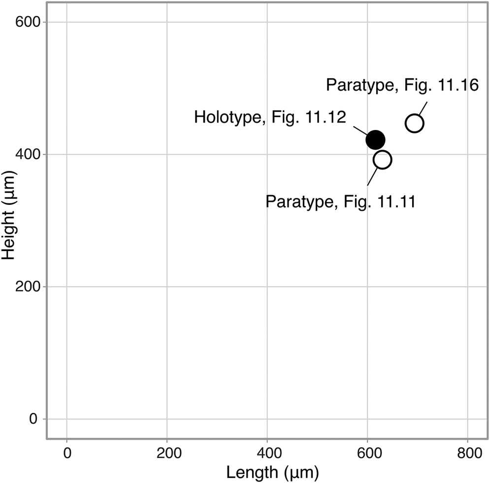


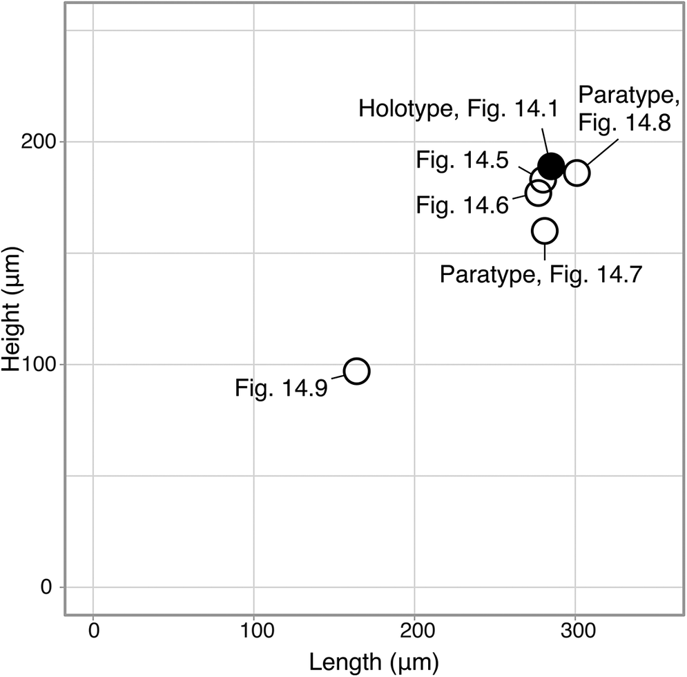


 Open access

