INTRODUCTION
Canine babesiosis is a serious tick-borne disease in dogs; it is caused by eight Babesia species, namely Babesia canis, Babesia conradae, Babesia gibsoni, Babesia rossi, Babesia sp., Babesia vitalii, Babesia vogeli and Babesia vulpes (Irwin, Reference Irwin2009, Reference Irwin2010; Schnittger et al. Reference Schnittger, Rodriguez, Florin-Christensen and Morrison2012). The most common symptoms of canine babesiosis are: fever, lethargy, anorexia, haemolytic anaemia and icterus. Infection severity varies according to several factors such as genotype, body condition, age and immune state. For example, babesiosis lethality is higher in puppies than adult dogs (Brandao et al. Reference Brandao, Hagiwara and Myiashiro2003; Bourdoiseau, Reference Bourdoiseau2006; Irwin, Reference Irwin2010). Globalization and increased international trade coupled to increased travel and mobility of pets, urbanization and climate change induced a rapid extension of tick-borne infections in the world (Shaw et al. Reference Shaw, Michael, Richard and Breitschwerdt2001). These factors led to the emergence of new tick and Babesia species including emerging (B. vitalii) and non-specific species (B. caballi) (Schnittger et al. Reference Schnittger, Rodriguez, Florin-Christensen and Morrison2012). The molecular prevalence of B. vogeli in rural Tunisian dogs was estimated to 6·7% (12/180) by M'ghirbi and Bouattour (Reference M'ghirbi and Bouattour2008). In Egypt, only B. vogeli was detected in dogs (Salem and Farag, Reference Salem and Farag2015). Canine babesiosis caused by B. vogeli has also been reported in Northern and Southern Africa (Zahler et al. Reference Zahler, Schein, Rinder and Gothe1998; Carret et al. Reference Carret, Walas, Carey, Grande, Precigout, Moubri, Schetters and Gorenfolt1999; Matjila et al. Reference Matjila, Penzhorn, Bekker, Nijhof and Jongejan2004).
Rhipicephalus sanguineus is the most frequent tick species infesting dogs in all bioclimatic zones of Tunisia (Bouattour, Reference Bouattour2002; Estrada-Peña et al. Reference Estrada-Peña, Bouattour, Camicas and Walker2004). In Southern Europe and in sub-Saharan Africa, B. vogeli, B. canis and B. rossi are transmitted by Dermacentor spp. and Haemaphysalis spp. respectively (Solano-Gallego and Baneth, Reference Solano-Gallego and Baneth2011; Schnittger et al. Reference Schnittger, Rodriguez, Florin-Christensen and Morrison2012).
The total population of dogs in Tunisia was estimated to 568 154 animals with a density of 3·5 km−2 (The World Organization for Animal Health, 2013) and clinical cases of canine babesiosis are often reported by pets’ practitioners (Chabchoub et al. Reference Chabchoub, Ghorbel, Landolsi and Arfaoui2001). Tropical theileriosis caused by Theileria annulata is a widespread disease of cattle in Tunisia; this protozoon is transmitted by Hyalomma ticks (Gharbi et al. Reference Gharbi, Rjeibi and Darghouth2014). To the best of our knowledge, only 2 studies from Turkey and Iran reported the presence of T. annulata in dogs (Bigdeli et al. Reference Bigdeli, Rafie, Namavari and Jamshidi2012; Aktas et al. Reference Aktas, Özübek, Altay, Ipek, Balkaya, Utuk, Kırbas, Şimsek and Dumanli2015).
Given the scarcity of epidemiological data on these species in Tunisian dogs, this study aimed to estimate the prevalence of piroplasm species in dogs and to compare the Tunisian genotype with others isolates.
MATERIALS AND METHODS
Dogs and blood samples
A total number of 200 vacutainer EDTA blood samples were collected from Tunisian dogs from Northern and Central Tunisia. The samples included male and female individuals of different ages, habitat and life styles.
Polymerase chain reaction (PCR)
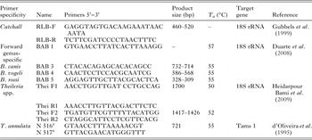
DNA extraction was performed from 150 µL of total blood, using Bio-Basic DNA extraction Kit (Markham Ontario, Canada) according to the manufacturer's instructions. Catchall primers (RLB-F and RLB-R) detecting both Theileria spp. and Babesia spp. DNA were used (Gubbels et al. Reference Gubbels, De Vos, Van Der Weide, Viseras, Schouls, De Vries and Jongejan1999). Forty PCR cycles were performed with a thermal cycler (ESCO Swift MaxPro) in a total reaction volume of 25 µL. In a second stage, each DNA positive sample was amplified using different sets of primers detecting specifically B. vogeli (BAB1/BAB4), B. canis (BAB1/BAB5) and B. rossi (BAB1/BAB3) (Duarte et al. Reference Duarte, Linhares, Romanowsky, Silveira and Borges2008) (Table 1).
Different primers used for detection by PCR of Babesia spp. and Theileria annulata from dog in this study

T a , annealing temperature.
a Primers used in sequencing reactions for the characterization of T. annulata.
Theileria annulata PCR amplifying a 721 bp fragment was performed with a set of specific primers of the gene encoding Tams1 of T. annulata (d'Oliveira et al. Reference d'Oliveira, Van Der Weide, Habela, Jacquiet and Jongejan1995) (Table 1). The PCR was carried out in 25 µL volume for each reaction consisting of 2·5 µL PCR buffer, 2 µL of the extracted DNA template, 0·4 mm of each dNTP, 0·5 µ m of each primer, 3 mm MgCl2 (25 mm), 0·05 U µL−1 of Taq DNA Polymerase (Vivantis) and 2 µL of the extracted DNA template. For all PCR runs, electrophoresis was performed in 1% agarose gel with ethidium bromide and visualized under UV light.
PCR–restriction fragment length polymorphism (RFLP)
A nested PCR detecting specific Theileria’s DNA of the 18S rRNA gene was performed. The primary PCR consisted of 25 cycles realised with 20 pg of outer primers (Thei F1 and Thei R1) in 30 µL volume. Each PCR product of 1 µL was used as template in a 30 cycles nested PCR in the same mixture as primary PCR with 2 different primers (Thei F2 and Thei R2) (Heidarpour Bami et al. Reference Heidarpour Bami, Haddadzadeh, kazemi, Khazraiinia, Bandehpour and Aktas2009) (Table 1). PCR products were electrophoresed in a 1·5% agarose gel and examined by UV transillumination.
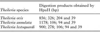
In order to differentiate T. annulata, T. lestoquardi and T. ovis, RFLP was performed with HpaII restriction enzyme (Fermentase) (Heidarpour Bami et al. Reference Heidarpour Bami, Haddadzadeh, kazemi, Khazraiinia, Bandehpour and Aktas2009) (Table 2). PCR product of 10 µL were mixed with 2 µL of 10× enzyme buffer and 10 U of the restriction enzyme then incubated at 37 °C for 2 h. Restriction digests were separated by electrophoresis on 2% agarose and analyzed with an UV transilluminator.
Restriction fragment length polymorphism pattern of different Theileria species after HpaII (Heidarpour Bami et al. Reference Heidarpour Bami, Haddadzadeh, kazemi, Khazraiinia, Bandehpour and Aktas2009)

DNA sequencing and phylogenetic analysis
Four selected B. vogeli and 4 T. annulata positive amplicons were purified with Wizard SV gel and PCR clean-up (Promega, Madison, USA). They were sequenced using the same primers as for PCR concerning B. vogeli and 2 other primers (N 516 and N 517) for T. annulata (Table 1) in an automatic sequencer (ABI 3730XL) and the conventional Big Dye Terminator cycle sequencing reaction kit (Applied Biosystems, Foster City, CA, USA) according to the manufacturer's protocol. ChromasPro 1·7·4 and MEGA 5·1 softwares served to make chromatograms and multiple sequence alignments, respectively (Tamura et al. Reference Tamura, Peterson, Peterson, Stecher, Nei and Kumar2011). Obtained DNA sequences were compared for similarity to sequences deposited in GenBank, using the BLAST programme (Altschul et al. Reference Altschul, Gish, Miller, Myers and Lipman1990). The 18S rRNA partial sequences of B. vogeli from Northern Tunisia (BvDNT) and from the Central (BvDCT) identified in the present survey were deposited in GenBank under accession numbers KT445940 and KT445941, respectively. The Tams1 partial sequences of T. annulata TaDgT1, TaDgT2 and TaDgT3 variants were deposited in the GenBank under accession numbers KX130955 to KX130957, respectively. Phylogenetic trees were constructed by the neighbour-joining method (Saitou and Nei, Reference Saitou and Nei1987). The evolutionary distances were computed using the Tamura-Nei method (Tamura and Nei, Reference Tamura and Nei1993) and are in the units of the number of base differences per site. Evolutionary analyses were performed using MEGA 5·1 software.
Statistical analyses
The infection prevalence percentages were compared with Epi Info 6 (Dean et al. Reference Dean, Arner, Sunki, Friedman, Lantinga, Sangam, Zubieta, Sullivan, Brendel, Gao, Fontaine, Shu, Fuller, Smith, Nitschke and Fagan2011) using Mantel Haenszel Chi-square test. Observed differences were considered to be significant when the P ≤ 0·05 (Schwartz, Reference Schwartz1993).
RESULTS
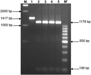
Twenty nine (29) out of the 200 (14·5% ± 0·05) samples were positive to catchall primers detecting Babesia and Theileria parasites. Among these, 25 dogs were positive to B. vogeli (12·5% ± 0·04); no infection by either B. canis or B. rossi was found. The enzymatic digestion profile by HpaII showed that the other 4 PCR products belong to T. annulata species (2% ± 0·02) (Fig. 1).
Digestion pattern of nested Theileria spp. 18S rRNA gene PCR products by HpaII. Lane M: 1Kb ladder; lane 1: Theileria spp. DNA; lanes 2–5: Theileria annulata (1178, 106 and 94 bp) and Lane M’: 100 bp ladder.

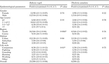
There were no differences in the molecular prevalences of B. vogeli for different age categories and sexes (P = 0·55). Babesia vogeli prevalence in working and companion dogs (25·8 ± 0·15 and 21·1% ± 0·13 respectively) were significantly higher than in guarding dogs (1·8% ± 0·03) (P = 0·01). The B. vogeli infection prevalence was significantly higher in Central Tunisia (26·5% ± 0·01) when compared with Northern Tunisia (9·6% ± 0·04) (P = 0·006) (Table 3).
Association between the presence of dogs’ DNA piroplasms and some risk factors

*Statistically significant.
a C.I., 95% confidence interval.
The sequencing of a partial sequence of 18S rRNA gene (533 bp) isolated from randomly selected 4 positive dog samples (2 from each sampling region) confirmed the B. vogeli infection. Alignment of these sequences revealed 2 variants with 3 distinct nucleotide positions (GenBank accession nos: KT445940 and KT445941).
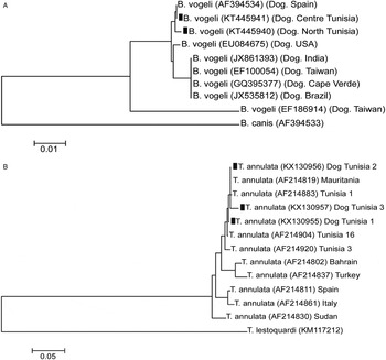
The variants from Northern and Central Tunisia (BvDNT and BvDCT respectively) were different and are 2 novel B. vogeli 18S rRNA genotypes. Babesia vogeli partial sequences from this study presented 99·5% identity between them and 3 different nucleotides. Indeed, the Northern (BvDNT) differed from the Central sequences (BvDCT) by a single deletion 55 (T) and 2 nucleotides substitutions at positions 48 (G/C) and 143 (T/G). Our sequences showed 99·4; 99·2 and 99% identity with B. vogeli from Taiwan (EF186914), USA (EU084675) and Spain (AF394534) respectively. Babesia vogeli partial sequences from this study presented 98·3–99·5% identity amongst each other and differed in 5 nucleotide positions (4 substitutions and 1 deletion). Phylogenetic study based on 533 bp fragment of 18S rRNA analysis was performed with sequences from this study and selected sequences isolated from mammals published in GenBank (Fig. 2A). Babesia vogeli sequences described in this study clustered with B. vogeli sequences from Spain; clearly differed with B. canis and B. vogeli from India, Taiwan, USA, Brazil and Cape Verde (Fig. 2A).
The tree was constructed using the neighbour-joining method (Saitou and Nei, Reference Saitou and Nei1987). The percentage of replicate trees in which the associated taxa clustered together in the bootstrap test (1100 replicates) was shown next to the branches. The evolutionary distances were computed using the Tamura-Nei method (Tamura and Nei, Reference Tamura and Nei1993) and are in the units of the number of base differences per site. Evolutionary analyses were conducted in MEGA5·1 (Tamura et al. Reference Tamura, Peterson, Peterson, Stecher, Nei and Kumar2011). GenBank accession numbers are given in parentheses. Species described in this study are indicated with a black square. (A) Partial sequence 18S rRNA gene phylogenetic tree of Babesia vogeli identified in dogs in the present survey and those deposited in GenBank. (B) Partial sequence Tams1 gene phylogenetic tree of Theileria annulata identified in dogs in the present survey and those deposited in GenBank.

Theileria annulata infections were validated by sequencing 664 bp of the Tams1 gene from the 4 positive dog samples. Phylogenetic analyses showed that 2 amplicons from Tunis have 100% identity (GenBank accession numbers KX130955). Sequences alignment revealed 3 distinct and novel genotypes; 2 from Tunis (GenBank accession numbers KX130955 and KX130956) and the third from Béja (KX130956), these amplicons have 98·7–99·7% identity (Table 4). Theileria annulata variants obtained in this study were 93–99% identity to the T. annulata genotypes published in the GenBank (Fig. 2B).
Nucleotide differences among Tams1 sequences from different strains of Theileria annulata

Nucleotides: T, Thymine; C, Cytosine; G, Guanine; A, Adenine.
a TaDgT1 and TaDgT2 variants were isolated from Tunis samples and TaDgT3 variant was isolated from Béja samples.
b GenBank accession number.
c Numbers represent the nucleotide position. Conserved nucleotide positions relative to the first sequence are indicated with asterisks.
DISCUSSION
Babesia vogeli was previously reported by Uilenberg et al. (Reference Uilenberg, Franssen, Perie and Spanger1989) in a dog suffering from concurrent babesiosis and ehrlichiosis, which had just returned to The Netherlands from Tunisia. M'ghirbi and Bouattour (Reference M'ghirbi and Bouattour2008) reported a lower molecular prevalence of B. vogeli (6·6%) in dogs from 4 Tunisian villages in different bioclimatic zones; the screening with the RLB assay detected only B. vogeli. The latter study also showed that the infection prevalence of B. vogeli in R. sanguineus was 0·6% (1/160).
Babesia vogeli has a large geographic distribution; it has been detected in dogs in tropical or subtropical areas of Northern, Eastern and Southern Africa (Uilenberg et al. Reference Uilenberg, Franssen, Perie and Spanger1989; Lewis et al. Reference Lewis, Penzhorn, Lopez-Rebollar and De Waal1996; Oyamada et al. Reference Oyamada, Davoust, Boni, Dereure, Bucheton, Hammad, Itamoto, Okuda and Inokuma2005), Europe (Criado-Fornelio et al. Reference Criado-Fornelio, Martinez-Marcos, Buling-Sarana and Barba-Carretero2003), Asia (Inokuma et al. Reference Inokuma, Oyamada, Davoust, Boni, Dereure, Bucheton, Hammad, Watanabe, Itamoto, Okuda and Brouqui2006), Australia (Hii et al. Reference Hii, Kopp, Thompson, O'Leary, Rees and Traub2012) and North and South America (Birkenheuer et al. Reference Birkenheuer, Levy and Breitschwerdt2003; Passos et al. Reference Passos, Geiger, Ribeiro, Pfister and Zahler-Rinder2005). More particularly in Europe, infection by B. vogeli seems to be widespread; it was reported in Portugal (Cardoso et al. Reference Cardoso, Costa, Tuna, Vieira, Eyal, Yisaschar-Mekuzas and Baneth2008), Spain (Criado-Fornelio et al. Reference Criado-Fornelio, Rey-Valeiron, Buling, Barba-Carretero, Jefferies and Irwin2007), France (Caccio et al. Reference Caccio, Antunovic, Moretti, Mangili, Marinculic, Baric, Slemenda and Pieniazek2002), Central and Southern Italy (Solano-Gallego et al. Reference Solano-Gallego, Trotta, Carli, Carcy, Caldin and Furlanello2008), Slovenia (Duh et al. Reference Duh, Tozon, Petrovec, Strasek and Avsic-Zupanc2004) and Albania (Hamel et al. Reference Hamel, Silaghi, Knaus, Visser, Kusi, Rapti, Rehbein and Pfister2009).
In the present study, exclusively B. vogeli and neither B. canis nor B. rossi was found. Despite frequent exchanges with many countries where B. canis is common (France, Spain…), no animal was infected by this species in our samples. The vector ticks of B. rossi (Haemaphysalis spp.) are not frequent in Tunisia and the level of animals’ exchange with Sub-Saharan countries where this Babesia species is reported is very low (South Africa, Nigeria and Sudan).
Babesia vogeli prevalences were similar in urban, suburban and rural areas. This is contrary to the findings in Brazil, where seroreactivity to B. vogeli was higher in urban (22%) when compared with rural dogs (11%) (da Costa et al. Reference da Costa, Costa, Labruna, Silveira, Moraes-Filho, Soares, Spolidorio and Guerra Rde2015). The same trend was reported by Spolidorio et al. (Reference Spolidorio, Minervino, Valadas, Soares, Neves, Labruna, Ribeiro and Gennari2013) in Brazil who reported anti-B. vogeli antibodies in 59·6 and 29·1% of urban and rural dogs, respectively (P < 0·05). Babesia vogeli molecular prevalence was significantly higher in the Centre compared with North and this could be due to different environmental conditions between the 2 zones (Harrus and Baneth, Reference Harrus and Baneth2005) or to different efficiencies of the tick control programmes.
The two B. vogeli sequences reported in this paper shared 99·5% identity between them. Our sequences presented 98–99% identity amongst isolates from Spain (AF394534), USA (EU084675) and Taiwan (EF186914). The phylogenetic tree showed that the B. vogeli sequences described in this study clustered with B. vogeli from Brazil, India, Spain, Taiwan, and USA but were clearly distinct from B. canis.
Tropical theileriosis (T. annulata infection) is a widespread tick-borne cattle disease in Tunisia (Gharbi et al. Reference Gharbi, Rjeibi and Darghouth2014). In this study, T. annulata DNA was detected in 4 dogs (2%) and was confirmed by sequencing. The analysis of T. annulata Tams1 sequences revealed 3 different variants all grouped in a single cluster. Theileria annulata variants obtained from Tunis (KX130955 and KX130956) differed by 2 nucleotide substitutions. However, the amplicon from Béja (located 105 km from Tunis); GenBank accession number (KX130957) differed from those of Tunis by 9 nucleotide substitutions. This finding is not surprising, since Hyalomma scupense could also feed on dogs (Gharbi and Darghouth, Reference Gharbi and Darghouth2014). In Turkey and Iran, T. annulata DNA was isolated in dogs with no clinical symptoms in 0·1 (1/757) and 0·7% (2/280) of the examined dogs, respectively (Bigdeli et al. Reference Bigdeli, Rafie, Namavari and Jamshidi2012; Aktas et al. Reference Aktas, Özübek, Altay, Ipek, Balkaya, Utuk, Kırbas, Şimsek and Dumanli2015). The sequencing was performed in order to: (i) confirm that the dogs were infected by T. annulata, (ii) verify that the dogs as non-sensitive host are selecting specific T. annulata genotypes and (iii) investigate the polymorphism of B. vogeli in Tunisia and compare our strains with others from different regions of the world.
Babesia vogeli and T. annulata DNAs were detected in dogs from several regions of Tunisia. Further studies are needed to know if T. annulata are detectable in Giemsa stained blood smears in infected dogs, which may cause confusion with Babesia piroplasms. If yes, this should be considered in the laboratories where Giemsa stained blood smears are performed routinely to detect Babesia spp. in infected dogs. Moreover, this paper raises an important question as to the role of dogs as reservoirs of T. annulata. This is an important issue in cattle farms where programmes are implemented to control bovine tropical theileriosis.
ACKNOWLEDGEMENTS
The authors thank Dr Moez Mhadhbi, Mr Bechir Guesmi, Mr Taoufik Lahmar and Mr Mohamed Jedidi for their support.
FINANCIAL SUPPORT
The work was funded by «Laboratoire d’épidémiologie des infections enzootiques des herbivores en Tunisie (Ministère de l'enseignement supérieur et de la recherche scientifique, Tunisia) and the Deutsche Forschungsgemeinschaft project ‘Molecular epidemiology network for promotion and support of delivery of live vaccines against Theileria parva and Theileria annulata infection in eastern and northern Africa’ (SE862/2–1), awarded to M. R. R.
ETHICAL AND REGULATORY GUIDELINES
The blood samples were collected by veterinarians and veterinary students. This study was conducted in accordance with relevant national and international guidelines on handling animals, taking care to respect animal welfare.