Case report
A 14-year-old previously healthy, fully vaccinated female presented to the emergency department with chest pain and dyspnoea on exertion for 2 days. She described the pain as a constant, non-radiating pressure on the left side of her chest, 8 out of 10 in severity. Two weeks prior she had mild upper respiratory infection symptoms, including cough, rhinorrhea, and fever, which had resolved. She denied headaches, vision changes, rashes, joint pain, palpitations, dizziness, or syncope. She took no medications and had no allergies. There was no recent travel. She played recreational basketball on occasion but denied being athletic or participating in competitive sports. There was no history of chest trauma. Family history was negative for CHD, early myocardial infarction, or sudden cardiac death.
In the emergency department, she was noted to have a heart rate of 110 beats per minute with otherwise unremarkable vital signs. On physical examination, she was an alert, thin-appearing female in no distress. She was stable on room air with symmetric chest rise and normal breath sounds. She had no tenderness to palpation over her chest. She had no chest wall deformity, swelling, or erythema. She had strong pulses, and her capillary refill was under 2 seconds. She had no organomegaly.
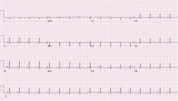
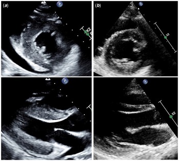
Her laboratory results were notable for elevated C-reactive protein of 4.7 mg/L (normal value <4.0 mg/L), troponin T of 0.26 ng/ml (normal value, ≤0.01 ng/ml), and Pro-Brain Natriuretic Peptide of 2954 pg/ml (normal value, ≤300 pg/ml). Her complete blood count and comprehensive metabolic panel, including liver enzymes, were normal. Her chest X-ray had no evidence of acute cardiopulmonary disease. A standard 12-lead electrocardiogram showed normal sinus rhythm with low-voltage QRS complexes (Fig 1). A transthoracic echocardiogram showed normal segmental anatomy with concentric left ventricular hypertrophy concerning for hypertrophic cardiomyopathy. In parasternal short axis, her end-diastolic septal thickness measured 16.4 mm, and her left ventricular posterior wall measured 13.1 mm (Fig 2a). Additionally, there was mildly decreased left ventricular systolic function with estimated ejection fraction of 45% (5/6 area × length method) and a moderate circumferential pericardial effusion. There was no systolic anterior motion of the mitral valve.
Electrocardiogram showed normal sinus rhythm with low-voltage QRS complexes.

Transthoracic echocardiogram images demonstrating concentric left ventricular hypertrophy on admission ( a ) with complete resolution at the 1-month follow-up visit ( b ).

Hospital course
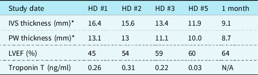
The patient was admitted for treatment and monitoring in the setting of myocarditis with additional concern for underlying hypertrophic cardiomyopathy. Her symptoms resolved, and she remained haemodynamically stable throughout her hospital course. On hospital day (HD) #1, she received 2 g/kg intravenous immunoglobulin, with prompt improvement in her left ventricular systolic function and resolution of her left ventricular hypertrophy and pericardial effusion. Her troponin also decreased to 0.03 ng/ml (Table 1), after which it was no longer followed. An infectious aetiology was suspected due to the patient’s recent respiratory symptoms. Peripheral blood culture collected on admission had no growth. A respiratory viral panel was negative, including testing for COVID-19 infection. Serological testing for Epstein–Barr virus and cytomegalovirus did not demonstrate active or recent infection with either virus. Adenovirus antibody titre was elevated at 1:64 (normal value, <1:8), consistent with recent adenovirus infection. Cardiac magnetic resonance was performed immediately prior to her hospital discharge on HD #5, which demonstrated normal left ventricular thickness, normal left ventricular systolic function (ejection fraction 57%; normal value, >55%), and a trivial pericardial effusion. There were multiple segments of left ventricular mid-myocardial and subepicardial delayed gadolinium enhancement, consistent with myocarditis.
Relevant echocardiographic measurements and troponin T trend.

HD = hospital day; IVS = interventricular septum; LVEF = left ventricular ejection fraction; PW = posterior wall.
* Measurement obtained in parasternal short axis.
At the patient’s 4-week follow-up visit, her electrocardiogram had normal QRS voltages. Her echocardiogram was also normal, including her left ventricular size and thickness (Fig 2b). Specifically, in parasternal short axis, her end-diastolic septal thickness measured 9.1 mm, and her left ventricular posterior wall measured 8.7 mm. There was no pericardial effusion. She reported no symptoms and appeared well. She returned to her normal routine, although she was restricted from competitive sports for a total of 3 months.
Discussion
Myocarditis is an inflammatory disease of the heart frequently associated with a viral infection. It has a wide clinical spectrum, from asymptomatic presentations to cardiogenic shock and death. The diagnosis is usually suspected in the setting of elevated inflammatory markers, evidence of myocardial damage, including troponin elevation, non-specific electrocardiogram changes, and imaging abnormalities. While there are no specific features of acute myocarditis on echocardiography, findings commonly include left ventricular dilation and systolic dysfunction with normal- or thin-appearing walls. Endomyocardial biopsy is the gold standard for diagnosis, but cardiac magnetic resonance has emerged as a less invasive and more sensitive alternative to confirm the diagnosis. Reference Canter and Simpson1,Reference Law, Lal and Chen2
Increased myocardial thickness is typically associated with fulminant myocarditis, an uncommon form of acute myocarditis, which is characterised by sudden and severe diffuse cardiac inflammation and by definition necessitates inotropic or mechanical circulatory support. Reference Kociol, Cooper and Fang3,Reference Tunuguntla, Jeewa and Denfield4 Our patient’s clinical presentation was consistent with non-fulminant myocarditis except that she developed reversible increased myocardial thickness. As a result, our case illustrates that left ventricular hypertrophy can also be a manifestation of non-fulminant myocarditis. It is important for providers to be cognizant of this rare manifestation and of the potential, in severe cases, for misdiagnosis as hypertrophic cardiomyopathy. Our patient presented with left ventricular hypertrophy meeting diagnostic criteria for hypertrophic cardiomyopathy, and we counselled the patient on the possibility that she had concomitant hypertrophic cardiomyopathy when she was admitted. A small number of case reports have described transient left ventricular hypertrophy with non-fulminant myocarditis in the paediatric age group. Reference Giafaglione, Morrison and Nandi5–Reference Kosutic8 However, most cases have been associated with influenza virus, and to our knowledge, this is the first case describing this finding with adenovirus, which is a more common cause of myocarditis. Reference Bowles, Ni and Kearney9
The pathophysiology of transient left ventricular hypertrophy in non-fulminant myocarditis is not fully understood but likely caused by myocardial oedema and/or cellular infiltration. Reference Law, Lal and Chen2 Interestingly, it appears to resolve quickly in most cases, as it did for this patient. Reference Giafaglione, Morrison and Nandi5 Further research is needed to improve our understanding of this phenomenon. It is possible that the rapid resolution observed in this case was due to early treatment with intravenous immunoglobulin, which has anti-inflammatory, antiviral, and immunomodulatory effects. Reference Law, Lal and Chen2,Reference Drucker, Colan and Lewis10 However, the efficacy of intravenous immunoglobulin in myocarditis remains controversial.
Conclusion
We present a case of non-fulminant myocarditis associated with transient concentric left ventricular hypertrophy. This phenomenon is rare in children, and this is the first case describing it with acute myocarditis associated with adenovirus. It is important to highlight that left ventricular hypertrophy occurring with myocarditis can be so striking that it can potentially be mistaken for an underlying cardiomyopathy. Clinicians should be aware of this possible manifestation and avoid making an incorrect diagnosis of hypertrophic cardiomyopathy. Our patient had normalisation of her left ventricular thickness over a short time frame, suggesting that reversible left ventricular hypertrophy has an excellent prognosis.
Financial support
This research received no specific grant from any funding agency, commercial, or not-for-profit sectors.
Conflicts of interest
None.
Ethical standards
The patient’s family provided written consent for this case report to be published.