Introduction
Heartworm disease is a zoonotic vector-borne disease caused by the filarial nematode Dirofilaria immitis. The domestic dog is the typical host and biological reservoir, although infection has been reported in other carnivores, including wild canids, domestic cats and other felids (McCall et al., Reference McCall, Genchi, Kramer, Guerrero and Venco2008). Furthermore, animal heartworm disease as a zoonotic disease constitutes a risk for the human population, since D. immitis is the causative agent of pulmonary dirofilariasis and can produce benign pulmonary nodules (Simón et al., Reference Simón, Siles-Lucas, Morchón, González-Miguel, Mellado, Carretón and Montoya-Alonso2012).
Heartworm disease is severe and life-threatening. The pathophysiological response is mainly due to the presence of adult worms in the pulmonary arteries and probably to the presence of microfilariae and the endosymbiotic bacterium Wolbachia pipientis, causing pulmonary thromboembolisms, pulmonary hypertension and right-sided congestive heart failure (McCall et al., Reference McCall, Genchi, Kramer, Guerrero and Venco2008).
Dirofilaria immitis is transmitted by different species of culicid mosquitoes, mainly from the genera Culex, Aedes and Anopheles. Thus, heartworm disease is mainly distributed in temperate and humid areas (Cancrini et al., Reference Cancrini, Magi, Gabrielli, Arispici, Tolari, Dell'Omodarme and Prati2006; Simón et al., Reference Simón, Siles-Lucas, Morchón, González-Miguel, Mellado, Carretón and Montoya-Alonso2012). In Europe, heartworm disease is endemic in Mediterranean countries, although some studies have shown that it is spreading towards areas previously considered free of the disease. The spread is probably due to the increasing number of infected animals travelling from endemic areas, the introduction of new species of mosquitoes able to act as vectors, climate change caused by global warming and the development of human activity in new areas (Genchi et al., Reference Genchi, Rinaldi, Mortarino, Genchi and Cringoli2009; Morchón et al., Reference Morchón, Carretón, González-Miguel and Mellado-Hernández2012).
The first epidemiological study carried out in Portugal reported that canine dirofilariosis was prevalent at 12–17% in several southern regions, finding the highest prevalence on the island of Madeira, where 30% of the studied dogs were microfilaraemic (Araújo, Reference Araújo1996). Subsequent studies confirmed the presence of canine dirofilariosis caused by D. immitis in Portugal (Seixas et al., Reference Seixas, Alho, Guerra and Madeira de Carvalho2011; Cardoso et al., Reference Cardoso, Mendao and Madeira de Carvalho2012). Alho et al. (Reference Alho, Landum, Madeira de Carvalho, Meireles, Gonçalves, Belo, Grandi, Kramer and Genchi2012) reported a canine prevalence of 20% in Setúbal, 13% in Santarém and 8.1% in Coimbra. However, there are no published epidemiological data regarding feline heartworm disease in Portugal. Therefore, the aim of the present study was to determine the prevalence of D. immitis infection in cats and dogs in several districts of central and northern Portugal.
Materials and methods
Study sites
According to the Köppen–Geiger climate classification, Portugal has a warm temperate climate with dry summers (type Cs). The climate of central and northern Portugal is divided into two different subtypes. These are Csa, having hot summers with the average temperature in the warmest month above 22°C, and Csb, having warm summers with the average temperature in the hottest month below or equal to 22°C, and with 4 months or more with the average temperatures above 10°C (AEMET, 2011).
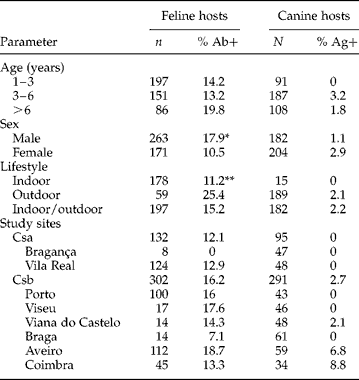
A total of 434 cats and 386 dogs were examined during the present study, including 132 cats and 95 dogs in the Csa climatic area, and 302 cats and 291 dogs in the Csb region (table 1). For the purpose of the present study, the Csa area was divided into the regions of Bragança (41°47′N/6°44′W) and Vila Real (41°18′N/7°45′W), while the Csb region was divided into the regions of Porto (41°7′N/8°36′W), Viseu (40°39′N/7°55′W), Viana do Castelo (41°42′N/8°50′W), Braga (41°33′N/8°26′W), Aveiro (40°38′N/8°39′W) and Coimbra (40°12′N/8°24′W).
Prevalences (%) of heartworm (Dirofilaria immitis) expressed as positive tests to antibodies (Ab) in feline hosts and antigens (Ag) in canine hosts, relative to host age, sex, lifestyle and geographical region.

n, Number of animals examined; Csa, hot summers with average temperatures above 22°C; Csb, warm summers with temperatures equal to or below 22°C. *P= 0.052; **P= 0.059.
Collection and examination of hosts
The 434 client-owned cats included in the present study were presented to veterinary clinics for routine examinations between September 2009 and July 2010. Of these, 60.6% were males and 39.4% were females, and 87.3% were domestic shorthair and 12.7% other breeds. The criteria for inclusion were being over 8 months of age, exposed for at least a season to mosquitoes, never having received treatment for heartworm disease and no previous history of heartworm infection. A complete record was kept for each animal, including identification by age, sex and breed; clinical history; indoor, outdoor or indoor/outdoor habitat; and demographic data, as well as feline immunodeficiency virus (FIV) and feline leukaemia virus (FeLV) status.
Blood samples were collected from the cephalic or jugular vein into dry tubes and centrifuged. Serum was kept at − 20°C until the tests were performed. Feline D. immitis infection was identified using serological techniques for anti-D. immitis and anti-Wolbachia antibody detection (Morchón et al., Reference Morchón, Ferreira, Martin-Pacho, Montoya, Mortarino, Genchi and Simón2004) with some modifications. In brief, the plates were coated with 0.8 μg of D. immitis somatic antigen and Wolbachia surface protein (WSP). Serum samples were prepared at 1/100 for anti-D. immitis serum antibodies and 1/40 for anti-WSP antibody detection. Horseradish peroxidase-labelled anti-feline IgG antibody (Kirkegaard and Perry Laboratories, Gaithersburg, Maryland, USA), was applied at 1/4000 dilution. The optical densities were measured in an Easy-Reader (Bio-Rad Laboratories, Hercules, California, USA) at 492 nm. Cut-off points of enzyme-linked immunosorbent assay (ELISA) D. immitis 0.8 and ELISA WSP 0.6 were obtained as arithmetic mean optical density ± 3 standard deviations of sera of clinically healthy cats.
The 386 client-owned dogs included in the present study were presented to veterinary clinics for routine health examination between November 2010 and June 2011. Of these, 47.2% were male and 52.8% were female. By breed, 42% were mongrel dogs, 11.1% Labrador retriever, 8% Portuguese warren hound, 5.9% boxer, 3.3% golden retriever, 2.8% cocker spaniel and 26.9% were other breeds. The criteria for inclusion included being over 6 months of age, exposed for at least a season to mosquitoes, never having received treatment for heartworm disease and no previous history of heartworm infection. A complete record was kept for each animal, including identification by age, sex and breed; clinical history; indoor, outdoor or indoor/outdoor habitat; and demographic data.
Blood samples were collected from the cephalic or jugular vein into 3-ml EDTA tubes and centrifuged. Serum was kept at − 20°C until tests were performed. Antigens of D. immitis were detected by using a commercial ELISA test kit (Uranotest Dirofilaria®, Urano Vet SL, Barcelona, Spain), following the manufacturer's instructions.
Data analysis
Data were analysed using SPSS Base 20.0 software for Windows (SPSS Inc./IBM, Chicago, Illinois, USA). Descriptive analysis of the considered variables was carried out considering the proportions of the qualitative variables. We performed χ2 and Fischer exact tests to compare proportions. In all cases, the significance level was established at P< 0.05.
Results and discussion
The seroprevalence of D. immitis in the feline population was 15%, with a greater prevalence of 17.9% in males compared with 10.5% in females (table 1). By age, seroprevalence was 14.2% for cats < 3 years, 13.2% for cats aged 3–6 years and 19.8% for those>6 years. Seroprevalence for those always kept outside the house was 25.4%, compared with 11.2% for cats always kept inside the house and 15.2% for cats that spent at least 1–50% of their time outdoors. By regions, the highest prevalences were 18.7% in Aveiro and 17.6% in Viseu compared with none in Bragança. Furthermore, there were no significant differences by breed, reproductive status or FIV and FeLV status between seropositive and seronegative cats. Of the clinically healthy cats, 11.5% were seropositive, compared with 18.2% of the cats showing any clinical signs. Of these, the seroprevalence of D. immitis in cats with respiratory signs was 24.4%, in cats with digestive symptoms was 2/4 (i.e. 50%) and cats with other clinical signs (neurological symptoms, weakness and loss of weight) presented a seroprevalence of 15.9%. No significant differences were found between clinically healthy and symptomatic cats.
The seroprevalence of D. immitis in the canine population was 2.1%. The highest prevalence of 8.8% was found in the Csb region of Coimbra, followed by 6.8% in Aveiro and 2.1% in Viana do Castelo. On the other hand, no dogs were found to be seropositive in the Braga, Vila Real, Bragança, Porto and Viseu regions. Similar results were observed for mongrel and pure-breed dogs. Only 1% of asymptomatic dogs were positive for D. immitis, compared with 5.2% of dogs demonstrating any clinical signs, and these differences were significant (P= 0.003). Of the symptomatic dogs, those with respiratory signs showed a prevalence of 17%.
The present study is the first epidemiological study of seroprevalence of D. immitis in cats in Portugal. As in canine hosts, feline heartworm disease has spread and increased during the past few years throughout the world (Simón et al., Reference Simón, Siles-Lucas, Morchón, González-Miguel, Mellado, Carretón and Montoya-Alonso2012). However, unlike the former, the diagnosis of feline dirofilariosis presents a challenge, mainly because cats usually present a low parasite burden, frequently leading to a negative antigen test. Another possible explanation that could lead to a negative antigen test may be due to the fact that the disease in cats is mainly caused by immature worms. Furthermore, feline patients appear to tolerate the infection better than dogs, clinical signs often being absent or minimal (Snyder et al., Reference Snyder, Levy, Salute, Gorman, Kubilis, Smail and George2000; Lee & Atkins, Reference Lee and Atkins2010).
One reason for the high feline seroprevalence found in the present study, when compared with canine prevalence, could be due to cats not receiving preventative treatment in a routine way, leading to the spread of D. immitis infection in this species. However, awareness of the disease in dogs, through control and prevention, may also influence the low prevalence found in this species in the same region (Lorentzen & Caola, Reference Lorentzen and Caola2008; Dunn et al., Reference Dunn, Levy, Colby and Michaud2011). Montoya-Alonso et al. (Reference Montoya-Alonso, Carretón, Corbera, Juste, Mellado, Morchón and Simón2011) reported similar conclusions in the endemic area of Gran Canaria, Spain, with a higher feline seroprevalence of D. immitis when compared with canine prevalence. On the other hand, cats exposed to infective L3 larvae are generally more resistant to infection than dogs (McTier et al., Reference McTier, Shanks, Watson, McCall, Genchi, Six, Thomas, Dickin, Pengo, Rowan and Jernigan2000). Since the present study was designed to detect antibodies of anti-D. immitis in feline serum, the results demonstrate exposure to the parasite rather than necessarily active infection. Cats living in the geographical areas studied present an important rate of exposure to D. immitis and, consequently, a high risk of developing heartworm disease. Consequently, both clinicians and cat owners should be alerted about the need for additional diagnostic tests and/or prophylactic measures. Further research using tests for heartworm antigens and/or echocardiography is required to confirm the diagnosis of adult D. immitis infection in seropositive cats.
The present results showed that 24.4% of cats showing respiratory signs and 50% of cats with digestive signs were seropositive for D. immitis. Heartworm infection in cats is basically pulmonary in nature, and most frequently symptoms are of respiratory or digestive origin, such as chronic coughing, laboured breathing and vomiting (Atkins & Litster, Reference Atkins, Litster and August2006; Litster & Atwell, Reference Litster and Atwell2008). When signs of infection are evident, they are usually developed during the arrival and death of immature heartworms in the pulmonary vasculature. These signs are often misdiagnosed as asthma or allergic bronchitis but are actually part of a syndrome known as heartworm-associated respiratory disease (Lee & Atkins, Reference Lee and Atkins2010; Garcia-Guasch et al., Reference Garcia-Guasch, Caro-Vadillo, Manubens-Grau, Carretón, Morchón, Simón and Montoya-Alonso2012). In light of the results, heartworm-associated respiratory disease should be a part of the differential diagnosis when a cat presents with respiratory signs in the geographical regions reported in this study.
Canine prevalence of D. immitis was 2.1% in Viana do Castelo, similar to the 2.9% reported by Cardoso et al. (Reference Cardoso, Mendao and Madeira de Carvalho2012) for the north of Portugal. In the present study, the highest prevalences of 8.8% and 6.8% were found in Coimbra and Aveiro, respectively. A previous study showed a similar prevalence in Coimbra of 8.1% (Alho et al., Reference Alho, Landum, Madeira de Carvalho, Meireles, Gonçalves, Belo, Grandi, Kramer and Genchi2012) while in Aveiro, Rendall-Rocha et al. (Reference Rendall-Rocha, Fonseca, Cardoso, Grandi, Kramer and Genchi2012) reported higher prevalences of heartworm disease, being 10% in asymptomatic dogs and rising to 44.8% in symptomatic animals. Contrary to this study, determination of the presence of circulating microfilariae by direct examination of fresh blood or by Knott test was not performed in the present study, and this could underestimate the real prevalence in those geographical regions with low prevalences of the infection, since low worm burdens in dogs could lead to a negative antigen test.
For cats and dogs, the present results suggest that both the Csa and Csb climatic areas provide favourable conditions for the development of the arthropod vectors and, thus, transmission of the disease.
The present study demonstrates the presence of feline D. immitis infection, and also completes the epidemiological map of canine heartworm disease, in central and northern Portugal. Heartworm disease should be included in the list of differential diagnosis of dogs and cats presenting with clinical signs compatible with this disease. The results also justify the implementation of adequate prophylactic plans in these regions.
Acknowledgements
The authors are grateful to all owners and veterinarians who kindly collaborated in this study. We also acknowledge Professor Luis Cardoso for statistical advice.
Financial support
The study has received no financial support from any public or private institution.
Conflict of interest
None.
Ethical standards
All owners gave their consent to participate in the study. The study was approved by the Ethical Committee of the Veterinary Medicine Service of the University of Trás-os-Montes e Alto Douro and was carried out in accordance with the current European legislation on animal protection.