INTRODUCTION
The infraorder Stenopodidea includes three families and twelve genera of small shrimps, commonly found associated with coral reefs, marine caves and rocky and muddy bottoms or deep waters of which many are commensals in Hexactnellid sponges (Williams, Reference Williams1984; Berggren, Reference Berggren1993; Ortiz et al., Reference Ortiz, Gómez and Lalana1994, Reference Ortiz, Lalana and Varela2007; Hendrickx, Reference Hendrickx2002; Alvarez et al., Reference Alvarez, Iliffe and Vilalobos2006; Burukovsky, Reference Burukovsky2009).
Until now, three species of Stenopodidea were recorded in the western South Atlantic Ocean: Microprosthema semilaeve (von Martens, 1872), Stenopus hispidus (Olivier, 1811) and Stenopus scutellatus Rankin, 1898, the last two belonging to the family Stenopodidae (Coelho & Ramos-Porto, Reference Coelho, Ramos-Porto and Young1998; Young & Serejo, Reference Young, Serejo, Dutra, Allen, Werner and McKenna2005; Coelho et al., Reference Coelho, de Almeida, Souza-Filho, de Bezerra and Giraldes2006). Five additional species of Stenopodidae (Odontozona libertae Gore, 1981; Odontozona striata Goy, 1981; Odontozona anaphorae Manning & Chace, Reference Manning and Chace1990; Odontozona edwardsi (Bouvier, 1908), and Richardina spinicincta A. Milne-Edwards, 1881) have been reported in the western North Atlantic, in the Caribbean, and in the Ascension Island (Goy, Reference Goy1982; Manning & Chace, Reference Manning and Chace1990; Criales, Reference Criales1997; Martin, Reference Martin2002; Saito & Takeda, Reference Saito and Takeda2003; Martin & Goy, Reference Martin and Goy2004; Becker et al., Reference Becker, Cordes, Macko and Fisher2009), resulting in a total number of eight species of Stenopodidae in the Atlantic.
Stenopus hispidus, the most abundant stenopodid in Brazil, is a large cleaner shrimp which removes ectoparasites, mucus, diseased and dead tissue, and undesirable food particles attached to many body parts of fish, such as gills, mouth and skin (Limbaugh, Reference Limbaugh1961; Côté, Reference Côté2000; Bshary & Grutter, Reference Bshary and Grutter2002). Adults of S. hispidus are common and have been recorded worldwide, from the Indo-Pacific, Red Sea, South Africa, and North and South Atlantic, usually in shallow waters to a depth of 210 m, on rocky shores and gravel bottoms (Williams, Reference Williams1984; Holthuis, Reference Holthuis1993; Coelho & Ramos-Porto, Reference Coelho, Ramos-Porto and Young1998). Stenopus scutellatus has a more restricted distribution in the South Atlantic, specifically in north-eastern Brazil, also in shallow waters to a depth of 180 m (Coelho & Ramos-Porto, Reference Coelho, Ramos-Porto and Young1998).
Information concerning larval development of Stenopodidea is still scarce and almost entirely restricted to European and Mediterranean species (Kurian, Reference Kurian1956; Barnich, Reference Barnich1996; Santos & González-Gordillo, Reference dos Santos and González-Gordillo2004). In general, information concerning stenopodidean larval development is mainly based on field-collected larvae (Martin & Goy, Reference Martin and Goy2004) because many species are rare or inhabit deep waters, and consequently are difficult to keep under laboratory conditions. Some authors have also focused on larval culture and growth of marine ornamental species of crustacean decapods, including Stenopodidae (Young, Reference Young1979; Gore, Reference Gore and Wenner1985; Fletcher et al., Reference Fletcher, Kötter, Wunsch and Yasir1995; Zhang et al., Reference Zhang, Lin and Creswell1997; Calado et al., Reference Calado, Lin, Rhyne, Araújo and Narciso2003). Recently, Saito & Takeda (Reference Saito and Takeda2003) focused on phylogenetic relationships between Spongicolidae species also including some larval characters.
The larval development of Stenopus hispidus was first studied toward the end of the 19th Century by Brooks & Herrick (Reference Brooks and Herrick1891) and Ortmann (Reference Ortmann and Hensen1893), the latter study based on larvae collected during the ‘Plankton Expedition’. Brooks & Herrick (Reference Brooks and Herrick1891) studied S. hispidus development from eggs hatched in a laboratory and according to Martin & Goy (Reference Martin and Goy2004) were the only researchers who did not use specimens collected in the plankton. Gurney (Reference Gurney1924), working on larvae obtained in the ‘Terra Nova Expedition’, described several of the older larval stages of S. hispidus. Also Gurney (Reference Gurney1936) described nine larval forms of Stenopodidae from the ‘Discovery Plankton Expedition’, including zoeae I, II, V, VI? and IX? of S. hispidus. Five years later, Lebour (Reference Lebour, Gurney and Lebour1941), working on field-collected larvae from Bermuda plankton, re-described the last larva and a post-larva of S. hispidus. Williamson (Reference Williamson1976), working on larvae collected during the ‘International Indian Ocean Expedition’, revealed some new information about larval development of S. hispidus. Until now, descriptions of larval development based upon laboratory-reared larvae are still unavailable. Attempts to settle larvae of S. hispidus in the laboratory have been made, without success (Castro & Jory, Reference Castro and Jory1983). Fletcher et al. (Reference Fletcher, Kötter, Wunsch and Yasir1995) successfully kept larvae for 210 days in a laboratory. Castro & Jory (Reference Castro and Jory1983) kept larvae through 35 days in a 1-l beaker. After that, all larvae died probably because of early induction of settlement.
The present study aimed to describe the larval stages of four species of Stenopodidae collected in the plankton of the western South Atlantic Ocean off the Brazilian coast.
MATERIALS AND METHODS
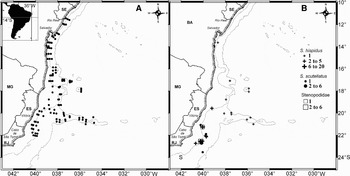
Larvae of Stenopodidae were sorted from plankton samples collected during six oceanographical cruises in the South Atlantic between May 1999 and October 2002. The first and most important comprised 114 oceanographical stations during the cruise ‘Bahia I’ of the RV ‘Thalassa’ (IFREMER) off the east coast of Brazil between Rio Real (11°S) and Cabo de São Tomé (22°S) (Figure 1). All the samples were taken using a bongo net with 333 µm and 500 µm mesh size and then immediately preserved in 4% buffered formaldehyde. The hauls were oblique, starting and ending at the surface. The maximum depth reached by the net was 200 m. Three other cruises occurred in Campos Basin (Rio de Janeiro State) in October 2001, May 2002 and August 2002 involving up to 20 stations in total and a similar sampling strategy (bongo net, 333 µm and 500 µm mesh size, oblique hauls up to 200 m). One cruise occurred off the Espírito Santo coast in February 2002 and the last in the Santos Basin (São Paulo State) in October 2002. In the laboratory, larvae of Stenopodidae were sorted from the samples, placed in 70% ethanol, and permanently stored in the collection of the Zoology Department, Federal University of Rio de Janeiro, Brazil (DZUFRJ). Total length (TL) and rostral length (RL) of the 52 larvae obtained were measured under a Zeiss Stemi SV6 stereoscope with a micrometer ruler (precision of 0.1 mm). TL was measured from the rostral tip to the posteriormost edge of the telson, excluding setae. RL was measured from the rostral tip to the point of eye-stalk insertion. Larval stages were established according to the degree of development of the larval appendages. Larval appendages were dissected and drawn under a Zeiss Axiostar Plus optical microscope equipped with phase contrast.
Study site in South America. (A) Sampling stations (small black circles); (B) distribution of Stenopus hispidus (plus), S. scutellatus (black circles), and Stenopodidae 1 and 2 (squares).

RESULTS
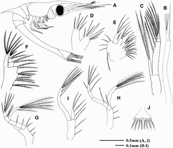
Stenopus hispidus (Olivier). First zoea. (A) Lateral view; (B and C) first and second antenna; (D and E) first and second maxilla; (F–H) first to third maxilliped; (I) first pereopod.

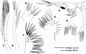
Stenopus hispidus (Olivier). Second zoea. (A) Lateral view; (B and C) first and second antenna; (D and E) first and second maxilla; (F) first maxilliped; (G) telson.

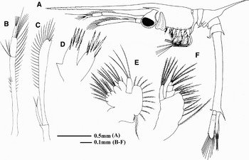
Stenopus hispidus (Olivier). Third zoea. (A) Lateral view; (B and C) first and second antenna; (D and E) first and second maxilla; (F) first maxilliped.

Stenopus hispidus (Olivier). Fourth zoea. (A) Lateral view; (B and C) first and second antenna; (D and E) first and second maxilla; (F–H) first to third maxilliped; (I–L) first to fourth pereopod.

Stenopus hispidus (Olivier). Sixth instar. (A) Lateral view; (B and C) first and second antenna; (D and E) first and second maxilla; (F–H) first to third maxilliped; (I–M) first to fifth pereopod.

Total length (TL), rostral length (RL) and sequence of appendage development in Stenopus hispidus (Olivier).

ST, stage; N, number of larvae measured; MP1-3, first to third maxilliped; P1-5, first to fifth pereopod; PL, pleopods; EU, endopod of uropod; T, telson (t, triangular; r, rectangular); +, present; -, absent; b, bud; c, chelate. Measurements in mm.
MATERIAL EXAMINED
Brazil, Bahia State, Revizee Program, Cruise ‘Bahia I’, RV ‘Thalassa’ (IFREMER), ‘Banco Rodger’, Station T5034, 17°05′06.00″S 037°35′07.08″W, 0–200 m, 1 zoea VI (DZUFRJ 914). ‘Banco Hotspur’, Station T5042, 17°46′08.40″S 035°52′40.44″W, 0–200 m, 1 zoea VII (DZUFRJ 903); Station T5044, 18°04′21.00″S 035°53′03.84″W, 0–200 m, 1 zoea II (DZUFRJ 944). ‘Arquipélago dos Abrolhos’, Station T5054, 18°45′55.08″S 037°56′57.48″W, 0–60 m, 1 zoea I. (DZUFRJ 422)—Espírito Santo State, Revizee Program, Cruise ‘Bahia I’, RV ‘Thalassa’, Station T5087, 20°02′01.32″S 038°06′52.20″W, 0–65 m, 1 zoea IV. (DZUFRJ 864); Station T5096, 20°06′05.76″S 037°27′02.88″W, 1 zoea I. (DZUFRJ 818); Station T5110, 20°59′49.20″S 035°28′16.32″W, 0–200 m, 1 zoea IV (DZUFRJ 1000). ‘Banco Jaseur’, Station T5109, 20°34′34.32″S 036°03′08.28″W, 0–200 m, 1 zoea II (DZUFRJ 937). Cruise ‘BC60’, SB ‘Astrogaroupa’, Station J1, 21°14′52.74″S 040°03′10.70″W, 0–200 m, 3 zoea III (DZUFRJ344), 2 zoea IV (DZUFRJ5, 350), 1 zoea VII (DZUFRJ7); Station J5, 22°43′34.40″S 040°41′36.30″W, 0–200 m, 3 zoea III (DZUFRJ357, 361), 2 zoea IV (DZUFRJ360, 544); Station M1, 21°14′53.94″S 040°03′03.77″W, 0–200 m, 4 zoea III (DZUFRJ364), 2 zoea IV (DZUFRJ48, 543); Station M5, 21°14′56.29″S 040°02′50.12″W, 0–200 m, 2 zoea III (DZUFRJ366, 545), 1 zoea IV (DZUFRJ368), 1 zoea VII (DZUFRJ52)—Rio de Janeiro State, Cruise ‘Barracuda-Caratinga’, SB ‘Astrogaroupa’, Station 34, 22°33′46″S 040°13′23″W, 0–200 m, 2 zoea I (DZUFRJ540), 1 zoea II (DZUFRJ461), 2 zoea III (DZUFRJ444); Station 38, 22°41′52″S 040°14′13″W, 0–200 m, 1 zoea II (DZUFRJ414); Station 40, 22°36′55″S 040°09′19″W, 0–200 m, 2 zoea IV (DZUFRJ467, 663). Cruise ‘Albacora Leste’, SB ‘Astrogaroupa’, Station 23, 0–200 m, 1 zoea VII (DZUFRJ512); Station 24, 0–200 m, 1 zoea V (DZUFRJ510); Station 26, 0–200 m, 1 zoea IV (DZUFRJ509), 1 zoea VI (DZUFRJ511). Cruise ‘Marlim Sul’, SB ‘Astrogaroupa’, Station Ca.2, 22°31′58.9″S 040°02′53.4″W, 0–200 m, 1 zoea V (DZUFRJ259); Station Cb.3, 22°31′40.9″S 040°02′39.6″W, 0–200 m, 1 zoea VII (DZUFRJ284).
DIAGNOSIS
First pereopod fully developed in the first stage; abdomen flexed about 90° on somite 3; rostrum long, about twice the carapace length and serrated in the first stage; postero-ventral margin of carapace without spine; supra-orbital spine present, except in the first stage; abdominal somite 6 longer than the other five together; ventral spine on abdominal somite 5 procurve; dorsal spine present on abdominal somites 3 and 5, that on third somite larger; pleural spines on abdominal somites 1 and 3; mandible exceptionally large, inserted in a cleft at the carapace; antennal scale four-segmented at the tip; telson triangular with a median cleft that bear 7+7 marginal plumose setae; second seta reduced to a thalassinid hair.
DESCRIPTION
First zoea ( Figure 2): TL = 4.1–4.3 mm; RL = 1.4–1.8 mm (N = 4); carapace (Figure 2A)—rostrum well developed, longer than antennae, toothed and slightly up-turned towards the end; posterior margin of carapace partly covering the first abdominal somite; eyes sessile; supra-orbital spine absent; abdomen—six somites, the sixth longer than the other five together and fused to the telson; first and third somites with pleural spines; fifth somite with single ventral procurved spine; first somite with small dorsal paired spine, and one lateral knob under the carapace; third and fifth somites with single dorsal spine, that on third somite larger; telson (Figure 2J)—triangular and toothed on its outer margin; posterior margin with V-shaped median cleft and 7+7 processes (outermost process an immovable spine, second process a small seta, and third to sixth processes long plumose setae); antennule (Figure 2B)—elongate with a one-segmented peduncle; exopod consisting of a small bulb with four aesthetascs terminally; endopod represented by a long simple seta with a bulbous base; antenna (Figure 2C)—peduncle small, shorter than endopod; exopod nearly cylindrical with four terminal joints and bearing nine plumose setae, six on the inner margin and three terminally; endopod short, approximately half the length of exopod, and with two long plumose setae; maxillule (Figure 2D)—consisting of two inner lobes, the proximal coxopod bearing three cuspidate and four plumodenticulate setae and the distal basipod bearing two cuspidate and three plumodenticulate setae; maxilla (Figure 2E)—protopod with four inner lobes, the first and second closest, and bearing 7+3+4+3 plumose setae; scaphognathite with five marginal plumose setae, the last stronger; endopod weakly two-segmented, with 1+3 plumose setae; first maxilliped (Figure 2F)—protopod with ten (2+3+3+2) plumose setae; exopod one-segmented with four terminal plumose setae; endopod three-segmented and bearing 2+2+3 plumose setae; second maxilliped (Figure 2G)—protopod with four (1+1+2) plumose setae; exopod one-segmented with one subterminal and four terminal plumose setae; endopod five-segmented with 1+1+0+2+5 plumose setae; third maxilliped (Figure 2H)—protopod with three (1+1+1) plumose setae; exopod one-segmented with one subterminal and four terminal plumose setae; endopod five-segmented with 1+1+0+1+5 plumose setae; first pereopod (Figure 2I)—protopod with three (1+1+1) plumose setae; exopod with six plumose setae; endopod one-segmented with one proximal, one median and two terminal plumose setae; second to fifth pereopods—no trace of any pereopod except the first; pleopods—absents; uropods—absents.
Second zoea ( Figure 3): TL = 5.3–7.1 mm; RL = 1.5–2.6 mm (N = 4); carapace (Figure 3A)—similar to the previous stage, except for the presence of stalked eyes and the appearance of the supra-orbital spine; abdomen—lateral knob on first abdominal somite now is uncovered by the carapace; telson (Figure 3G)—the telson has changed to form two divergent arms, smooth on the outer margin; setation as in first stage; antennule (Figure 3B)—peduncle two-segmented that bear 8+4 marginal and two apical plumose setae; exopod with five terminal aesthetascs; endopod as in first stage; antenna (Figure 3C)—peduncle short, one-segmented; exopod unsegmented, flattened, and broadened in a scale (scaphocerite) with 16 plumose setae on the margin; endopod one-segmented, lance-like, with two terminal plumose setae; maxillule (Figure 3D)—coxopod with three cuspidate and four plumodenticulate setae and basipod with four cuspidate and two plumodenticulate setae; proximal region of basipod with a spined process; maxilla (Figure 3E)—protopod with 8+3+4+4 plumose setae; scaphognathite with eight marginal plumose setae, the last larger than the others; endopod with three terminal plumose setae; jirst maxilliped (Figure 3F)—protopod with 11 (2+3+3+3) plumose setae; exopod and endopod similar to first stage; second maxilliped—endopod five-segmented with 1+1+0+2+7 plumose setae; third maxilliped—protopod and exopod unchanged; endopod five-segmented with 1+1+0+2+7 plumose setae; first pereopod—protopod with three (1+1+1) plumose setae; exopod with two subterminal and four terminal plumose setae; endopod one-segmented with one proximal, two median and three terminal plumose setae; second pereopod—small bud; third to fifth pereopods—absents; pleopods—absents; uropods—absents.
Third zoea ( Figure 4): TL = 7.9–9.9 mm; RL = 2.7–3.3. mm (N = 14); carapace (Figure 4A)—extending posteriorly in a more or less straight line from the rostrum; rostrum nearly twice the length of carapace, downturned and serrated in the tip; supra-orbital spine stout; abdomen—sixth abdominal somite has become separated from the telson and has a pleural spine turned forwards; lateral knob more pronounced on first somite and engaged on posterior margin of carapace; telson—the telson became rectangular; antennule (Figure 4B)—peduncle two-segmented, bearing 13+4 plumose setae on inner margin, and five terminal; exopod with five terminal aesthetascs; endopod as a long seta with a bulbous base; antenna (Figure 4C)—scaphocerite with 26 plumose setae on the inner margin; endopod with two terminal plumose setae; maxillule (Figure 4D)—coxopod with three cuspidate and five plumodenticulate setae; basipod with four cuspidate and four plumodenticulate setae; spined process enlarged; maxilla (Figure 4E)—protopod bearing 8+3+4+5 plumose setae; scaphognathite with 12 marginal plumose setae; endopod unsegmented with three terminal plumose setae; first maxilliped—protopod with 13 plumose setae; exopod similar to third stage, endopod with 3+2+4 plumose setae; second maxilliped—exopod with three subterminal and four terminal plumose setae; endopod with 2+1+0+3+7 plumose setae; third maxilliped—exopod with four subterminal and four terminal plumose setae; endopod with 2+1+0+2+8 plumose setae; first pereopod—protopod with three (1+1+1) plumose setae; exopod with three subterminal and four terminal plumose setae; endopod as in second stage; second pereopod—protopod with no seta; exopod with two subterminal and four terminal plumose setae; endopod one-segmented, with two terminal plumose setae; third pereopod—protopod with no seta; exopod with two subterminal and three terminal plumose setae; endopod one-segmented, with one subterminal and two terminal plumose setae; fourth and fifth pereopods—present as small buds; pleopods—absents; uropods—exopod flattened, longer than telson and with 18 marginal plumose setae; endopod present as a small bud.
Fourth zoea ( Figure 5): TL = 10.0–14.4 mm; RL = 2.8–4.8 mm (N = 13); carapace (Figure 5A)—unchanged; abdomen—similar to third stage; lateral knob more pronounced on first somite and engaged on posterior margin of carapace; telson—unchanged; antennule (Figure 5B)—peduncle two-segmented, bearing 14+4 plumose setae on inner margin, and five terminal; exopod unchanged; endopod unchanged; antenna (Figure 5C)—scaphocerite with 30 marginal plumose setae; endopod with three terminal plumose setae; maxillule (Figure 5D)—coxopod with four cuspidate and six plumodenticulate setae; basipod with four cuspidate and four plumodenticulate setae; spined process unchanged; maxilla (Figure 5E)—protopod with 8+3+4+5 plumose setae; scaphognathite with 12 marginal plumose setae; endopod unsegmented with three terminal plumose setae; first maxilliped (Figure 5F)—protopod with 13 plumose setae; exopod similar to third stage, endopod with 3+2+4 plumose setae; second maxilliped (Figure 5G)—exopod with two subterminal and four terminal plumose setae; endopod with 2+1+0+3+7 plumose setae; third maxilliped (Figure 5H)—protopod with two plumose setae; exopod with two subterminal and four terminal plumose setae; endopod with 2+1+0+2+8 plumose setae; first pereopod (Figure 5I)—protopod with no setae; exopod with two subterminal and four terminal plumose setae; endopod small, one-segmented and with four plumose setae; second pereopod (Figure 5J)—protopod with no setae; exopod with two subterminal and four terminal plumose setae; endopod one-segmented, with three plumose setae; third pereopod (Figure 5K)—protopod with no seta; exopod with two subterminal and three terminal plumose setae; endopod one-segmented, with three plumose setae; fourth and fifth pereopods (Figure 5L)—present as small buds; pleopods – absents; uropods—protopod with a spinous process; exopod with 32 marginal plumose setae; endopod almost the same length as exopod, with 28 marginal plumose setae.
Fifth instar: TL = 15.3–16.9 mm; RL = 4.7–5.0 mm (N = 2); larva very similar to previously; carapace–unchanged; abdomen—unchanged; telson—unchanged; antennule—unchanged; antenna—scaphocerite with 38 plumose setae on the inner margin; endopod with two terminal plumose setae; maxillule—coxopod with four cuspidate and six plumodenticulate setae; basipod with four cuspidate and six plumodenticulate setae; spined process enlarged; maxilla—scaphognathite with 26 marginal plumose setae; first maxilliped—protopod with 20 plumose setae; exopod and endopod similar to previous stage; second maxilliped—endopod with 3+2+0+4+7 plumose setae; third maxilliped—endopod with 2+1+0+3+8 plumose setae; first to third pereopod—similar to fourth stage, but larger; fourth and fifth pereopods—one-segmented and longer than in fourth stage; still no trace of exopods; pleopods—absents; uropods—exopod with 44 marginal plumose setae; endopod with 46 marginal plumose setae.
Sixth instar ( Figure 6): TL = 20.3–20.7 mm; RL = 5.9–6.1 mm (N = 2); carapace (Figure 6A)—rostrum smooth; abdomen—unchanged; telson—similar to fifth stage; antennule (Figure 6B)—unchanged, except by two additional plumose setae (16+4); antenna (Figure 6C)—scaphocerite with 46 plumose setae on the inner margin; maxillule (Figure 6D)—coxopod with 24 plumose setae; basipod with five cuspidate and 14 plumose setae; maxilla (Figure 6E)—scaphognathite with 34 marginal plumose setae; first maxilliped (Figure 6F)—enlarged; protopod with an outer epipod and 21 plumose setae; exopod unchanged; endopod three-segmented with 2+2+4 plumose setae; second maxilliped (Figure 6G)—protopod with five plumose setae; exopod unchanged; endopod with 3+4+0+3+9 plumose setae; third maxilliped (Figure 6H)—protopod with one plumose seta; endopod with 3+1+0+2+13 plumose setae; first pereopod (Figure 6I)—exopod with 14 plumose setae; endopod chelate, with one subterminal and two terminal simple setae; second pereopod (Figure 6J)—exopod with 15 plumose setae; endopod chelate, with two terminal simple setae; third pereopod (Figure 6K)—exopod with eight plumose setae; endopod chelate, with no seta; fourth and fifth pereopods (Figures 6L and 6M)—uniramous and longer than in fifth stage; pleopods—absents; uropods—similar to previously, but with additional setae.
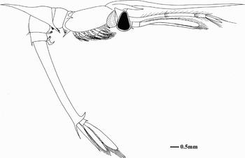
Seventh instar ( Figure 7): TL = 22.5–28.5 mm; RL = 7.1–9.3 mm (N = 5); all larvae damaged during the sampling. Setation could be described with certainty for only few appendages; carapace—unchanged; abdomen—unchanged; telson—unchanged; antennule—exopod with two terminal plumose setae; antenna—endopod longer than scaphocerite, with two terminal plumose setae; maxillule—spined process with one terminal seta; maxilla—scaphognathite with 43 marginal plumose setae; first maxilliped—protopod with 30 plumose setae; endopod with 3+2+5 plumose setae; second maxilliped—exopod with 24 marginal plumose setae; endopod with 3+5+0+3+10 plumose setae; third maxilliped—exopod with 60 marginal plumose setae; first pereopod—unchanged; second pereopod—unchanged; third pereopod—endopod chelate, with one subterminal and two terminal plumose setae; fourth and fifth pereopods—longer than in sixth stage; pleopods—first to fifth pleopods present as long bifid buds; uropods– unchanged.
Stenopus hispidus (Olivier). Seventh instar, lateral view.

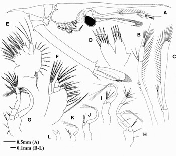
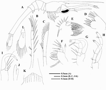
Stenopus scutellatus Rankin. Second zoea. (A) Lateral view; (B and C) first and second antenna; (D and E) first and second maxilla; (F–H) first to third maxilliped; (I), first pereopod; (J) telson; (K) posterior margin of telson.

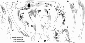
Stenopus scutellatus Rankin. Third zoea. (A) Lateral view; (B and C) first and second antenna; (D and E) first and second maxilla; (F–H) first to third maxilliped; (I) first pereopod; (J) second pereopod; (K) telson; (L) uropod.

Stenopus scutellatus Rankin. Fifth instar, lateral view.

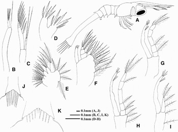
Stenopus scutellatus Rankin. Seventh instar. (A) Lateral view; (B and C) first and second maxilla; (D and E) first maxilliped; (F and G) second and third maxilliped; (H–K) first to fourth pereopod.

Total length (TL), rostral length (RL) and sequence of appendage development in Stenopus scutellatus Rankin.

ST, stage; N, number of larvae measured; MP1-3, first to third maxilliped; P1-5, first to fifth pereopod; PL, pleopods; EU, endopod of uropod; T, telson (t, triangular; r, rectangular); +, present; -, absent; b, bud; c, chelate. Measurements in mm.
MATERIAL EXAMINED
Brazil, Espírito Santo State, Revizee Program, Cruise ‘Bahia I’, RV ‘Thalassa’, Station T5079, 21°03′57.35″S 040°14′22.56″W, 0–80 m, 1 zoea II (DZUFRJ 986)—Rio de Janeiro State, Cruise ‘Roncador’, SB ‘Astrogaroupa’, Station 2.2, 21°53′24″S 039°45′00″W, 0–200 m, 1 zoea VII (DZUFRJ 328). Cruise ‘Barracuda/Caratinga’, SB ‘Astrogaroupa’, Station 34, 22°33′36″S 040°13′12″W, 0–200 m, 1 zoea II (DZUFRJ538), 1 zoea III (DZUFRJ537); Station 40, 22°37′12″S 040°09′36″W, 0–200 m, 1 zoea II (DZUFRJ665), 1 zoea III (DZUFRJ664). Cruise ‘Marlin Sul’, SB ‘Astrogaroupa’, Station P9-R3, 23°31′48″S 040°03′00″W, 0–200 m, 1 zoea II (DZUFRJ493)—São Paulo State, Cruise ‘B. Santos’, Station P16, 25°48′2″3S 043°41′47″W, 0–200 m, 1 zoea V (DZUFRJ598).
DIAGNOSIS
Rostrum smooth, longer than antenna; postero-ventral margin of carapace with a conspicuous spine; mandible exceptionally large, inserted in a cleft at the carapace; small dorsal spine present on abdominal somite 5; pleural spines on abdominal somites 1, 3 and 4; abdominal somite 6 longer than the other five together; ventral spine on abdominal somite 5 procurve; telson without lateral spines.
DESCRIPTION
Second zoea ( Figure 8): TL = 2.8–3.9 mm; RL = 0.7–1.3 mm (N = 4); carapace (Figure 8A)—rostrum well developed, longer than antennae and smooth; posterior margin of carapace partly covering the first abdominal somite and with a pronounced spine pointed back; eyes stalked; small supra-orbital spine present; abdomen—first, third, and fourth abdominal somites with pleural spines; fifth abdominal somite with procurved ventral spine and dorsal spine; sixth abdominal somite still fused with telson; telson (Figure 8J, K)—triangular and with two divergent arms; outer margin smooth; posterior margin with 7+7 processes, outermost an immovable spine; second process a small simple seta; third to sixth processes long plumose setae; seventh a small plumose seta; antennule (Figure 8B)—peduncle unsegmented; exopod as small bulbous with four terminal aesthetascs; endopod a long seta with broad base; antenna (Figure 8C)—peduncle small; exopod flattened, with four-segmented apices and bearing nine plumose setae; endopod bulbous-like with two long simple setae; maxillule (Figure 8D)—coxopod with three cuspidate and four plumose setae and basipod with two cuspidate and three plumose setae; maxilla (Figure 8E)—endites with 9+3+4+4 plumose setae; scaphognathite with six marginal plumose setae, the last larger than the others; endopod with three terminal plumose setae; first maxilliped (Figure 8F)—protopod with 15 plumose setae; exopod one-segmented with four plumose setae; endopod three-segmented with 3+2+4 plumose setae; second maxilliped (Figure 8G)—protopod with no setae; exopod one-segmented with six plumose setae; endopod five-segmented with 1+1+0+0+3 plumose setae; third maxilliped (Figure 8H)—protopod with three (1+1+1) plumose setae; exopod one-segmented with six plumose setae; endopod five-segmented with 2+1+0+1+3 plumose setae; first pereopod (Figure 8I)—protopod with two (1+1) plumose setae; exopod with six plumose setae; endopod one-segmented with four plumose setae; second to fifth pereopods—absents; pleopods—absents; uropods—absents.
Third zoea ( Figure 9): TL = 4.9–5.0 mm; RL = 1.6–1.8 mm (N = 2); carapace (Figure 9A)—supra-orbital spine enlarged; abdomen—sixth abdominal somite became separated from telson; telson (Figure 9K)—outer spine absent; antennule (Figure 9B)—peduncle two-segmented with 4+2 plumose setae on inner margin; exopod and endopod similar to previous stage; antenna (Figure 9C)—peduncle small; exopod flattened, unsegmented and bearing 13 marginal plumose setae; endopod longer than previously; maxillule (Figure 9D)—coxopod with three cuspidate and six plumose setae and basipod with four cuspidate and two plumose setae; spinous process absent; maxilla (Figure 9E)—endites with 8+2+4+4 plumose setae; scaphognathite with eight marginal plumose setae, the last larger than the others; endopod with three terminal plumose setae; first maxilliped (Figure 9F)—protopod with ten plumose setae; exopod one-segmented with four plumose setae; endopod three-segmented with 3+2+4 plumose setae; second maxilliped (Figure 9G)—protopod with three plumose setae; exopod one-segmented with six plumose setae; endopod five-segmented with 2+1+0+2+4 plumose setae; third maxilliped (Figure 9H)—protopod with one plumose setae; exopod one-segmented with six plumose setae; endopod five-segmented with 2+1+0+2+4 plumose setae; first pereopod (Figure 9I)—protopod with one plumose setae; exopod with two plumose setae; endopod one-segmented with one proximal, one median and one terminal plumose setae; second pereopod (Figure 9J)—biramous but small; third to fifth pereopods—absents; pleopods—absents; uropods (Figure 9L)—biramous, the exopod larger and bearing 12 marginal plumose setae; endopod as a small bud.
Fifth zoea ( Figure 10): TL = 8.9 mm; RL = 2.9 mm (N = 1); larva similar to previously, but bigger. Appendages not well preserved to be dissected; carapace—unchanged; abdomen—first, third, fourth, and sixth abdominal somites with pleural spines; procurved ventral spine present on fifth abdominal somite; dorsal spine present on fifth and sixth abdominal somites.
Seventh instar ( Figure 11): TL = 16.9 mm; RL = 5.1 mm (N = 1); carapace (Figure 11A)—unchanged; abdomen—first, third, fourth, and sixth abdominal somites with pleural spines; first somite with two dorso-lateral spines pointed forward, the larger engaged to the carapace; fifth abdominal somite with procurved ventral spine and dorsal spine; antennule—peduncle three-segmented; exopod and endopod the same length; antenna—peduncle small; exopod flattened, unsegmented and bearing 23 marginal plumose setae; endopod as long as exopod; maxillule (Figure 11B)—coxopod with seven cuspidate and nine plumose setae and basipod with four cuspidate and seven plumose setae; maxilla (Figure 11C)—endites with 15+4+7+7 plumose setae; scaphognathite with 32 marginal plumose setae; endopod with two terminal plumose setae; first maxilliped (Figure 11D, E)—protopod with 23 plumose setae and one outer epipod; exopod one-segmented with six plumose setae; endopod three-segmented with 3+2+4 plumose setae; second maxilliped (Figure 11F)—protopod with one plumose setae; exopod one-segmented with six plumose setae; endopod five-segmented with 3+2+0+3+7 plumose setae; third maxilliped (Figure 11G)—protopod with one plumose setae; exopod one-segmented with six plumose setae; endopod five-segmented with 2+1+1+2+10 plumose setae; first pereopod (Figure 11H)—protopod with no setae; exopod with four plumose setae; endopod chelate with four simple setae; second pereopod (Figure 11I)—similar to first, but with three simple setae; third pereopod (Figure 11J)—chelate with two simple setae; fourth pereopod (Figure 11K)—uniramous, longer than third pereopod and with no seta; fifth pereopod—absent; pleopods—biramous but small; uropods—protopod spined at the tip; exopod and endopod about the same length.
Stenopodidae 1. Fifth zoea? (A) Lateral view. Stenopodidae 2. First zoea. (B) Lateral view.

MATERIAL EXAMINED
Brazil, Espírito Santo State, Cruise ‘BC60’, SB ‘Astrogaroupa’, Station J1C, 21°14′52.74″S 040°03′10.70″W, 0–200 m, 1 zoea VII? (DZUFRJ41).
DIAGNOSIS
Rostrum smooth, longer than antenna; postero-ventral margin of carapace with no spine; mandible exceptionally large, inserted in a cleft at the carapace; small dorsal spine present on abdominal somite 3, 5 and 6; pleural spines on abdominal somites 1, 2 and 3 pointed forwards; abdominal somite 6 twice longer than the other five together; ventral spine on abdominal somite 5 absent.
DESCRIPTION
Fifth (?) zoea ( Figure 12A): TL = 9.7 mm; RL = 3.3 mm (N = 1); carapace—rostrum smooth, well developed, longer than antennae; posterior margin of carapace almost completely covering the first abdominal somite; eyes stalked; supra-orbital spine present; abdomen—first, second, and third abdominal somites with pleural spines pointed forwards; third, fifth, and sixth somites with dorsal spine, that on third somite stronger; fifth abdominal somite with no ventral spine; first to third pereopods—well-developed; fourth and fifth pereopods—as small buds.
MATERIAL EXAMINED
Brazil, Rio de Janeiro State, Cruise ‘Barracuda/Caratinga’, SB ‘Astrogaroupa’, Station 34, 22°33′46″S 040°13′23″W, 0–200 m, 1 zoea I (DZUFRJ539).
DIAGNOSIS
Rostrum serrated, longer than antenna; postero-ventral margin of carapace with no spine; mandible exceptionally large, inserted in a cleft at the carapace; small dorsal spine present on abdominal somite 3; pleural spines on abdominal somites 1, 2 and 3; small postero-ventral spines pointed backwards on abdominal somites 4 and 5; ventral spine on abdominal somite 5 straight.
DESCRIPTION
First zoea ( Figure 12B): TL = 3.5 mm; RL = 1.1 mm (N = 1); carapace—rostrum well developed, longer than antennae and serrated laterally; posterior margin of carapace almost completely covering the first abdominal somite; eyes sessile; supra-orbital spine absent; abdomen—first, second, and third abdominal somites with pleural spines; fourth and fifth abdominal somites with small postero-ventral spines pointed backwards; small dorsal spine present on third abdominal somite; ventral spine on fifth abdominal somite straight and small; sixth abdominal somite still fused with telson; telson—triangular and with two divergent arms; outer margin smooth; posterior margin with 7+7 plumose setae, the second reduced; first pereopod—well-developed; second pereopod—as small bud; third to fifth pereopods—absents.
DISCUSSION
To date, two species of Stenopodidae have been recorded in the western South Atlantic—Stenopus hispidus and Stenopus scutellatus (Coelho & Ramos-Porto, Reference Coelho, Ramos-Porto and Young1998; Young & Serejo, Reference Young, Serejo, Dutra, Allen, Werner and McKenna2005; Coelho et al., Reference Coelho, de Almeida, Souza-Filho, de Bezerra and Giraldes2006; Fernandes et al., Reference Fernandes, Almeida, Peixoto, Souza and Bonecker2006; Gregati et al., Reference Gregati, Pinheiro and Cobo2006). Three species of Microprosthema, a genus included in the closely related family Spongicolidae, also occur in the area (Coelho & Ramos-Porto, Reference Coelho, Ramos-Porto and Young1998; Fernandes & Bonecker, Reference Fernandes and Boneckerin press). According to Martin & Goy (Reference Martin and Goy2004) and Fernandes & Bonecker (Reference Fernandes and Boneckerin press), the larvae of Microprosthema can be separated from those of Stenopus by the following diagnostic characters: abbreviated development, presence of dorsal groove on carapace, rostral length smaller than or equal to antennules, absence of dorsal spine on third somite, and absence of procurved ventral spine on fifth somite. With the exception of Stenopodidae 1 and 2, whose larvae have no procurved spine on fifth somite, none of the larvae described herein exhibit these characters. In addition, the vast majority of the larvae found in the study area show all the characters assumed previously by Lebour (Reference Lebour, Gurney and Lebour1941) and Williamson (Reference Williamson1976) as being diagnostic for the genus Stenopus: absence of a postero-ventral spine on the carapace, presence of a median dorsal spine on third somite, presence of a median ventral spine on fifth somite (frequently procurved) and presence of a pleural spine on sixth somite. All these characters are seen throughout larval development of Stenopus.
Stenopodidea has a range of developmental patterns from abbreviated to extended larval development (Felder et al., Reference Felder, Martin, Goy and Wenner1985; Seridji, Reference Seridji1990). Direct development was confirmed for Spongicola japonica and Spongicoloides koehleri (Felder et al., Reference Felder, Martin, Goy and Wenner1985; Saito & Konishi, Reference Saito and Konishi1999), while Microprosthema semilaeve exhibit abbreviated development (Seridji, Reference Seridji1990). According to Lebour (Reference Lebour, Gurney and Lebour1941), the development of S. hispidus goes through at least nine larval instars. Although the total number of larval stages is not known for Stenopus spinosus, Seridji (Reference Seridji1990) suggested more than five zoeal stages. In addition, Fletcher et al. (Reference Fletcher, Kötter, Wunsch and Yasir1995) kept larvae in a laboratory for up to 210 days, indicating extended larval development for Stenopus species when compared to other Stenopodidea, such as Microprosthema semilaeve (Seridji, Reference Seridji1990; Martin & Goy, Reference Martin and Goy2004).
The larval development of S. hispidus is known mainly from plankton specimens (Gurney, Reference Gurney1924, Reference Gurney1936, Reference Gurney1939, Reference Gurney1942; Lebour, Reference Lebour, Gurney and Lebour1941; Williamson, Reference Williamson1976). Gurney's first stage of S. hispidus (Gurney, Reference Gurney1936) and that described here fully agree in the number and position of dorsal, ventral and pleural spines. However, Gurney (Reference Gurney1936) showed an outer seta on the antennal scale, an outer spinous process on the maxillule, fewer seta on the endites of second maxilla (7+2+3+3 against 7+3+4+3), endopod of second maxilla missing one median seta, one more seta on the first segment of the endopod of second maxilliped, and one more plumose setae on the endopod of first pereopod. Intra-specific variation was stressed by Seridji (Reference Seridji1990) concerning pleural spines in S. spinosus larvae described by Bourdillon-Casanova (Reference Bourdillon-Casanova1960) and Williamson (Reference Williamson1976), which are more conservative characters than number of setae. This kind of variation was also observed in M. semilaeve larvae (Martin & Goy, Reference Martin and Goy2004). Anger (Reference Anger2001) considered that specific differences are frequently restricted to small morphological details, such as the setation of mouthparts. Similarly, S. hispidus development seems to include some intra-specific or regional variation, as revealed by the appearance of the spined process, which was observed since the first stage by Gurney (Reference Gurney1936) and since the second stage in the present paper. The present study, actually the third larval description of Stenopus hispidus, agrees with genus and species diagnosis made previously by Gurney (Reference Gurney1936) and Lebour (Reference Lebour, Gurney and Lebour1941).
Ten additional species of Spongicolidae (Microprosthema manningi, M. looensis, M. granatense, M. jareckii, M. inornatum, Spongicola cubanicus, Spongiocaris hexactinellicola, S. goyi, Spongicoloides koehleri and S. tabachnicki) and six of Stenopodidae (Stenopus spinosus, Odontozona liberate, O. striata, O. anaphorae, O. edwardsi and Richardina spinicincta) have been reported in the western North Atlantic, in the Caribbean, and in Ascension Island (Goy, Reference Goy1982; Goy & Felder, Reference Goy and Felder1988; Manning & Chace, Reference Manning and Chace1990; Berggren, Reference Berggren1993; Ortiz et al., Reference Ortiz, Gómez and Lalana1994; Criales, Reference Criales1997; Martin, Reference Martin2002; Saito & Takeda, Reference Saito and Takeda2003; Martin & Goy, Reference Martin and Goy2004; Ortiz et al., Reference Ortiz, Lalana and Varela2007; Burukovsky, Reference Burukovsky2009), increasing the total number of species of Stenopodidea in the Atlantic Ocean to nineteen. None of these fourteen additional species was already registered in north-eastern Brazil, but at least ten of the 52 larvae collected in the study site do not fit the diagnosis of Stenopus or Microprosthema larvae because of the following characters: presence of a postero-ventral spine on the carapace margin; absence of ventral procurved spine on fifth abdominal somite; presence of pleural spines on second abdominal somite; and small dorsal spine on third somite. According to Williamson (Reference Williamson1976) the presence of postero-ventral spine in carapace could be diagnostic for Odontozona larvae. Three species of Odontozona were registered in adjacent areas, but never in Brazil. The presence of such spine on carapace and also the pleural spine on fourth abdominal somite in the seven larvae ascribed to S. scutellatus suggest the diagnosis proposed before by Williamson (Reference Williamson1976) to the genus Odontozona should be revised.
According to Gurney (Reference Gurney1936), the presence of a median ventral spine on fifth somite is regarded as probably diagnostic for larvae of Stenopus. This procurved spine is shared with most Odontozona larvae. The larvae of Stenopodidae 1 and Stenopodidae 2 have also dorsal spine on third abdominal somite, but it is stronger in its base and smaller in length than in the larvae of S. hispidus. In addition, both larvae have pleural spines on first to third abdominal somite and no ventral procurved spine on fifth somite. Other species of Stenopodidea were registered in the study site or in adjacent areas by many authors (Goy, Reference Goy1982; Manning & Chace, Reference Manning and Chace1990; Ortiz et al., Reference Ortiz, Gómez and Lalana1994; Criales, Reference Criales1997; Coelho & Ramos-Porto, Reference Coelho, Ramos-Porto and Young1998; Young & Serejo, Reference Young, Serejo, Dutra, Allen, Werner and McKenna2005; Coelho et al., Reference Coelho, de Almeida, Souza-Filho, de Bezerra and Giraldes2006; Fernandes et al., Reference Fernandes, Almeida, Peixoto, Souza and Bonecker2006; Ortiz et al., Reference Ortiz, Lalana and Varela2007; Fernandes & Bonecker, Reference Fernandes and Boneckerin press), mainly S. scutellatus, M. semilaeve and Microprosthema sp. (sensu Fernandes & Bonecker, in press). The larvae of Microprosthema and those of Stenopodidae 1 and 2 share only few similarities, such as the same small ventral spine on fifth somite and also pleural spines on first to third somite. On the other hand, they can be distinguished from each other by rostral length, dorsal groove on carapace, sequence of appendages development and size. In general, the larvae named Stenopodidae 1 and 2 resemble those of the genus Stenopus. The larvae of Stenopodidae 1 and Gurney's Stenopid VI (Gurney, Reference Gurney1936) share the same general appearance, first to third abdominal somite with large forwardly directed pleural spines, fourth and fifth somites with no pleural spines, third somite produced into a strong dorsal spine, fifth and sixth somites with dorsal spines, and absence of procurved ventral spine on fifth somite. In addition, both larvae were collected in the same area in the western South Atlantic Ocean.
ACKNOWLEDGEMENTS
We would like to thank Dr Maria Criales for the manuscripts sent, review and remarks; Dr Joseph W. Goy, Dr Niel Bruce, and MSc. Daniela Bartholo for helpful suggestions; and LIZI–UFRJ staff for collected specimens. This study was supported by the Executive Committee, Interministerial Commission for Marine Resources (SECIRM), Ministry of Environment (MMA), Sectoral Plan for Marine Resources (REVIZEE), and also by the ‘Centro de Pesquisas e Desenvolvimento Leopoldo Américo Miguez de Melo’ (CENPES)—Petrobras.