Introduction
Goldenhar syndrome refers to a wide spectrum of congenital abnormalities in the head and neck which originate from developmental disturbance of the first and second branchial apparatus.Reference Mahore, Dange, Nama and Goel1, Reference Vinay, Reddy, Uloopi, Madhuri and Sekhar2 Hypoplasia or aplasia of the zygoma, ear, parotid gland, mandible and masticatory muscles are usually present in this syndrome. Hemifacial microsomia often occurs secondary to the anomalies.Reference Tiner and Quaroni3
Congenital salivary fistulae due to abnormal development of an accessory parotid gland have been documented in several articles.Reference Gadodia, Seith, Sharma and Thakar4–Reference Hah, Kim, Sung and Kim8 Their association with Goldenhar syndrome has not been fully studied. A congenital cheek fistula of an accessory parotid gland should be considered indicative of Goldenhar syndrome.
We present two such cases for further documentation.
Case report
Case one
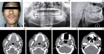
A 16-year-old male was referred to our hospital because of a fistula on his left cheek, which had been discovered soon after birth (Figure 1a). Discharge of clear, serous fluid was reported by the patient, which significantly increased in quantity during eating. No purulent excretion had been noticed.
(a) Clinical photograph of case one showing the left-sided fistula skin opening (arrow) and mandibular asymmetry. (b) Orthopantogram showing hypoplasia of the left mandibular ramus (arrow). (c) X-ray sialogram showing a narrow Stensen's duct (red arrow) and the main parotid gland (yellow arrow). (d), (e) Computed tomography fistulograms showing the fistulous tract (red arrows) extending from the skin opening posteriorly, with a salivary mass (green arrows) located at the anterior border of the masseter muscle (yellow arrows); note also the abnormal morphology of the masseter and its indirect attachment to the ramus, and the abnormal fat tissue seen between the left masseter muscle and the ramus (white arrow). (f), (g) Computed tomography sialograms of the left parotid gland, showing leakage of the contrast medium outside Stensen's duct; note the narrowed Stensen's duct (arrow in part f) and leakage of contrast medium superior to the duct (arrow in part g). R = right; L = left

Physical examination showed a skin opening located approximately 2 cm lateral to the left commissure. No swelling or redness of the facial skin was found. Asymmetry of the lower face was noted, with the chin slightly shifted to the left (Figure 1a).
A spiral computed tomography (CT) scan, performed without contrast medium, revealed a cord-like soft tissue mass extending from the skin to the lateral side of the left masseter muscle. The left parotid gland was of relatively normal size (Figure 1c and 1d). Hypoplasia of the left masseter muscle and the left mandibular ramus was noted (Figure 1b and 1d). The left masseter muscle was separated from the ramus by an abnormal collection of fat tissue (Figure 1d).
Computed tomography fistulography was performed, involving injection of contrast medium (iopamidol, 370 mg I/ml) into the skin opening, and showed the soft tissue cord to be a fistulous tract ending in a mass of attenuated salivary tissue (Figure 1d and 1e). No contrast medium was seen within the left parotid gland. An X-ray sialogram showed a narrowed left Stensen's duct (Figure 1c). A CT sialogram of the left parotid gland via the normal orifice showed leakage of the contrast medium outside Stensen's duct (Figure 1f and 1g). No communication was found between the fistula and the Stensen's duct system.
Resection of the fistulous tract and an accessory parotid gland involved an intraoral incision together with excision of an ellipse of skin containing the fistula orifice.
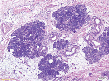
Histopathological examination showed that the surgical specimen contained abundant glandular tissue (Figure 2).
Photomicrograph of the surgical specimen, showing abundant glandular acini and ducts. (H&E; original magnification ×100)

Thus, the cheek fistula was considered to have arisen from an accessory parotid gland. A diagnosis of Goldenhar syndrome was established, based on hypoplasia of the ipsilateral mandibular ramus and masseter muscle, Stensen's duct abnormality, and a congenital salivary fistula from an accessory parotid gland.
Case two
A 12-year-old girl was referred to our hospital with a fistula on her right cheek, discovered after birth. The fistula constantly discharged clear liquid, more so while eating. No history of infection was reported by the patient.
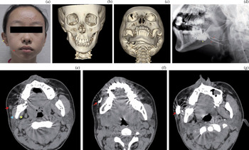
Clinical examination revealed that the skin opening of the fistula was located approximately 1 cm lateral to the commissure on the right side (Figure 3a). No purulent secretion or skin erythema was found. Facial asymmetry was noted, with the chin shifted to the right (Figure 3a and 3c). Prominent deformity of the right tragus was also noted (Figure 3a).
(a) Clinical photograph of case two, showing the fistula skin opening lateral to the right commissure of the lip (red arrow, with a small naevus below); note the ipsilateral tragal deformity (yellow arrow) and the chin shifted towards the right. (b), (c) Volume-rendered images showing hypoplasia of the right mandibular ramus and mandibular asymmetry. (d) Combined right parotid gland sialogram and fistulogram, showing duplication deformity of Stensen's duct (red arrows; sialography) and opacification of the accessory parotid gland (yellow arrow; fistulography via the cheek fistula opening). (e) Axial computed tomography scan without contrast medium, showing a small mass (red arrow) located anterior and lateral to the masseter muscle; note also the blue and yellow arrows indicate the masseter and internal pterygoid muscle separately. (f), (g) Computed tomography fistulography scans showing the fistulous tract extending posteriorly from the skin opening to the lateral side of the masseter muscle (arrow in part f); the salivary mass shown in part (e) is enlarged and filled with contrast (arrow in part g).

Sialography of the right parotid gland showed a duplication deformity of Stensen's duct (Figure 3d).
Computed tomography performed without contrast medium showed a cord-like mass extending from the skin, with a large area of attenuated salivary tissue lying anterior and lateral to the masseter muscle (Figure 3e). Computed tomography fistulography via the skin fistula orifice showed that this attenuated area corresponded with the fistulous tract (Figure 3f and 3g). Volume-rendered images showed hypoplasia of the right mandibular ramus (Figure 3b and 3c). The right main parotid gland was small. The right masseter muscle and internal pterygoid muscle were thin compared with those on the left side (Figure 3e to 3g).
The patient refused any surgical treatment, but was followed up closely.
Discussion
Approximately during the fourth week of embryonic development, the ectodermal lining of the stomodeum or primitive mouth gives rise to buds or branches which form solid cords with round ends, and which subsequently develop into ducts and acini. Accessory parotid glands are derived from a similar pattern of branching and glandular proliferation, arising anterior to and separate from the main parotid tube.Reference Currarino and Votteler9 Aberrant buds which lose their communication with the main parotid gland may give rise to an abnormal, separate, accessory parotid gland.
The normally developed accessory parotid gland is a flattened nodule of salivary tissue separated from the main parotid gland, which lies superficial to the masseter muscle and connects to Stensen's duct via one or occasionally two (or more) ducts; such accessory parotid glands are present in approximately 21 per cent of healthy people.Reference Currarino and Votteler9 Congenital malformation and acquired tumours of accessory parotid glands have been documented.Reference Gadodia, Seith, Sharma and Thakar4, Reference Moon, Han, Kim, Sung, Chang and Choo5, Reference Zhao, Wang and Qi7–Reference Frommer12 Fistulae deriving from abnormal development of an accessory parotid gland in patients with Goldenhar syndrome are rare.Reference Gadodia, Seith, Sharma and Thakar4, Reference Moon, Han, Kim, Sung, Chang and Choo5, Reference Hah, Kim, Sung and Kim8
Goldenhar syndrome occurs due to abnormal embryogenesis of the first and second branchial apparatus, structures which give rise to most of the important facial structures.Reference Mukherji, Fatterpekar, Castillo, Stone and Chung13 The first branchial arch contributes to the formation of the maxilla, zygoma, temporal bone, mandible, malleus, incus and masticatory muscles. The second branchial arch gives rise to portions of the ossicles, the styloid process, the hyoid bone and the muscles of facial expression. The terms oculo-auriculo-vertebral syndrome and first and second branchial arch syndrome have also been used to refer to this condition.
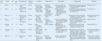
In addition to the two cases presented, five cases of congenital salivary fistula of an accessory parotid gland have been previously documented (Table I).Reference Gadodia, Seith, Sharma and Thakar4–Reference Hah, Kim, Sung and Kim8 The prominent clinical sign in all cases was a punctate skin orifice lateral to the commissure of the lips, with salivary discharge. This condition usually causes only minor cosmetic and functional morbidity, and patients may delay their treatment until their teenage years. Although the diagnosis of Goldenhar syndrome had not been clearly presented in previous articles, tragal deformityReference Gadodia, Seith, Sharma and Thakar4, Reference Moon, Han, Kim, Sung, Chang and Choo5, Reference Hah, Kim, Sung and Kim8 and parotid gland malformationsReference Moon, Han, Kim, Sung, Chang and Choo5 have been documented, which are considered important markers for Goldenhar syndrome.Reference Miller and Metry14
Reported cases of congenital salivary fistula of accessory parotid gland

*Patient refused surgery. No = number; y = years; ext = external; M = male; L = left; lat = lateral; CT = computed tomography; ant = anterior; F = female; R = right; +ve = positive; approx = approximately
Mandibular hypoplasia on the ipsilateral side was notable in the present two cases, and contributed to diagnosis. In our second patient, a duplication deformity of Stensen's duct revealed developmental disorder of the main parotid gland duct system. Thus, we suggest that congenital salivary fistula should be included in the spectrum of disorders which make up Goldenhar syndrome, due to its origin from an abnormal accessory parotid gland.
Computed tomography assisted fistulography and sialography can identify abnormal duct systems and aid in both the diagnosis and clinical management of such patients. From our experience and our review of the literature, the fistulous tracts do not usually communicate with the main parotid gland system.
• Congenital salivary fistula from an accessory parotid gland has been described in Goldenhar syndrome
• This study reports two further cases
• This abnormality should be added to the description of this syndrome
• Such cases often have deformity of the tragus, Stensen's duct, the main parotid gland and the mandibular ramus
• Computed tomography fistulography and sialography are valuable for diagnosis
• These patients' fistulous tracts did not communicate with the Stensen's duct system
As congenital salivary fistula of an accessory parotid gland is a very rare clinical entity, there is a paucity of data on its treatment. Surgical excision of the lesion should be radical to prevent recurrence, and should be performed with aesthetic considerations, as patients are usually children or adolescents. We recommend surgical excision of the accessory parotid gland, using an intra-oral approach, and of the fistula and skin orifice, via a small skin excision, in order to maximise both therapeutic and aesthetic outcomes. Translocation of the fistula to the oral cavity using Delore's method has also been documented: a tunnel is made through the cheek and the orifice is implanted into the oral buccal mucosa.Reference Yamasaki, Tashiro and Watanabe6 The more conservative treatment of chemocauterisation with botulinum toxin has also been reported to have a satisfactory outcome.Reference Hah, Kim, Sung and Kim8