Pseudoaneurysm of the ascending aorta is a rare late complication of ascending aortic surgery.Reference Kouchoukos, Wareing, Murphy and Perrillo 1 It is well reported in the literature following graft replacement of the ascending aorta in adults.Reference Dossche, Schepens, Morshuis, de la Rivière, Knaepen and Vermeulen 2 Mortality of pseudoaneurysm surgical repair has been reported to be as high as 43%.Reference Dawson, Clarke and Banning 3 Percutaneous closure using a septal occluder device has been previously reported in adults.Reference Bashir, Quaife and Caroll 4 – Reference Kanani, Neilan, Palacios and Garasic 6 We report a novel percutaneous-transthoracic technique using an Amplatzer occluder device (AGA Medical Corporation, Golden Valley, Minnesota, United States of America) for closure of an ascending aortic pseudoaneurysm in a 9-year-old Lebanese girl diagnosed with relapsing polychondritis and Takayasu's arteritis.
Case report
A 7-year-old Lebanese girl with relapsing polychondritis was hospitalised for management of malignant pyoderma gangrenosum. She was started on intravenous steroids with partial improvement of her skin lesions. Diagnostic evaluation with computed tomography angiography revealed a large right axillary aneurysm compressing the right brachial nerve plexus, as well as aneurysmal dilation of the ascending aorta, right common carotid, and right subclavian arteries. Transthoracic echocardiography revealed marked dilation of the ascending aorta (aortic root = 3.6 cm, z-score = 6), mild aortic insufficiency, and normal systolic function. She underwent resection of the ascending aortic aneurysm and replacement by a 20 mm interposition Dacron graft. Pathology findings were consistent with Takayasu's arteritis.
Our patient developed significant cushingoid features secondary to several months of steroid therapy. Therefore, she was shifted to infliximab therapy with marked improvement in her skin lesions and decreasing frequency of her relapsing polychondritis symptoms. However, over the next 2 years, she developed progressive aneurysmal dilation involving the transverse arch and the thoracic descending aorta. At the age of 9 years, she presented to the emergency room with sudden onset of severe respiratory distress. She was intubated and haemodynamically stabilised. Computed tomographic angiography (Fig 1a–c) showed evidence of a pseudoaneurysm measuring 5.2 × 2.7 × 5.4 cm arising from the anterior wall of the ascending aorta at the level of the proximal suture line of the Dacron graft, and causing significant narrowing of the right pulmonary artery, the lower trachea, and proximal bronchi at the same level.
( a ) Three-dimensional view of the pseudoaneurysm (P) arising from the ascending aorta (AA). ( b ) Computed tomographic angiogram view of the P in the coronal section. ( c ) Computed tomographic angiogram view in the transverse section showing the P arising from the AA with the neck of the defect (arrow) visualised.

Owing to the condition of the patient and the extremely high risk of surgical intervention, and after discussion of the risks and benefits of the traditional operative repair with the family, we decided to proceed with a high-risk attempt of closing the pseudoaneurysm with an Amplatzer septal occluder device after a consent form was obtained.
The procedure
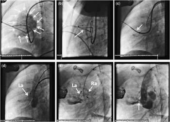
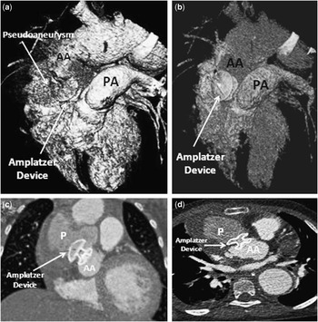
Bilateral femoral arterial access was obtained. Angiography revealed marked dilation of the ascending and thoracic descending aorta, and abdominal aorta. The large pseudoaneurysm arising from the ascending aorta measured 9.5 mm at its orifice. A 5 Fr Judkins right catheter was advanced into the large pseudoaneurysm and a 0.035′′ exchange wire (AGA Medical Corporation, Golden Valley, Minnesota, United States of America) was advanced through the Judkins catheter into the pseudoaneurysm. Difficulties were encountered to advance and position the Amplatzer device sheath over the exchange wire into the pseudoaneurysm. Therefore, the pseudoaneurysm was entered percutaneously through the anterior chest wall. Under fluoroscopy and ultrasound guidance, a needle was inserted and placed into the pseudoaneurysm (Fig 2a). Over a guide wire that was advanced through the needle, a 4 Fr sheath was introduced percutaneously and placed into the pseudoaneurysm. A snare was advanced through the 4 Fr sheath and snared the exchange wire that was introduced from the aorta into the pseudoaneurysm achieving a femoral arterial-pseudoaneurysm-transthoracic loop (Fig 2b) that was exteriorised to provide a rail along which the Amplatzer device delivery sheath was easily advanced into the pseudoaneurysm (Fig 2c). Owing to the fact that the pseudoaneurysm orifice measured 9.5 mm in its largest dimension based on computed tomography and angiography, we decided to utilise a 12 mm Amplatzer atrial septal defect occluder device (AGA Medical Corporation), which was delivered with the distal (left atrial) disc into the pseudoaneurysm and the proximal (right atrial) disc into the aortic side (Fig 2d and e). The delivery cable tracked smoothly without difficulty. Post-deployment ascending aorta angiogram revealed almost complete occlusion of the pseudoaneurysm with a tiny leak and no obstruction along the left ventricular outflow tract (Fig 2f). Following the deployment of the Amplatzer device, the 4 Fr percutaneous sheath was removed and haemostasis was achieved with no complications. Subsequent computed tomographic angiography (Fig 3a–c) confirmed the position of the device occluding the pseudoaneurysm. There was no evidence of bleeding following the procedure with stable serial haemoglobin levels.
( a ) Left lateral angiographic projection showing that the pseudoaneurysm (arrows) entered percutaneously through the anterior chest wall and a guidewire crossing from the aorta into the pseudoaneurysm. ( b ) A transthoracic snare retrieves the guidewire (arrow) from the pseudoaneurysm. ( c ) Femoral arterial-pseudoaneurysm-anterior chest wall loop is achieved and a rail is provided to deliver the ASD Amplatzer delivery sheath (arrow). ( d ) and ( e ) Amptatzer atrial septal defect occluder device delivered with left atrial (La) disc in the pseudoaneurysm and the right atrial (Ra) disc in the aorta. ( f ) Left lateral angiogram of ascending aorta revealing occlusion of the pseudoaneurysm with a small residual leak (arrow).

Post Amplatzer device deployment computed tomography angiography confirming the position of the device between the pseudoaneurysm (P) and the ascending aorta (AA). ( a ) and ( b ) Three-dimensional views. ( c ) Coronal view. ( d ) Transverse view. PA = pulmonary artery.

The patient developed progression of multi-organ system failure 1 week after the procedure, which was unrelated to the procedure, and that eventually progressed to cardiopulmonary arrest and death.
Discussion
Takayasu's arteritis is a chronic large-vessel arteritis commonly involving the aorta and its major branches.Reference Numano, Okawara, Inomata and Kobayashi 7 It is a rare disease in children younger than 10 years of age.Reference Bolin, Moodie, Fraser, Guirola, Warren and Eldin 8 Pseudoaneurysms of the ascending aorta may occur in 1–6% of patients following aortic reconstruction surgery.Reference Mulder, van Bockel, Maas, van den Akker and Hermans 9 These false aneurysms, as seen in our case, usually arise from the proximal suture line of the composite graft.Reference Dawson, Clarke and Banning 3 Treating these pseudoaneurysms is necessary as they may undergo rupture, fistulisation, or thrombosis. Traditionally, surgery has been the treatment of choice.Reference Mulder, van Bockel, Maas, van den Akker and Hermans 9 However, these re-do operations carry a high risk of morbidity and mortality, which may reach up to 43%.Reference Dawson, Clarke and Banning 3 Transcatheter approach using septal occluder device is well described in the literatureReference Bashir, Quaife and Caroll 4 – Reference Kanani, Neilan, Palacios and Garasic 6 as an alternative to surgery in selected patients with multiple comorbidities. Our patient had severe Takayasu's disease with an overall poor general physical condition for traditional surgical repair. Transcatheter occluder device closure of an abdominal aortic pseudoaneurysm has been previously described in a 15-year-old child by Li et al.Reference Li, Kou and Chen 10
To the best of our knowledge, the use of septal occluder device to exclude pseudoaneurysms from the ascending aorta has not been described before in the paediatric population. We also report the novel utilisation of a transthoracic approach to create a femoral arterial-pseudoaneurysm-anterior chest wall loop, which allows easier deployment and stabilisation of the delivery sheath into the pseudoaneurysm. Our patient is the youngest to be reported using septal occluder device exclusion of an ascending aortic pseudoaneurysm and the first in the literature through a percutaneous femoral arterial-transthoracic approach.
Although we have experienced success in exclusion of the pseudoaneurysm by the septal occluder device with no immediate complications, practice of this approach shall be considered with extreme caution and only in selective cases where surgical option carries a high risk of morbidity and mortality.
Conclusion
Percutaneous femoral arterial-transthoracic exclusion of ascending aortic pseudoaneurysms using a septal occluder device is a novel approach that might be considered as an alternative to surgery in selective cases of the paediatric population.
Disclosure
The authors have nothing to disclose with regard to commercial support.